File:Bryce1908 plate08.jpg

Original file (1,047 × 1,200 pixels, file size: 151 KB, MIME type: image/jpeg)
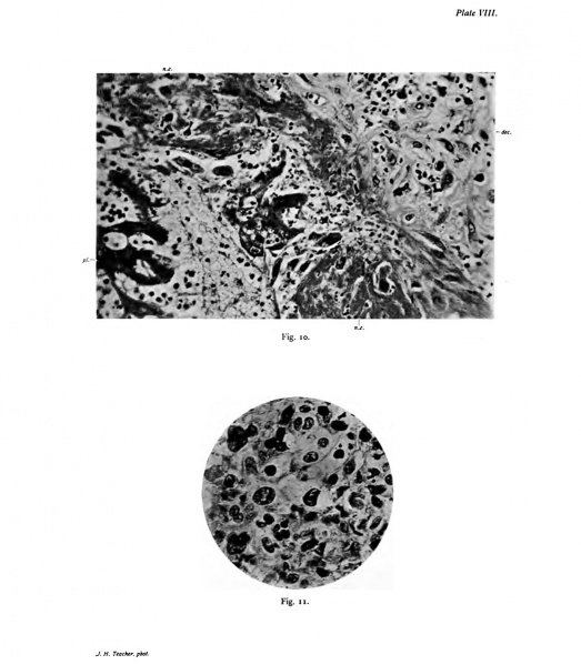
Plate 8.
Fio. 10. Section showing a Mass of Vacuolated Plasmodium Invading the Decidua. Photograph. X 350 D. pL, Plasmodium; dec., decidua; n.^., necrotic zone of decidua.
The mass of plasmodium shows a number of small vacuoles. It lies in a l)ay of the necrotic zone, and between the two are several large free, probably maternal, cells. the decidua is crowded with leucocytes. The decidual cells adjoining the necrotic zone show the early stages of degeneration ; further out they are more normal.
Fig. 11. Tangential Section of the Opposite Pole of the Cyto-trophoblastic Sphere from that figuied in Fig. 7, Plate VII. Photograph. x 350 d.
The nuclei are very irregular. The well-defined central cells belong to the innermost layer of the cyto-trophoblast.
- Bryce 1908 Human Ovum: Plate 1 | Plate 2 | Plate 3 | Plate 4 | Plate 5 | Plate 6 | Plate 7 | Plate 8 | Plate 9 | Plate 10
| Historic Disclaimer - information about historic embryology pages |
|---|
 |
File history
Click on a date/time to view the file as it appeared at that time.
| Date/Time | Thumbnail | Dimensions | User | Comment | |
|---|---|---|---|---|---|
| current | 08:50, 3 November 2013 | | 1,047 × 1,200 (151 KB) | Z8600021 (talk | contribs) |
You cannot overwrite this file.
File usage
The following page uses this file: