File:Bryce1908 plate04.jpg
Original file (997 × 1,200 pixels, file size: 156 KB, MIME type: image/jpeg)
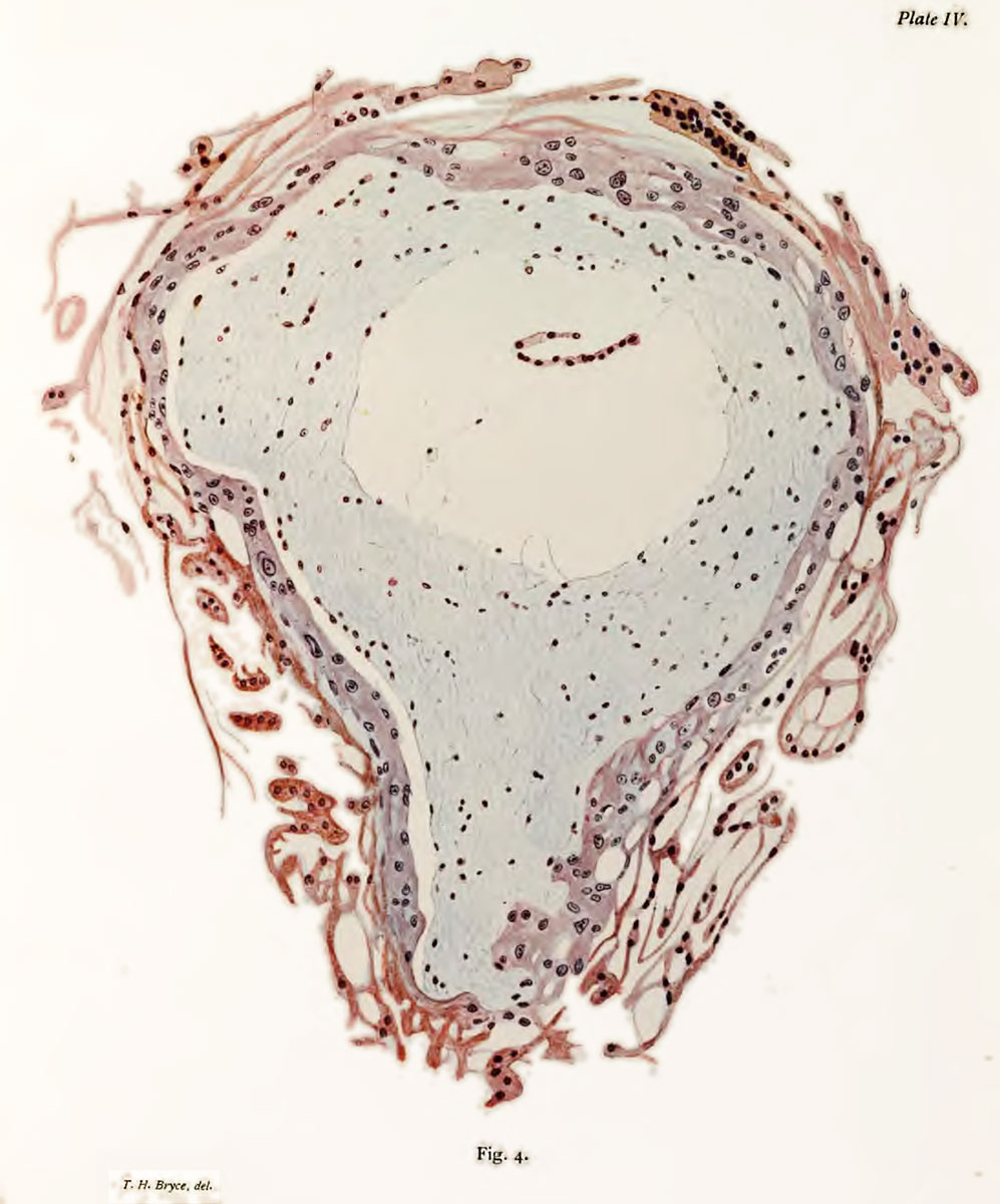
Plate 4. Section of Blastocyst
Fig. 4. Section of Blastocyst. x200 d.
The cyto-trophoblast appears as a blue-pink lamella with irregular nuclei. Directly continuous with it are strands of plasmodiuni ; only the central strands of the plasmodium are represented. The mesoblast is shown as a mass of mucous tissue. The delicate blue reticulum which forms its base is largely a precipitation product ; in it are numerous small rounded or spindle-shaped cells, which form a very loose syncytial tissue. The mesoblast has shrunk away from the cyto-trophoblast on the left, and in the centre is the retraction cavity containing a portion of the torn amnio-embryonic vesicle.
- Bryce 1908 Human Ovum: Plate 1 | Plate 2 | Plate 3 | Plate 4 | Plate 5 | Plate 6 | Plate 7 | Plate 8 | Plate 9 | Plate 10
| Historic Disclaimer - information about historic embryology pages |
|---|
 |
File history
Click on a date/time to view the file as it appeared at that time.
| Date/Time | Thumbnail | Dimensions | User | Comment | |
|---|---|---|---|---|---|
| current | 08:25, 3 November 2013 | | 997 × 1,200 (156 KB) | Z8600021 (talk | contribs) | ==Plate 4.== {{Historic Disclaimer}} |
You cannot overwrite this file.
File usage
The following page uses this file: