File:Brown022.jpg
From Embryology
Size of this preview: 468 × 599 pixels. Other resolution: 625 × 800 pixels.
Original file (625 × 800 pixels, file size: 93 KB, MIME type: image/jpeg)
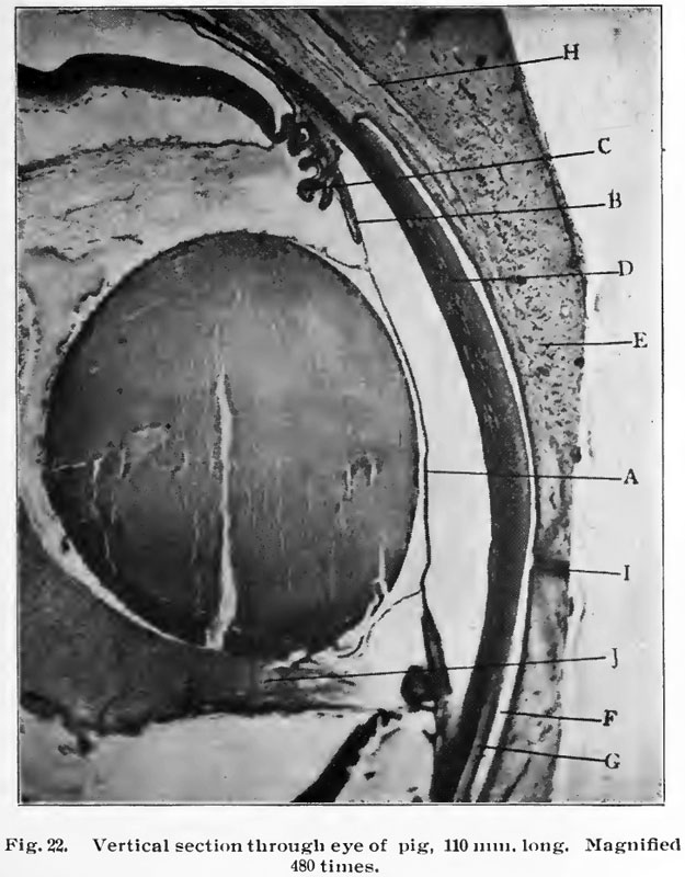
Fig. 22. Vertical section through eye of pig 110 mm long
Magnified 480 times.
- A - shows the pupilary membrane stretching across the pupilary space, and in it may be seen Httle white areas. These are the branches of the hyaloid artery which furnishes the nutrition to the lens during its development, and it will be remembered that this artery atrophies before birth and that the pupilary membrane disappears, ostensibly being absorbed.
- B - is shown the iris growing out from the ciliary bodies.
- C and D - shows the cornea and in it is shown the lacuna (small lakes), which are minute openings between the layers of the lamina propria (proper layer)
- E - shows the lid with its developing structures.
- F - shows the conjunctival sack.
- G - shows the ocular conjunctiva and just back of it the anterior portion of Tenon's space.
- H - shows the levator palpebra superioris (the lifter of the upper lid).
- I - shows the lids held together by the cement substance.
- J - shows the vitreous body (glass-like body).
| Historic Disclaimer - information about historic embryology pages |
|---|
 |
Reference
Brown EJ. The embryology anatomy and histology of the eye. (1906) Chicago: Hazlitt & Walker.
Cite this page: Hill, M.A. (2024, April 26) Embryology Brown022.jpg. Retrieved from https://embryology.med.unsw.edu.au/embryology/index.php/File:Brown022.jpg
- © Dr Mark Hill 2024, UNSW Embryology ISBN: 978 0 7334 2609 4 - UNSW CRICOS Provider Code No. 00098G
File history
Click on a date/time to view the file as it appeared at that time.
| Date/Time | Thumbnail | Dimensions | User | Comment | |
|---|---|---|---|---|---|
| current | 15:14, 14 February 2011 | | 625 × 800 (93 KB) | S8600021 (talk | contribs) | {{Template:Brown 1906 Figures}} Category:Pig |
You cannot overwrite this file.
File usage
The following 2 pages use this file: