File:Berkeley1905 fig05.jpg
Original file (1,000 × 1,029 pixels, file size: 172 KB, MIME type: image/jpeg)
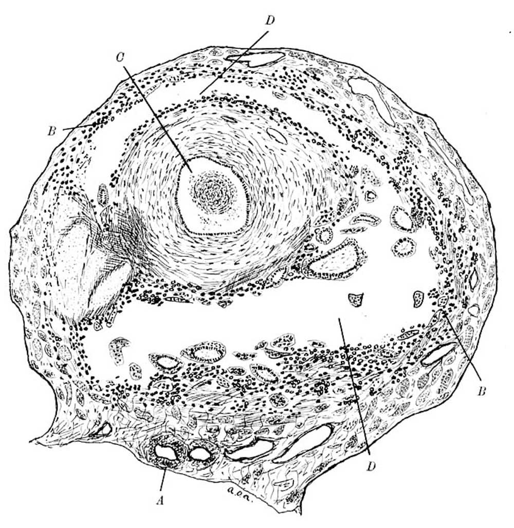
Fig. 5. Transverse section of the pregnant tube
A. Mesoaalpinx. B. B. “Cell Sheet " C. Lumen of Tube diatended,nnd containing a mu: of fibrin. D. D. Gestation uc.
Fig. 5 represents a transverse section of the pregnant tube about 1 mm. nearer the uterus.
The gestation sac is now large, and is seen to be surrounding the circular muscle coat in a very remarkable way.
It is evident that the ovum has split the longitudinal coat from the circular coat and is lying between them.
The wall of the ac is composed of fibrinised muscle tissue deeply infiltrated with trophoblast cells.
Within the cavity lie many villi, covered by-the usual double layer of epithelium. At a few spots this can be seen to -be continuous with the layers of trophoblast cells, which constitute the “cell-sheet.”
No villi are found amongst the maternal tisues.
Reference
Berkeley C. and Bonney V. Tubal Gestation - A Pathological Study. (1905) Brit. J. Obst. and Gyn. 7(2): 78-96.
Cite this page: Hill, M.A. (2024, April 26) Embryology Berkeley1905 fig05.jpg. Retrieved from https://embryology.med.unsw.edu.au/embryology/index.php/File:Berkeley1905_fig05.jpg
- © Dr Mark Hill 2024, UNSW Embryology ISBN: 978 0 7334 2609 4 - UNSW CRICOS Provider Code No. 00098G
File history
Click on a date/time to view the file as it appeared at that time.
| Date/Time | Thumbnail | Dimensions | User | Comment | |
|---|---|---|---|---|---|
| current | 10:50, 5 November 2017 | | 1,000 × 1,029 (172 KB) | Z8600021 (talk | contribs) | |
| 10:50, 5 November 2017 | 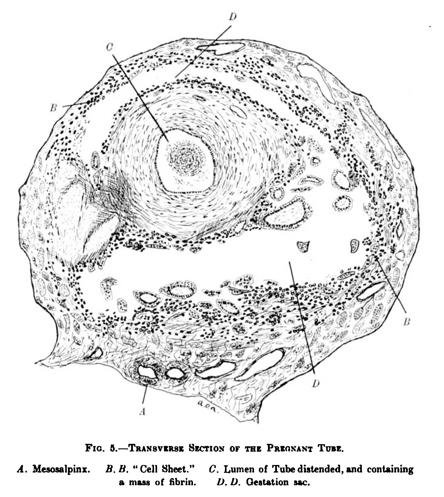 | 1,470 × 1,646 (293 KB) | Z8600021 (talk | contribs) |
You cannot overwrite this file.
File usage
The following page uses this file: