File:Axolotl brain ventricular zone proliferative activity.jpg

Original file (815 × 1,000 pixels, file size: 192 KB, MIME type: image/jpeg)
Axolotl brain ventricular zone proliferative activity
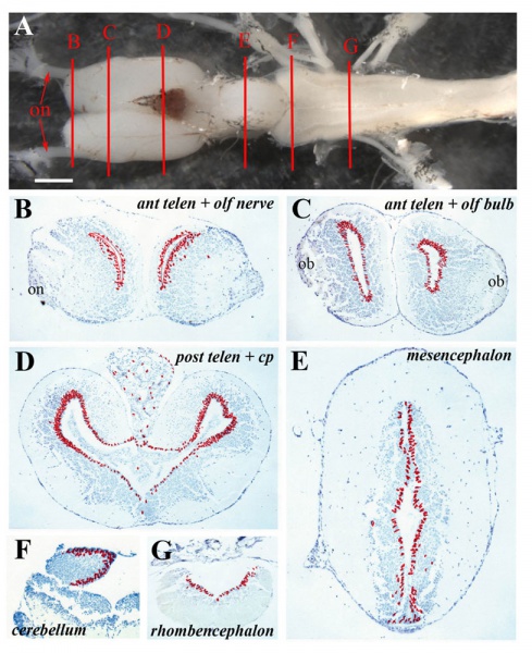
Ventricular zones with variable proliferative activity can be detected throughout the axolotl central nervous system.
A Structure of the axolotl brain showing the levels at which sections ventricular zone proliferation was analyzed.
B = anterior telencephalon and olfactory nerve, C = anterior telencephalon and olfactory bulb, D = posterior telencephalon, E = mesencephalon, F = cerebellum, G = rhombencephalon; on: olfactory nerve. Scale bar = 1 mm.
B – G Cumulative BrdU+ cell numbers on five sections from each level depicted in A on which individual labeled cells are marked with a red dot.
Regions of the brain are marked.; on: olfactory nerve; ob: olfactory bulb; cp: choroid plexus.
- Links: Axolotl Development
Reference
<pubmed>23327114</pubmed>
Copyright
© 2013 Maden et al.; licensee BioMed Central Ltd.
This is an Open Access article distributed under the terms of the Creative Commons Attribution License (http://creativecommons.org/licenses/by/2.0), which permits unrestricted use, distribution, and reproduction in any medium, provided the original work is properly cited.
Figure 1. 1749-8104-8-1-1-l.jpg
File history
Click on a date/time to view the file as it appeared at that time.
| Date/Time | Thumbnail | Dimensions | User | Comment | |
|---|---|---|---|---|---|
| current | 00:12, 26 November 2013 |  | 815 × 1,000 (192 KB) | Z8600021 (talk | contribs) | ==Axolotl brain ventricular zone proliferative activity== Ventricular zones with variable proliferative activity can be detected throughout the axolotl central nervous system. (A) Structure of the axolotl brain showing the levels at which sections ven... |
You cannot overwrite this file.
File usage
The following page uses this file: