File:Autophagy types.jpg
Original file (1,000 × 625 pixels, file size: 57 KB, MIME type: image/jpeg)
Types of Autophagy and their Morphology
Information from ASCB
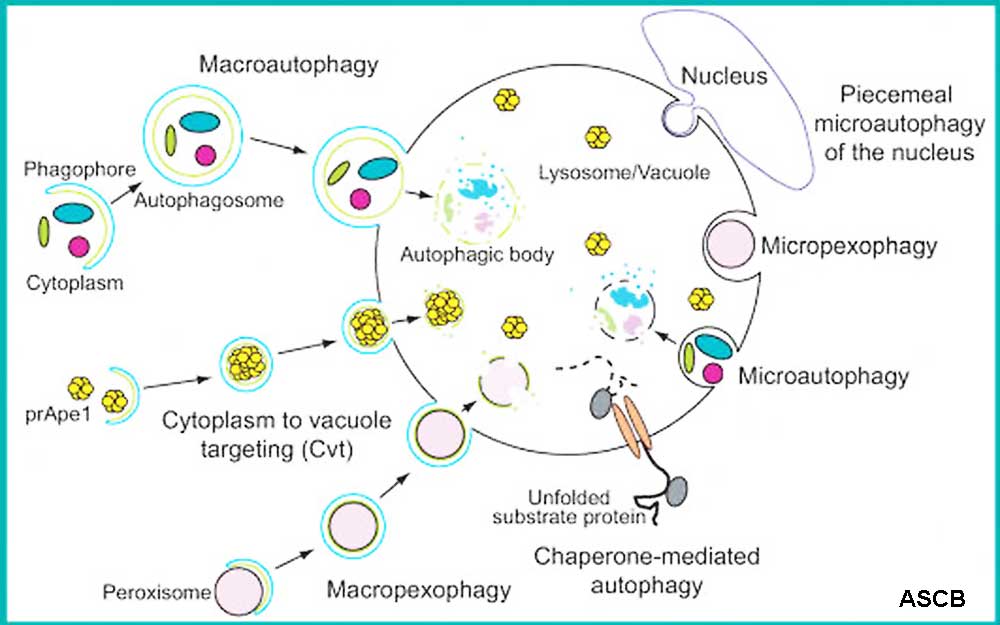
Autophagy is a process of self-eating at the subcellular level. It is defined as a collection of processes typically involving degradative delivery of a portion of the cytoplasm to lysosomes or the plant or fungal vacuole that does not involve direct transport through the endocytic or vacuolar protein sorting, Vps, pathways.
The three principle types of autophagy are macroautophagy, microautophagy and chaperone-mediated autophagy.
During nonspecific macroautophagy, cytoplasm is sequestered within cytosolic vesicles, and delivered to the lysosome or vacuole where it is degraded and the resulting macromolecules released back into the cytosol for reuse. This process begins with the formation of the phagophore, the initial sequestering structure. In yeast, the phagophore may be nucleated at the phagophore assembly site or pre-autophagosomal structure (PAS). This is the site where most of the autophagic protein machinery, the autophagy-related (Atg) proteins reside, at least transiently. The phagophore expands (http://cellimages.ascb.org/u?/p4041coll12,351; http://cellimages.ascb.org/u?/p4041coll12,326), probably by vesicular addition, to form a double-membrane autophagosome (http://cellimages.ascb.org/u?/p4041coll12,311). The outer membrane of the autophagosome fuses with the lysosome or vacuole limiting membrane. In fungi and plants in particular, the large size of the vacuole allows the release of the inner single-membrane of the autophagosome into the organelle lumen, where it is now termed an autophagic body (http://cellimages.ascb.org/u?/p4041coll12,307). This inner vesicle is broken down by vacuolar hydrolases to allow access to the cargo, which is typically degraded. The resulting macromolecules are then released back into the cytosol for reuse (http://cellimages.ascb.org/u?/p4041coll12,331).
In mammalian cells, the phagophore (http://cellimages.ascb.org/u?/p4041coll12,330) can be detected by indirect immunofluorescence or immunoelectron microscopy using antisera to HsAtg18/WIPI-1 or to the tagged protein (http://cellimages.ascb.org/u?/p4041coll12,321; http://cellimages.ascb.org/u?/p4041coll12,322; http://cellimages.ascb.org/u?/p4041coll12,323). Mammalian autophagy clearly converges with endocytosis and the autophagosome may fuse with an endosome or multivesicular body (http://cellimages.ascb.org/u?/p4041coll12,328; http://cellimages.ascb.org/u?/p4041coll12,334; http://cellimages.ascb.org/u?/p4041coll12,336); this may also be the case in fungi, but the endocytic compartments are less well defined. Subsequent of an autophagosome or an amphisome with the lysosome generates an autolysosome, sometimes referred to as an autophagolysosome (http://cellimages.ascb.org/u?/p4041coll12,329; http://cellimages.ascb.org/u?/p4041coll12,327).
In contrast to macroautophagy, microautophagy involves direct uptake at the surface of the lysosome/vacuole, although it still requires the same protein machinery. During microautophagy, the limiting membrane sequesters cytoplasm by invagination, protrusion and/or septation. During microautophagy in fungi, the Atg proteins localize to the sequestering arms of the vacuole, and to the micropexophagic membrane apparatus (MIPA). The MIPA (http://cellimages.ascb.org/u?/p4041coll12,312) is a specialized structure that may be critical in the final step of microautophagy to allow scission of the sequestering membrane from the limiting membrane of the vacuole.
Chaperone-mediated autophagy is only characterized in mammalian cells. This process involves direct translocation of unfolded proteins across the lysosome membrane through the action of the cytosolic and lysosomal chaperone hsc70, and the lysosomal membrane protein LAMP-2A (http://cellimages.ascb.org/u?/p4041coll12,342;http://cellimages.ascb.org/u?/p4041coll12,332).
Specific and non-specific autophagy
Macroautophagy was first characterized as a non-specific response to the hormone glucagon in mammalian cells, and to starvation in yeast. Macroautophagy occurs at a basal level, allowing a routine turnover of cytoplasm that is critical in maintaining normal physiology. In addition to non-specific macro- and microautophagy, however, there are many types of specific autophagy. For example, one role of autophagy is to enable cellular remodeling in response to changing nutrient conditions; autophagy is the primary method for organelle turnover. The best example of selective organelle degradation is seen with the autophagic removal of peroxisomes, termed pexophagy. When fungi are grown on carbon sources that require peroxisome function, these organelles proliferate. If the cells are then shifted to alternate carbon sources the peroxisomes are rapidly and specifically degraded. This turnover can occur through either micro- (http://cellimages.ascb.org/u?/p4041coll12,310) or macropexophagy. Other examples of selective organelle degradation include mitophagy (http://cellimages.ascb.org/u?/p4041coll12,315; http://cellimages.ascb.org/u?/p4041coll12,308) and piecemeal microautophagy of the nucleus (http://cellimages.ascb.org/u?/p4041coll12,296; http://cellimages.ascb.org/u?/p4041coll12,348; http://cellimages.ascb.org/u?/p4041coll12,297). The cytoplasm to vacuole targeting (Cvt) pathway is the only characterized autophagy-like biosynthetic process that uses the Atg proteins to selectively deliver resident hydrolases to the yeast vacuole (http://cellimages.ascb.org/u?/p4041coll12,313; http://cellimages.ascb.org/u?/p4041coll12,309).
Health and disease
By allowing cells to adapt to changing conditions, autophagy plays an important role in development (http://cellimages.ascb.org/u?/p4041coll12,347). Autophagy is also involved in various aspects of health and disease. For example, autophagy function in tumor suppression, but can also be used by cancer cells to protect them against certain anti-cancer treatments. Basal and possibly induced autophagy protect against some types of neurodegeneration (http://cellimages.ascb.org/u?/p4041coll12,293; http://cellimages.ascb.org/u?/p4041coll12,294; http://cellimages.ascb.org/u?/p4041coll12,295), but autophagosomes may also provide a site for the formation of some types of toxic amyloid proteins. Another function of macroautophagy is seen in the immune response. For example, some types of microbes are sequestered within autophagosomes and subsequently delivered to the lysosome where they are killed (http://cellimages.ascb.org/u?/p4041coll12,306; http://cellimages.ascb.org/u?/p4041coll12,298; http://cellimages.ascb.org/u?/p4041coll12,346; http://cellimages.ascb.org/u?/p4041coll12,345; http://cellimages.ascb.org/u?/p4041coll12,335). On the other hand, some microbes have evolved to evade autophagy (http://cellimages.ascb.org/u?/p4041coll12,299; http://cellimages.ascb.org/u?/p4041coll12,300; http://cellimages.ascb.org/u?/p4041coll12,301; http://cellimages.ascb.org/u?/p4041coll12,302; http://cellimages.ascb.org/u?/p4041coll12,303; http://cellimages.ascb.org/u?/p4041coll12,304; http://cellimages.ascb.org/u?/p4041coll12,305) or even to subvert this process to provide a replicative niche. The role of autophagy in myopathies and in lysosomal storage diseases is less clear because much of the work has relied on tissue samples, making it unclear whether autophagy is induced as a cytoprotective response, or if excessive autophagy or the accumulation of autophagosomes contributes to the symptoms of the disease (http://cellimages.ascb.org/u?/p4041coll12,338; http://cellimages.ascb.org/u?/p4041coll12,339; http://cellimages.ascb.org/u?/p4041coll12,340). Macroautophagy and chaperone-mediated autophagy decline with age, and one hypothesis is that this leads to a decline in longevity. For example, lifespan may in part be affected by the accumulation of oxidatively damaged proteins and organelles (http://cellimages.ascb.org/u?/p4041coll12,344).
Title - Autophagy 1. Introduction
Author(s) - Daniel J Klionsky
Materials & Methods - Generated using Adobe Illustrator.
References
Klionsky DJ, Emr SD. Autophagy as a regulated pathway of cellular degradation. Science [serial online]. 2000;290:1717-1721. Available with subscription at: http://www.sciencemag.org/cgi/content/full/290/5497/1717
Reggiori F, Klionsky DJ. Autophagy in the eukaryotic cell. Euk Cell [serial online]. 2002;1:11-21. Available at: http://ec.asm.org/cgi/content/full/1/1/11
Shintani T, Klionsky DJ. Autophagy in health and disease: A double-edged sword. Science [serial online]. 2004;306:990-995. Available with subscription at: http://www.sciencemag.org/cgi/content/full/306/5698/990
Yorimitsu T, Klionsky DJ. Autophagy: Molecular machinery for self-eating. Cell Death Diff [serial online]. 2005;12:1542-1552. Available at: http://www.nature.com/cdd/journal/v12/n2s/full/4401765a.html
Klionsky DJ. The molecular machinery of autophagy: Unanswered questions. J Cell Sci [serial online]. 2005;118:7-18. Available at: http://jcs.biologists.org/cgi/content/full/118/1/7
Diagram originally appeared in: Huang J, Klionsky DJ. Autophagy and human disease. Cell Cycle [serial online]. 2007;6:1837-1849. Available with subscription at: http://www.landesbioscience.com/journals/cc/article/4511 Citation Klionsky DJ. Autophagy 1. Introduction. ASCB Image & Video Library. 2008 CYT-123. Available at: http://cellimages.ascb.org/u?/p4041coll12,314 Click the link to rate and comment on this resource. (Rate the resource from 1 (high) to 5 (low) and type your comments after the rating).
Reference
Section Editor: Daniel J Klionsky (University of Michigan), Autophagy Section Editor Publication Date - May 2008 Resource Type - Still image; diagram Digital Format - jp2 Original Date: 5/25/2007 Original Format eps
Copyright
Terms for non-commercial use: http://cellimages.ascb.org/cdm4/terms.php Publisher: The American Society for Cell Biology
© [2007 Daniel J Klionsky]. All rights reserved. Reprinted under license from The American Society for Cell Biology.
File history
Click on a date/time to view the file as it appeared at that time.
| Date/Time | Thumbnail | Dimensions | User | Comment | |
|---|---|---|---|---|---|
| current | 13:49, 8 May 2018 | | 1,000 × 625 (57 KB) | Z8600021 (talk | contribs) |
You cannot overwrite this file.
File usage
The following 2 pages use this file: