File:Atwell1926 textfigA.jpg
Original file (869 × 700 pixels, file size: 191 KB, MIME type: image/jpeg)
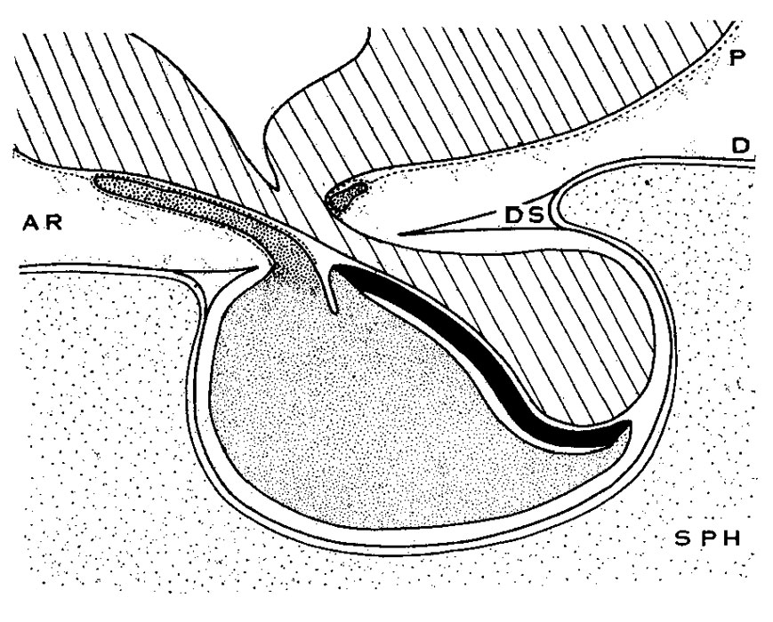
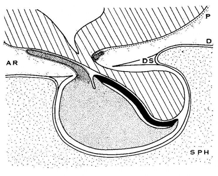
Text fig. A Diagrammatic sagittal section of hypophysis
Showing relation of pars tuberalis to the meninges. Text figure A represents the relations of the hypophysis to the dura mater and of the pars tuberalis to the pia mater. No attempt has been made to represent the pia inside the sella, either according to the theoretical View advocated by Steudell and others or according to the recent view of Hughson.
Lined, brain floor and pars neuralis; fine stipple, pars anterior proprior; coarse stipple, pars tnberalis; solid black, pars interniedia. SPH, sphenoid bone; P, pia lnator; D, (lura; DS, diaphragma sellae; AR, arachnoid spaces.
| Historic Disclaimer - information about historic embryology pages |
|---|
 |
Reference
Atwell WJ. The development of the hypophysis cerebri in man, with special reference to the pars tuberalis. (1926) Amer. J Anat. 37: 139-193.
Cite this page: Hill, M.A. (2024, April 26) Embryology Atwell1926 textfigA.jpg. Retrieved from https://embryology.med.unsw.edu.au/embryology/index.php/File:Atwell1926_textfigA.jpg
- © Dr Mark Hill 2024, UNSW Embryology ISBN: 978 0 7334 2609 4 - UNSW CRICOS Provider Code No. 00098G
File history
Click on a date/time to view the file as it appeared at that time.
| Date/Time | Thumbnail | Dimensions | User | Comment | |
|---|---|---|---|---|---|
| current | 16:59, 9 November 2016 | | 869 × 700 (191 KB) | Z8600021 (talk | contribs) | |
| 16:58, 9 November 2016 | 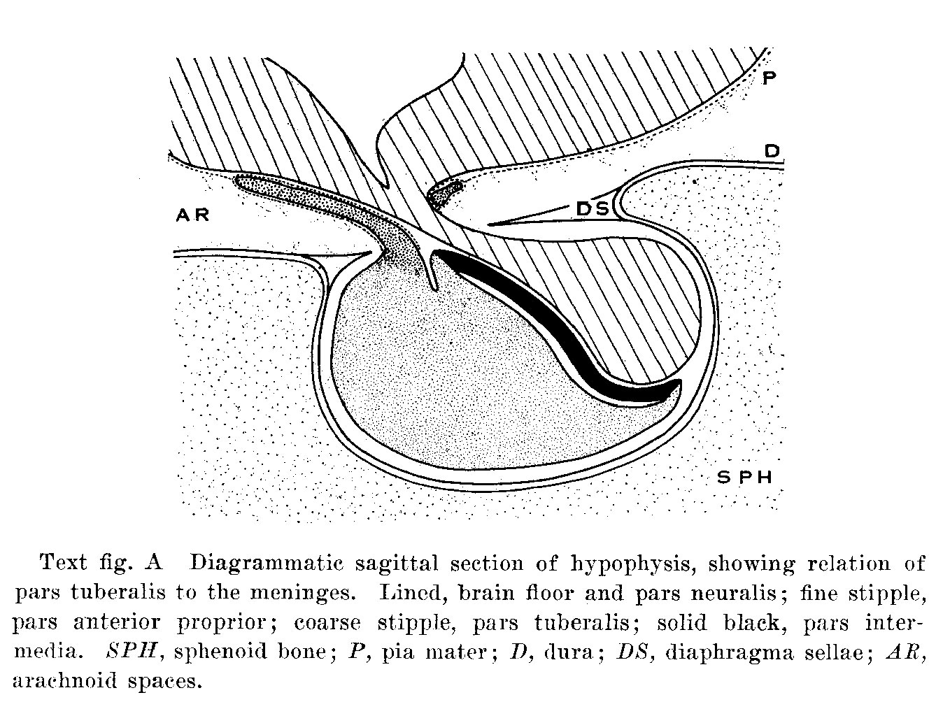 | 1,338 × 1,024 (308 KB) | Z8600021 (talk | contribs) | ==Text fig. A Diagrammatic sagittal section of hypophysis== Showing relation of pars tuberalis to the meninges. Lined, brain floor and pars neuralis; fine stipple, pars anterior proprior; coarse stipple, pars tnberalis; solid black, pars interniedia... |
You cannot overwrite this file.
File usage
The following page uses this file: