File:Altschule1930 fig03.jpg
From Embryology
Size of this preview: 513 × 600 pixels. Other resolution: 1,000 × 1,169 pixels.
Original file (1,000 × 1,169 pixels, file size: 98 KB, MIME type: image/jpeg)
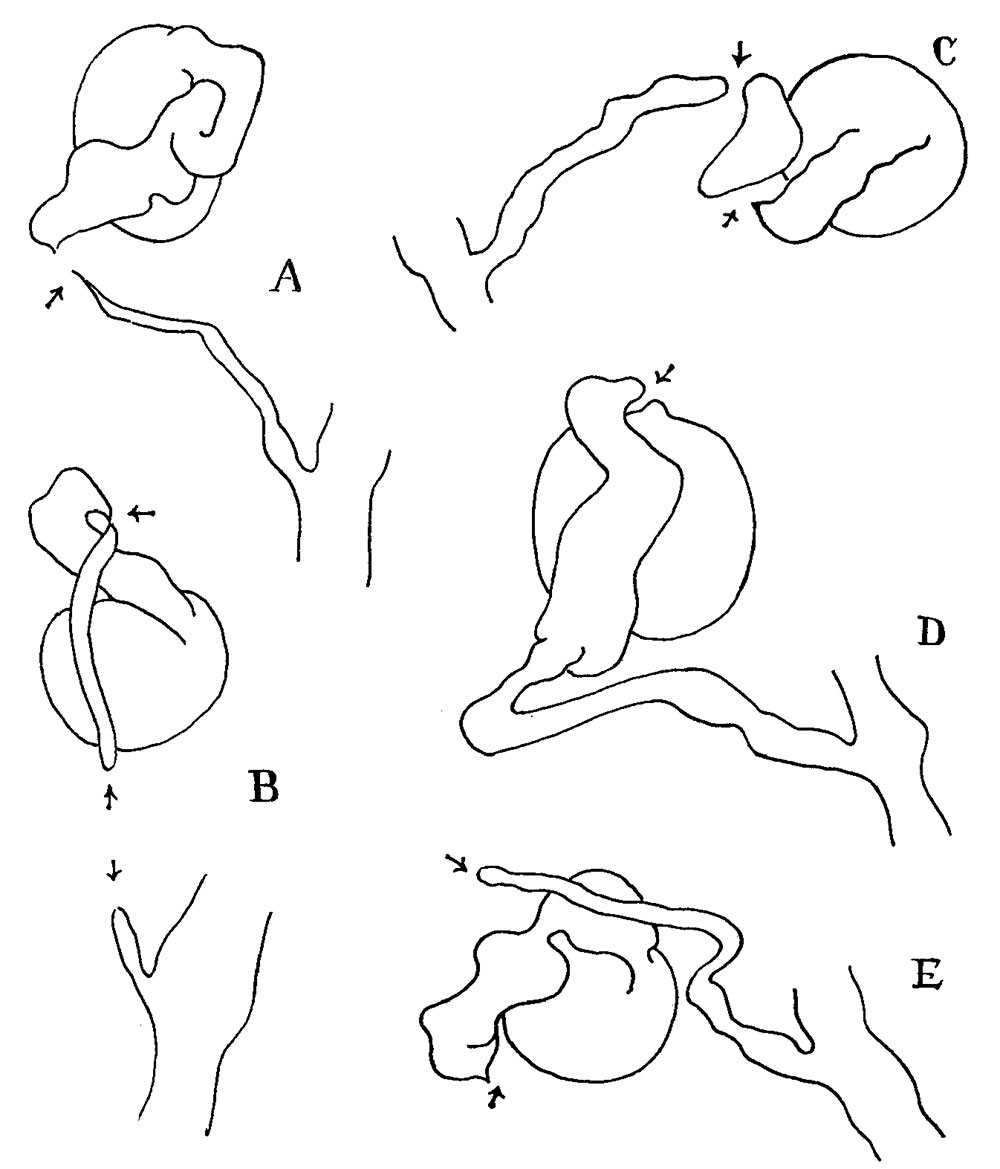
Fig. 3 Reconstructions of broken mesonephric tubules
A and E are broken immediately below the junction of secreting and collecting tubules. B is reduced to :2. lumenless cord at the junction of the secreting and collecting tubules. In addition, the collecting tubule itself is broken somewhat lower down. C is broken at the junction of the secreting and collecting tubules as well as in the secreting tubule itself. D is broken at the junction of the glomerulus and secreting tubule.
File history
Click on a date/time to view the file as it appeared at that time.
| Date/Time | Thumbnail | Dimensions | User | Comment | |
|---|---|---|---|---|---|
| current | 13:39, 26 February 2017 | | 1,000 × 1,169 (98 KB) | Z8600021 (talk | contribs) | |
| 13:39, 26 February 2017 | 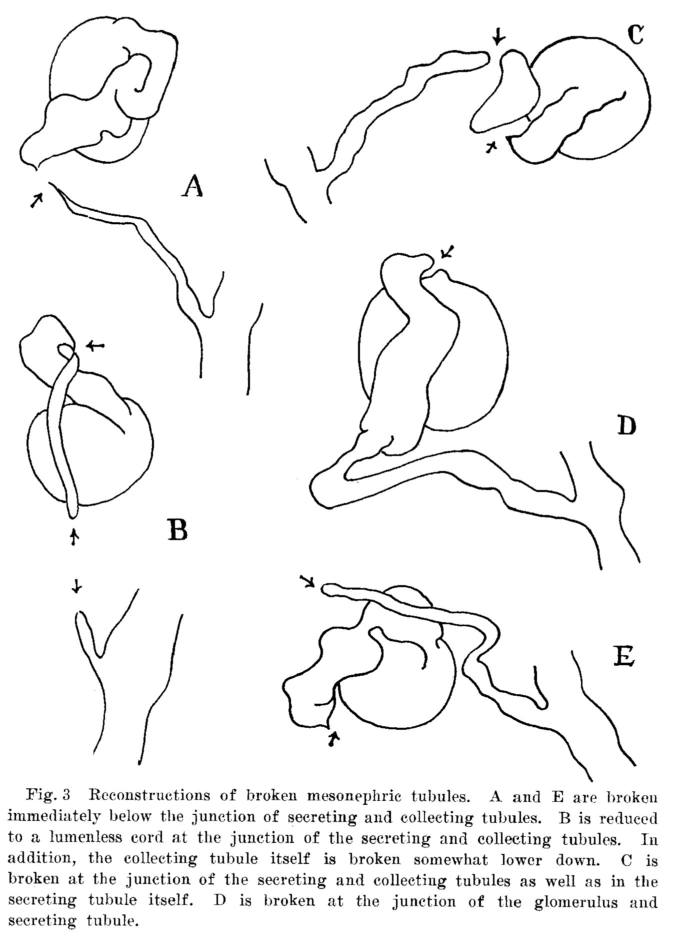 | 1,346 × 1,874 (272 KB) | Z8600021 (talk | contribs) |
You cannot overwrite this file.
File usage
The following page uses this file: