Book - The Nervous System of Vertebrates (1907) Figures
| Embryology - 19 Apr 2024 |
|---|
| Google Translate - select your language from the list shown below (this will open a new external page) |
|
العربية | català | 中文 | 中國傳統的 | français | Deutsche | עִברִית | हिंदी | bahasa Indonesia | italiano | 日本語 | 한국어 | မြန်မာ | Pilipino | Polskie | português | ਪੰਜਾਬੀ ਦੇ | Română | русский | Español | Swahili | Svensk | ไทย | Türkçe | اردو | ייִדיש | Tiếng Việt These external translations are automated and may not be accurate. (More? About Translations) |
Johnston JB. The Nervous System of Vertebrates. (1907) Blakiston's Son & Co., London.
| Historic Disclaimer - information about historic embryology pages |
|---|
| Pages where the terms "Historic" (textbooks, papers, people, recommendations) appear on this site, and sections within pages where this disclaimer appears, indicate that the content and scientific understanding are specific to the time of publication. This means that while some scientific descriptions are still accurate, the terminology and interpretation of the developmental mechanisms reflect the understanding at the time of original publication and those of the preceding periods, these terms, interpretations and recommendations may not reflect our current scientific understanding. (More? Embryology History | Historic Embryology Papers) |
List of Illustrations
1. Outlines of the spinal cord with the dorsal and ventral roots in Petromyzon and in a mammal 14
2. The brain of Heptanchus 15
3. The medulla oblongata of the lake sturgeon (Acipenser rubi cundus), to show the longitudinal zones 16
4. Two views of the brain of the buffalo fish, Carpiodes velifer (Raf.); (i) from above; (2) from the right side. From C. Judson Herrick after C. L. Herrick 17
5. Simple diagrams of the branchial nerves of lower vertebrates as seen from the left side 19
6. A diagram of the lateral line canals and pit organs together with the nerves which supply them, in a ganoid fish (Amia calva). After E. Phelps Allis. 21
7. A sketch of the brain of Chimaera monstrosa from the left side to show especially the position of the nerve roots 23
8. The outline of the brain ventricles as seen from above; A, of a cyclostome fish, Lampetra Wilderi; B, of a selachian, Mustelus canis; C, of a young specimen of a bony fish, Coregonus albus; D, of a tailed amphibian, Necturus maculatus 25
9. A diagram of one side of the forebrain in Mustelus to show what is believed to be the primitive relations of the wall and ventricle 26
10. The outline of the ventricles in man 26
11. The mesial surface of the right half of the brain of a selachian, Squalus acanthias ' ' 29
12. A sketch of the brain of a cyclostome fish, Lampetra Wilderi, as seen from the left side 3
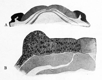
13. Sections of the neural plate and folds in amphibia, Amblystoma tigrinum and A. punctatum
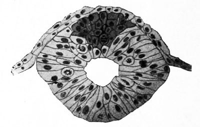
14. Transverse section of the neural tube of Amblystoma puncta tum just after closing
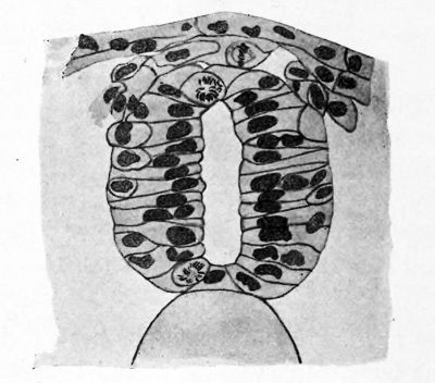
15. Transverse section through the neural tube, neural crest and ectoderm of Amblystoma at a later stage than that shown in Fig. 14
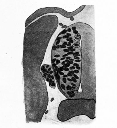
16. Same as Fig. 15, later stage
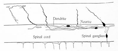
17. A part of the spinal cord of an i8-day Catostomus embryo, showing the giant ganglion cells
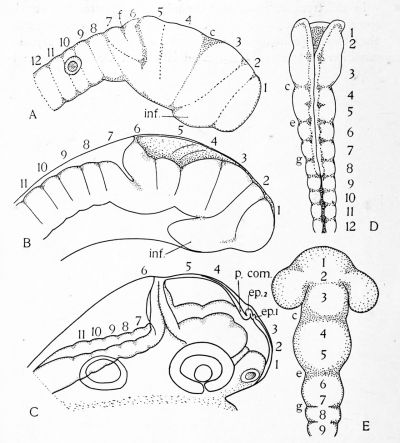
18. Figures of the brain of a selachian embryo (Squalus acanthias) to show the history of the neuromeres. After Locy.
19. Figures to show the history of the neuromeres in a bony fish and in the chick. After Hill
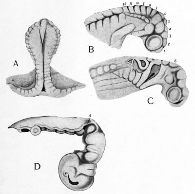
20. The brain of a pig embryo of 12 mm. from the right side. From Minot as drawn and revised by Dr. F. T. Lewis. 41
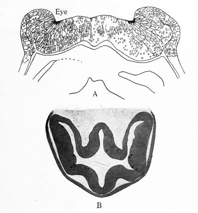
21. Transverse sections through the region of the optic vesicle in embryos of: A, Torpedo ocellata; B, Gallus domesticus; C, Cavia cobaya. After Froriep
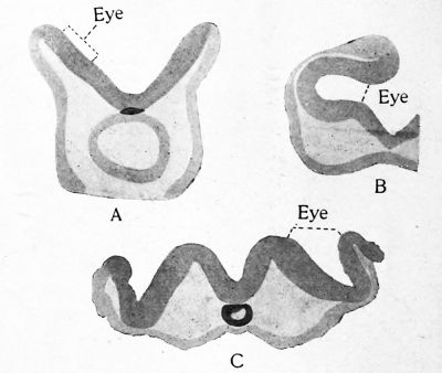
22. The development of the optic vesicles in Amblystoma punc tatum
23. A part of a transverse section of the inferior lobe of the stur geon, stained by the Golgi method 47
24. A horizontal section of the nucleus praeopticus of the sturgeon, by the Golgi method 48
25. A part of a transverse section of the optic lobe of the sturgeon, by the Golgi method 49
26. The ganglion of the IX nerve in Amblystoma punctatum at the time of formation of the central processes 51
27. A few cells of the trigeminal ganglion of Amblystoma puncta tum with the fibers of the ramus mandibularis growing out from them 51
28. A, a diagram of the head of Petromyzon at a stage when the neural crest is segmented into the anlages of the cranial ganglia. B, a similar diagram at a later stage when all the cranial nerves are to be recognized. After Koltzoff. 52
29. Three diagrams of the head of Squalus acanthias to show the differentiation of the neural crest into the cranial ganglia. After Neal 53
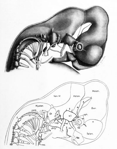
30. A reconstruction of the peripheral nerves in a four weeks human embryo, 6.9 mm. long. From Streeter 54
31. Reconstruction of peripheral nerves in four weeks human embryo, 7.0 mm. long. From Streeter 55
32. Reconstruction of peripheral nerves in six weeks human em bryo, 17.5 mm. long. From Streeter 56
33. Three stages in the development of the acustico-lateral system in the sea bass. From H. V. Wilson 57
34. A transverse section through the nasal sac of an embryo of a bony fish at about the time of hatching, to show the origin of the fibers of the olfactory nerve 61
35. Two figures representing the formation of unipolar cells in the spinal ganglion of the dog embryo. After Van Gehuchten 62
36. A median sagittal section of the head of an embryo Ambly stoma punctatum 67
37. Diagrams representing the development of the buccal cavity, hypophysis and nasal pit in Amphioxus and Petromyzon. After Legros 68
38. A diagram representing the segmentation in a generalized vertebrate head 71
39. Several types of nerve cells from the central and peripheral nervous system of vertebrates 78
40. A unipolar nerve cell from the brain of Carcinas to illustrate Bethe's experiment 80
41. Diagrams intended to show several forms of reflex chains in the nervous system of vertebrates 83
42. A portion of the subepithelial nervous network in the palate of the frog. From Prentiss 86
43. A portion of the nervous network about the walls of a small vessel in the palate of the frog. From Prentiss 87
44. Two ganglion cells of the nervous network in the intestinal wall of the leech Pontobdella, showing neurofibrillae passing through the cells. From Bethe after Apathy 88
45. A diagram of the component elements in the spinal cord and the nerve roots of a trunk segment, to illustrate the relations of the four functional divisions of the nervous system 97
46. Diagrams to represent the extent and arrangement of the functional divisions in the brain of a selachian 100
47. A diagram to show the arrangement of the two afferent divis ions in the brain of man 101
48. General cutaneous endings in the skin of a cyclostome, Lam petra Wilderi 105
49. A diagrammatic representation of the general cutaneous com ponents of a trunk segment 106
50. A simple diagram of the general cutaneous components in the cranial and spinal nerves of a fish 107
51. A reconstruction of the cranial nerves of Petromyzon dorsatus to show the arrangement and distribution of the several systems of nerve components 108
52. The principal sensory collaterals in the spinal cord of the new-born rat. From Cajal 109
53. Some cells of the dorsal horn in the chick embryo of five days. From Cajal in
54. Transverse section of the substance of Rolando of the cervical cord in the new-born cat. From Cajal 112
55. Cells in the dorsal horn of the cord in a chick embryo of nineteen days incubation. From Cajal 113
56. Transverse section through the spinal V tract and the sub stance of Rolando in a new-born rabbit. From Cajal 114
57. A, transverse section of the spinal cord of Lampetra to show the ceils of the dorsal horn 115
58. A, transverse section of the tuberculum acusticum of Lam petra; B, a sagittal section of the cerebellum of the same animal 116
59. A diagram representing the centers and fiber tracts related to the general cutaneous components in fishes 118
60. A diagram representing the general cutaneous centers and fiber tracts in the human brain 120
61. A large and a small neuromast from a sucker embryo at about the time of hatching 124
62. A diagram of the lateral line canals and nerves in Amia. After E. Phelps Allis 125
63. A reconstruction of the chief rami of the cranial nerves in a bony fish, Menidia, to show the arrangement of the several systems of components. After C. Judson Herrick 129
64. Transverse section of the brain of the sturgeon at the level of the VII and VIII nerves 131
65. Transverse section of the brain of the sturgeon at the level of the V nerve 132
66. Transverse section of the brain of Scy Ilium to show the fold ing of the cerebellar crest and tuberculum acusticum 133
67. Transverse section of the acusticum of the sturgeon to show acusticum cells and a Purkinje cell 134
68. A section through the same region as in Fig. 67, to show cell forms intermediate between the acusticum and Purkinje cells. 136
69. A diagram to show the central endings of the special cutaneous components in fishes 138
70. A diagram to show the central ending of the vestibular and cochlear nerves and of the optic tract in man and the chief secondary tracts related to them 140
71. A, a section through the retina of a mouse embryo of 15 mm.;
B, a section through the retina of a chick embryo of fourteen days incubation; C, a section of the retina of a dog. From Cajal 144
72. Cells from the retina of the chicken. From Cajal 145
73. The tectum opticum of the sturgeon 147
74. A section of the optic lobe of a bird. From Cajal 149
75. A series of diagrams intended to illustrate the origin and mode of formation of the optic vesicle in vertebrates 151
76. A sketch showing the relations of the two epiphyses in verte brates 152
77. A diagrammatic representation of the general visceral sensory components in a trunk segment 155
78. A transverse section through the region of Clarke's column of the thoracic cord of a new-born dog. From Cajal 156
79. A reconstruction of the cranial nerve components in a tailed amphibian, Amblystoma. After Coghill 158
80. A simple diagram of the visceral nerves of the head in fishes. 159
81. Four transverse sections through the medulla oblongata of the frog to show the position and ending of the fasciculus communis and nucleus commissuralis 160
82. Transverse section through the medulla oblongata of a mouse at the level of the nucleus commissuralis. From Cajal . . 161
83. Transverse section through the medulla oblongata of a mouse four days old. From Cajal 162
84. A, a transverse section through the medulla oblongata of the sturgeon at the level of the X nerve; B, at the level of the IX nerve 163
85. Sense organs of bony fishes. A, a taste bud from the oesopha gus of Catostomus at the time of hatching; B, a taste bud from the pharynx of the same embryo; C, two neuromasts from the skin of the same embryo 165
86. A taste organ from the pharynx of the ammocoetes of Petro myzon dorsatus 166
87. A taste bud from the skin of an adult Lampetra 167
88. A projection of the cutaneous branches of the communis root of the right facial nerve in a bony fish, Ameiurus. From C. Judson Herrick 168
89. A parasagittal section through the brain of the spotted sucker, Minytrema melanops, to show the gustatory centers and tracts. From C. Judson Herrick 170
90. A diagram of the gustatory paths in the brain of the carp as seen from the left side. From C. Judson Herrick . . . . 172
91. Part of a sagittal section of the brain of a newly hatched bony fish 173
92. A diagram representing the centers and tracts related to the visceral sensory components in fishes 174
93. A transverse section through the olfactory epithelium of a bony fish at the time of hatching 177
94. An oblique section through the forebrain of Lampetra . . . 178
95. A horizontal section through the olfactory bulb of the sturgeon 179
96. A spindle cell and two granules from the olfactory bulb of the sturgeon 180
97. An olfactory glomerulus from the brain of the sturgeon . . 181
98. Cells with short neurites in the olfactory bulb. From Cajal. 182
99. A transverse section of the forebrain of the sturgeon at the level of the anterior commissure 184
100. An outline of the median sagittal section of the forebrain of Lampetra 185
101. A diagram of the olfactory conduction paths in the sturgeon. 187
102. A diagrammatic representation of the somatic motor compo nents of a trunk segment 191
103. An early stage in the formation of the pectoral fin and brachial plexus in a selachian, Spinax. After Braus ... 192
104. The formation of the cervical plexus in a selachian, Hexan chus. After Fiirbringer 193
105. A transverse section through the nucleus of origin of the III nerve in Lampetra 195
106. A diagrammatic representation of the visceral efferent com ponents in a trunk segment 199
107. Diagram to show the central relations of the IX, X and XI nerves in mammals according to Onuf and Collins . . . 201
108. A diagram of the sympathetic system and the arrangement of its neurones in a mammal. Chiefly after Huber . . . 209
109. Diagram illustrating the spinal representation of the sympa thetic nerve in a mammal. From Onuf and Collins ... 210 no. A diagram to illustrate Langley J s " axone reflex " .... 213 in. Tract cells in the spinal cord of the trout. After Van Ge huchten 219
112. The relations of the cerebellum, brachium conjunctivum and gustatory tracts in selachians (Scyllium) 226
113. Transverse section through the cerebellum of the sturgeon . 227
114. Transverse section through the cerebellum and secondary gustatory nucleus of the sturgeon 228
115. Transverse section of the brain of the sturgeon at the junc tion of cerebellum and midbrain 229
116. Transverse section through the midbrain of the sturgeon . . 230
117. The relations of the cerebellum, brachium conjunctivum and gustatory tracts in a ganoid fish (Acipenser) . . . . 231
118. A, the left lateral aspect of the brain of a pouch specimen of Dasyurus viverrinus; B, median sagittal section through the cerebellum of the same brain. After G. Elliot Smith . 232
119. A diagram representing the fundamental and more constant secondary fissures of the mammalian cerebellum spread out in one plane. After G. Elliot Smith 234
120. The mesial surface of the right half of the brain of Squalus acanthias 236
121. A diagrammatic transverse section of one fold of the cere bellum. From Koelliker 238
122. Net-like fiber ending in the human cerebellum. After Cajal. 241
123. Two schemes to show the course of impulses in the cerebellar cortex. From Cajal 242
124. Two transverse sections through the cerebellum of Scyllium. 244
125. A transverse section through the cerebellum of Necturus . . 246
126. A transverse section through the deep gray nuclei of the cerebellum of man 2 47
127. Outline transverse sections through the mesencephalon of a cyclostome, a 'selachian, a ganoid, a bony fish, an amphibian and a mammal 255
128. Transverse section of the diencephalon of the rat at the level of the body of Luys. From Cajal 257
129. Transverse section of the dorso-mesial portion of the posterior corpus quadrigeminum of the new-born dog. From Cajal. 259
130. Transverse section of the anterior corpus quadrigeminum of the rabbit of eight days. From Cajal 260
131. Lower part of the corpus geniculatum laterale of a new-born cat. From Cajal 262
132. Nucleus of posterior commissure in Lampetra 266
133. Transverse section through the corpora mammillaria of the sturgeon 270
134. Transverse section through the inferior lobes of the sturgeon. 272
135. Transverse section through the posterior commissure of the sturgeon 273
136. Scheme of the connections of the mammillary tracts, nucleus habenulae and the nucleus dorsalis thalami. From Cajal. 274
137. Sagittal section of the tuber cinereum of a new-born rat. From Cajal 275
138. Sagittal section of the tuber cinereum of the rat of eight days. From Cajal 276
139. A scheme to show the embryological relations of the nucleus habenulae and the inferior lobes in fishes 277
140. Nucleus habenulae of the sturgeon 278
141. Transverse sections through the nucleus habenulae and the facial lobe of a young Amia . . . . 279
142. Transverse section of the habenular nuclei in the dog. From Cajal 280
143. The efferent tract to the saccus vasculosus in the sturgeon.. 283
144. A general scheme of the saccus apparatus 284
145. A diagram of the fiber tracts in the forebrain of a cyclostome (Lampetra) 294
146. An oblique section through the inferior lobes and forebrain of Lampetra 296
147. A diagram of the fiber tracts in the forebrain of a selachian (chiefly after Kappers) 299
148. A diagram of the fiber tracts in the forebrain of a bony fish (chiefly after Goldstein) 302
149. A transverse section of the brain of the sturgeon behind the anterior commissure 303
150. A diagram of the fiber tracts in the forebrain of a tailed amphibian (Necturus) 306
151. Simple diagrams to show the history of the epistriatum in fishes and amphibia 308
152. Sketches of transverse sections through (A) the caudal part of the epistriatum in amphibia and (B) the corresponding structure in Ornithorhynchus. B after G. Elliot Smith . 310
153. A sagittal section of the forebrain and interbrain of a chick embryo of 7.0 days. After Minot 312
154. Part of a transverse section of the cortex of a chameleon. After Cajal 313
155. Transverse section through the forebrain of Lacerta at the level of the anterior commissure. After Cajal 314
156. A transverse section through the right lateral lobe of the forebrain of Lacerta. After Cajal 315
157. A diagram of the mesial surface of the hemisphere of a reptile to show the extent of the hippocampus and related structures. 316
158. A mesial sagittal section of the brain of an embryo of Sphe nodon punctatum. From G. Elliot Smith 317
159. The mesial surface of the right half of the brain of Ornitho rhynchus. From G. Elliot Smith 318
1 60. The mesial surface of the cerebral hemisphere of a marsupial (Phascolarctos). From G. Elliot Smith 319
161. Portion of a transverse section through the brain of a Moni tor (Hydrosaurus). From G. Elliot Smith 320
162. Transverse section of the cerebral hemispheres of Ornitho rhynchus. From G. Elliot Smith 321
163. Plan of cerebral hemispheres, lamina terminalis and optic thalami in horizontal section. From G. Elliot Smith . . 322
164. Sagittal section of the commissural and precommissural regions of the hemisphere of Ornithorhynchus. From G. Elliot Smith 323
165. Ventral surface of the brain of Ornithorhynchus. After G. Elliot Smith 324
166. Transverse section of the brain of a rat of four days through
the anterior commissure. From Cajal 325
167. Diagram of the mesial surface of (A) the hemisphere of a monotreme and (B) that of a mammal to show the extent and relations of the hippocampus 326
168. Schemes to explain the expansion of the corpus callosum and the formation of the septum pellucidum. After figures by G. Elliot Smith 328
169. Scheme of cerebral commissures and the margin of the cor tex of the human brain. From G. Elliot Smith .... 330
170. Scheme of the structure and connections of the hippocampus. From Cajal 332
171. Structure of the cerebral cortex 339
172. Diagram showing the probable course of impulses in the cells of the cerebral cortex. After Cajal 340
173. Scheme of long association tracts in the cerebral cortex. After Cajal 342
174. Scheme of commissural and projection fibers of the cortex. After Cajal 343
175. The primordial areas in the cerebral hemispheres, lateral surface. From Flechsig 346
176. The primordial areas in the hemispheres, mesial surface. From Flechsig . . .' 347
177. The primordial areas and border zones of the association fields, lateral surface. From Flechsig 350
178. The primordial areas and border zones of the association fields, mesial surface. From Flechsig . . . 350
179. Diagram of lateral surface of hemisphere showing localiza tion of functions 351
180. Diagram of mesial surface of hemisphere showing localiza tion of functions 351
Johnston JB. The Nervous System of Vertebrates. (1907) Blakiston's Son & Co., London.
| Historic Disclaimer - information about historic embryology pages |
|---|
| Pages where the terms "Historic" (textbooks, papers, people, recommendations) appear on this site, and sections within pages where this disclaimer appears, indicate that the content and scientific understanding are specific to the time of publication. This means that while some scientific descriptions are still accurate, the terminology and interpretation of the developmental mechanisms reflect the understanding at the time of original publication and those of the preceding periods, these terms, interpretations and recommendations may not reflect our current scientific understanding. (More? Embryology History | Historic Embryology Papers) |
Cite this page: Hill, M.A. (2024, April 19) Embryology Book - The Nervous System of Vertebrates (1907) Figures. Retrieved from https://embryology.med.unsw.edu.au/embryology/index.php/Book_-_The_Nervous_System_of_Vertebrates_(1907)_Figures
- © Dr Mark Hill 2024, UNSW Embryology ISBN: 978 0 7334 2609 4 - UNSW CRICOS Provider Code No. 00098G

