Book - The Frog Its Reproduction and Development 13
| Embryology - 28 Apr 2024 |
|---|
| Google Translate - select your language from the list shown below (this will open a new external page) |
|
العربية | català | 中文 | 中國傳統的 | français | Deutsche | עִברִית | हिंदी | bahasa Indonesia | italiano | 日本語 | 한국어 | မြန်မာ | Pilipino | Polskie | português | ਪੰਜਾਬੀ ਦੇ | Română | русский | Español | Swahili | Svensk | ไทย | Türkçe | اردو | ייִדיש | Tiếng Việt These external translations are automated and may not be accurate. (More? About Translations) |
Rugh R. Book - The Frog Its Reproduction and Development. (1951) The Blakiston Company.
| Historic Disclaimer - information about historic embryology pages |
|---|
| Pages where the terms "Historic" (textbooks, papers, people, recommendations) appear on this site, and sections within pages where this disclaimer appears, indicate that the content and scientific understanding are specific to the time of publication. This means that while some scientific descriptions are still accurate, the terminology and interpretation of the developmental mechanisms reflect the understanding at the time of original publication and those of the preceding periods, these terms, interpretations and recommendations may not reflect our current scientific understanding. (More? Embryology History | Historic Embryology Papers) |
Chapter 13 - The Mesodermal Derivatives
The Epimere
(Segmental or Vertebral Plate)
The Somites
These dorsally located blocks of metamerically arranged mesoderm are differentiated from the anterior to the posterior. The first ones to be completed are located just posterior to the auditory capsule. A total of 13 pairs is formed from the head to the base of the tail. In the tail of the 6 mm. tadpole there may develop as many as 32 pairs of these transient somites, making a total of 45 pairs by the 6 mm. stage. The two most anterior or occipital pairs of somites become indistinguishable and the larval tail somites are lost during the process of metamorphosis. This leaves a total of 11 pairs of somites from which develop most of the striated muscles and the primary skeleton of the body. In the head and pharyngeal region the mesoderm is in the form of loose mesenchyme.
Each somite develops in a characteristic manner, having an outer thin layer of cells known as the dermatome or cutis plate and a mesial mass of cells known as the myotome or muscle segment. Between these is a cavity, the myocoel, which becomes elongated dorso-ventrally as the cutis plate extends to the body wall and appendages. Originally this myocoel is continuous with the splanchnocoel or body cavity. The myocoel eventually becomes obliterated. From the mesial margin of the somite, sclerotomal cells are proliferated off to take up their positions as a continuous skeletogenous sheath around the spinal cord and notochord, and between the segmental myotomes. This embryonic sheath gives rise to the cartilage and finally the bone of the axial skeleton. A total of nine anterior well-formed vertebrae develop in this manner, alternating with the myotomes. A tenth vertebra is modified into a urostyle or posteriorly elongated bony projection. The sclerotome of the two potential posterior vertebrae fuses to form the cartilage and then the bony urostyle which encloses the posterior end of the notochord.
The dermatome or cutis plate gives rise to the dermal layer of the dorsal and lateral skin, to connective tissue between the myotomes, and to the musculature of the limbs. The dermal layer of the ventral body wall arises from the somatopleure, so that while the dermis becomes continuous it originates from two sources.
The myotomes or muscle segments of the early larva arise as rather solid aggregations of cells, concentrated at the level of the spinal cord and notochord. They enlarge at the expense of the contained myocoels. Before the time of hatching, these myotomal cells become elongated and their muscle fibrillae orient their axes in the longitudinal direction of the embryo. This indicates a shift in the axes from the original direction in the early myotomes. The myotomes become separated from each other by septa or myocommata (connective tissue sheets). These are derived from the cutis plate, and assume a "V" shape with the apex of the "V" pointing posteriorly. These myotomes become the layer of striated or voluntary (voluntarily controlled) muscles of the back, limbs, and dorsal body wall. The fibers of many of these muscles are arranged ultimately in a variety of directions to provide for complete body control.
The muscles of the heart, blood vessels, and viscera arise from the hypomeric splanchnopleure and are known as smooth or involuntary muscles.
Limb buds develop from the accumulation of loose mesenchyme of adjacent somites surrounded by ectoderm. Within this blastema of somatic mesoderm there arise muscle and bone, developing from the body outwardly. The forelimb muscles develop before those of the hindlimbs, although the hindlimbs are the first to emerge during the process of metamorphosis.
The sclerotome arises at loose cells which are proliferated off from the ventro-medial portion of the myotome of the somite. They will give rise to the entire axial skeleton, as described below. By this time (5 mm. stage) the somites are entirely separate from the lateral plate mesoderm.
The Vertebral Column
The development of the notochord has been described already. Its origin cannot be attributed exclusively to any of the three germ layers. Its cells remain few in number, become flattened antero-posteriorly, and are highly vacuolated. The notochord then becomes surrounded by (1) the primary or elastic sheath derived from the notochordal cells, (2) the secondary or fibrous sheath, and (3) eventually an outer skeletogenous sheath which is of mesodermal origin. This latter connective tissue sheath encapsulates the nerve cord and also extends laterally between the myotomes by the 15 mm. stage. The notochord finally disappears in the frog. It is partially replaced by and partially converted into material of the centrum of each vertebra. Neural arches and transverse processes develop outwardly from the cartilaginized skeletogenous sheath.
The vertebral column of the frog consists of ten vertebrae of which the last is modified into an elongated rod-like bone known as the urostyle, mentioned above. The first vertebra is the cervical atlas, followed by seven abdominal vertebrae and finally the ninth or sacral vertebrae. The urostyle takes the place of the caudal vertebra. The skeletogenous sheath from the sclerotome encloses the spinal cord and notochord by the time of hatching, and by the 15 mm. stage some of this sheath has given rise to cartilage. It is within this cartilage that the vertebrae develop. The formation of this cartilage, due to the accumulation of the sclerotomal cells between the myotomes, is segmented in a manner which alternates with the developing muscle segments. However, at all levels the spinal cord and notochord will be enclosed in this double ring of connective tissue which is being transformed progressively into cartilage and finally into bone. Around the notochord it is known as the perichondrium and around the spinal cord as the vertebral cartilaginous arch.
Ossification begins, and the cartilage gradually is displaced by an inward growth of true bony cells known as osteoblasts. Eventually the notochord itself becomes invaded by these bone-forming cells and is transformed into the bony centrum of the vertebra. Each centrum becomes concave anteriorly, for the reception of the convex projection of the more anterior centrum. At the level of the lateral myotomal mass, connective tissue invades the notochord to form the intervertebral discs or ligaments of hyaline cartilage. Each disc splits into an anterior and posterior part, and each becomes ossified and later fuses with the corresponding part of the adjacent centrum. There are connecting ligaments which arise from the original sclerotome along both the dorsal and the ventral faces of the centra. They alternate in position with the myotomal segments.
The ossified cartilage which surrounds the spinal cord is known as the neural arch. Both the anterior and the posterior margins of each neural arch bear a pair of short zygapophyses which are processes that articulate and tend to join successive vertebrae. Spinal nerves emerge from the spinal cord through intervertebral foramina, between the sides of the succeeding neural arches.
Dorsal to each potential vertebra surrounding the spinal cord is a single median cone of ossification which becomes the spinous or neural process of the vertebra. The successive neural spines are connected by ligaments. The paired transverse and bony processes arise laterally, at right angles, from the cartilage of the centrum and become continuous with the minute bony ribs. No transverse processes or anterior zygapophyses are developed on the most anterior vertebra, the atlas. This vertebra is modified to articulate with the occipital condyles of the skull. The transverse processes of the ninth vertebra are elongated and are directed obliquely posterior to provide attachment for the ilium of the pelvic girdle. The tenth vertebra is greatly modified into the single tubular urostyle and is derived from the sclerotome (skeletogenous material) of the last two somites of the body.
The Skull
(Neurocranium of the Adult)
The skull of the adult frog, defined as that part of the skeleton which surrounds and supports the brain and special sense organs, is composed of relatively more cartilage than is the skull of most higher vertebrates. The cranial cartilages are an exception to the general rule as to origin, and are derived from ectodermal neural crests.
The frog skull represents a type intermediate between the skulls
of lower fishes and higher reptiles. It is made up of the cranium,
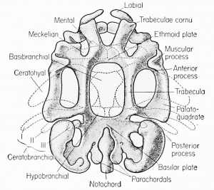
which arises from the cartilage and is therefore known as the chondrocranium; the sense capsules; a portion of the notochord; and a visceral skeleton, which includes the jaws and the hyoid apparatus and membrane and dermal bones. There are cartilage (endochondral) bones which arise by the ossification of cartilage and there are membrane (dermal) bones which develop from (dermis) superficial membranes without the intervention of a cartilage phase. These latter bones are sheet-like and can be stripped from the cartilage of the true cranium. The bony floor of the cranium arises largely from the notochord in conjunction with a pair of cartilaginous (sclerotomal) rods known as the parachordals at about the 7 mm. stage. These are arranged longitudinally on either side of the notochord with which they become fused to form the parachordal plate or floor of the cranium. The basilar plate is immediately articulated with the notochord and is united with the parachordals on either side. The cartilaginous elements of the skull appear by the time of hatching and form what is known as the chondrocranium. These elements are not displaced by bone until about the 30 mm. body length stage.
Anterior to the parachordal plate, and continuous with it, is another pair of rods of cartilage and then bones. These are the trabeculae (or trabecular cartilages) which join mesially to form the ethmoid or intranasal plate. The anterior space between the trabeculae is the basicranial fontanelle within whose cavity the infundibulum temporarily lodges.
The sense capsules are simultaneously formed. Each auditory capsule forms from mesenchyme which surrounds the developing inner ear. It consists of mesotic and occipital cartilages. The occipital cartilages form the side and roof of the auditory capsule and skull and the mesotic forms the floor. Between the occipital cartilages of the two sides is the large posterior opening for the spinal cord to the brain, known as the foramen magnum. Anteriorly the trabeculae form the orbital bones around the eyes. The anterior extremity of these cartilages are the trabecular cornu which form the bony olfactory capsules. Anteriorly they fuse with the labial or suprarostral cartilages.
Some of the parts of the cranium are covered by thin plates which originate in the dermis and not from the sclerotome and are therefore called dermal bones. Many of these cover open spaces of the chondrocranium and line the mouth. They appear very early and include the fronto-parietals, nasals, premaxillae, maxillae, quadratojugals, squamosals, parasphenoids, vomers, dentaries, and palatines. The main cartilage bones are the exoccipitals, prootics, stapes, ethmoids, parts of the pterygoquadrates, articulars, hyoids, branchials, and mento-Meckelians.
The Visceral Arches
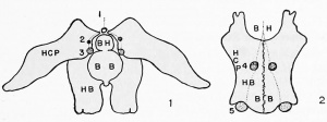
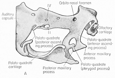
These arches give rise to the visceral skeleton (splanchnocranium) but arise as six pairs of vertical condensations of mesenchyme lateral to the embryonic pharynx. After the mouth opens each of these arches develops cartilage. The most anterior pair, the mandibular arches, give rise to the dorsal palatoquadrate bone and to the ventral Meckel's cartilage. The palatoquadrate (pterygoquadrate) cartilage arises from the maxillary (dorsal) portion of this first visceral arch and joins the posterior end of the trabeculae. The palato portion gives rise to the bulk of the upper jaw and the quadrate portion to the annulus tympanicus of the middle ear. The Meckel's cartilage gives rise to the core of the lower jaw or the dentary bone. The large ceratohyal cartilages appear posterior to the Meckelian cartilage in the hyomandibular arch. The basicranial (copula) is an unpaired cartilage found between the two large ceratohyals. The more anterior basihyal is slow to develop cartilage but can be identified by the U-shaped thyroid gland which appears directly ventral to it. The jaws are suspended to the skull by a cartilaginous bone which develops from membranes.
Portions of the second visceral (hyoid) arch give rise to the hyoid cornu. Parts of the third and fourth visceral arches (first and second
branchials) give rise (at the 9 mm. stage) to the plate-like hypobranchial cartilage apparatus of the adult. The four pairs of slender
ceratobranchials extend posteriorly from the hypobranchials. None
of the arches remain as such by the time of metamorphosis, the only
remnants being the derivatives just listed. The frog tadpole does not
possess true teeth, but does have ectodermally covered horny "jaws"
and so-called teeth. Oral papillae, appearing as teeth, constantly are
wearing away and bear no relation to the bones with which the
more functional teeth of the post-metamorphic frog will be associated.
At metamorphosis dermal papillae appear on the upper jaw, associated with the vomers, and are known as tooth germs. The overlying
epidermis is then involved in transforming the papillae into pseudoteeth, with a minute amount of dentin and enamel. The teeth are for
holding rather than for macerating living food, and never are developed as highly as in reptiles or mammals.
The Appendicular Skeleton
The pectoral girdle consists of the scapuia, coracoid, and precoracoid, the last two to be replaced by the clavicle. Except for the clavicle, which is dermal, this girdle arises from ossified cartilage that is calcified only at the base, and articulates with the elongated scapula. The epicoracoid (or precoracoid) and the xiphisternum remain as cartilage while the single sternum, paired coracoids, and single episternuni become ossified. The sternum arises from the fusion of a longitudinal pair of cartilages which never join the ribs but remain both anterior and posterior to the pectoral girdle. The humerus, or upper portion of the forelimb, articulates with the glenoid cavity of the pectoral girdle.
The pelvic girdle is a V-shaped fused mass of bone which supports the hindlimbs and is associated with the urostyle. The paired anterior ends of the pelvic girdle (ilium) are united with the enlarged transverse process of the ninth or sacral vertebra. Other bones of this girdle, arising from cartilage, are the ischium and pubis. The pubis alone does not ossify. The ilium forms a cup, the acetabulum, which receives the head of the femur of the hindlimb. The bones of both anterior and posterior girdles and limbs arise by the ossification of preformed cartilage (i.e., endochondral in origin). There is evidence that appendicular muscles of some amphibia may develop in situ from somatic mesoderm.
The Mesomere
(Intermediate Cell Mass)
Early in embryonic development the upper level of the lateral
plate mesoderm becomes separated off from the ventral sheets of
mesoderm, to give rise to the excretory and parts of the reproductive
systems. This is known as the intermediate cell mass or mesomere
because of its position relative to the other masses of mesoderm. It
is also known as the nephrotome because it gives rise to excretory
units, both the larval pronephros and the functional mesonephros of
the frog. The derivatives of this mesoderm will now be described;
the description of the development of the hypomere and its derivatives, the coelom and circulatory system, will be deferred until later
in the book.
The Pronephros or Head Kidney

The somatic wall of the nephrotomal region at the level of the second, third, and fourth somites thickens and projects laterally between the hypomere and the body ectoderm. This longitudinal bulge is known as the pronephric shelf, because it is due to the development of the pronephros or head kidney and can be seen readily from the exterior. This is noticed first from the external view at the tail bud stage (2.5 mm. body length) as the pronephric ridge or bulge just posterior to the gill plate. Within the lateral extensions of these nephrotomal masses there appear cavities, the nephrocoels, which run together longitudinally to form a common tube. This tube and its lumen grow posteriorly along the lateral border of the nephrotomes, to join the cloaca and be known as the paired pronephric or segmental ducts. Actually the duct is not in any way segmented, but it will receive segmental kidney tubules. Posterior to the fourth somite this duct develops independently of the nephrotomal tissue and becomes joined to the cloaca at the 4.5 mm. stage.
Three pairs of pronephric tubules, one at the level of each of the somites II, III, IV, appear between the segmental duct and the coelom. The original connection of the nephrotomes with the somites is lost in favor of a nephrogenic cord. Finally each tubule acquires an opening, the nephrostonie, into the adjacent coelom. This opening
shortly acquires cilia and becomes funnel-shaped, by the time it reaches the 5 mm. body length stage. The pronephric tubules lengthen and become coiled (convoluted) and at the 6 mm. stage are embedded in the mesenchyme and sinuses of the developing posterior cardinal vein. The entire pronephric mass is surrounded by the connective tissue of the pronephric capsule, derived from the adjacent somatic mesoderm and myotome.
Between each nephrotome and the dorsal aorta, suspended into the body cavity and surrounded by splanchnic mesoderm, a very rudimentary capillary network known as the glomus arises from the dorsal aorta. This appears to be an abortive attempt to develop a glomerulus characteristic of the adult mesonephric kidney, and occurs in response to inductive influences from the non-functioning pronephric tubules.
The pronephros is structurally much like the functional kidney of the frog, except that it is much less complex. It acquires a rich vascular supply, both arterial and venous, but has no outlet. It is doubtful that it ever functions as an excretory organ, but it attains its maximum development at about the 12 mm. stage. Along with the
glomus the elongated and coiled tubes of the pronephros constitute a formidable mass of tissue which projects into the dorsal body cavity and is surrounded largely by peritoneal epithelium. As the pronephric mass expands, it fills the posterior cardinal sinus and is bathed in venous blood. As the lungs enlarge and grow against the pronephros, the splanchnic mesoderm covering the lungs and the pronephros is brought together to form a trap or pocket which is merely a reduced
coelomic chamber into which the temporary nephrocoels open. This is the pronephric chamber. It remains open both anteriorly and posteriorly into the lung chamber. A pronephric capsule is formed by an overgrowth of myotomic mesenchyme and an upfolding of somatic mesoderm from the lateral plate. Thus a connective tissue sheath is formed around the embryonic head kidney.
By the 11 mm body length stage the pronephroi are large and conspicuous bodies with blind tubular outgrowths; but by the 20 mm. stage they have begun to degenerate and neither the tubules at this level nor the related glomi remain. The mesonephric kidney develops
rapidly and takes over the increasingly important excretory functions.
The Mesonephros
(Wolffian Body)
The nephrotomal mass posterior to the pronephros and mesial to the segmental duct gives rise to the mesonephros or Wolffian body which is the functional kidney of the adult frog. It extends from the level of somites VII through XII and begins to develop at the 8 to 10 mm. stage.
In this region, as in the more anterior regions, there are segmental nephrotomes which merge to form a pair of continuous nephrotomal masses from the seventh to the twelfth somites. The mesonephros is therefore of both somatic and splanchnic mesodermal origin. At the level of each of these somites there develop several nephrotomes, each with a separate nephrocoel. The nephrocoels at this level are known as the mesonephric vesicles which become convoluted and constricted so as to give rise to primary, secondary, and tertiary units, in that consecutive order. It is obvious, therefore, that the kidney units of the mesonephros are more complicated than those of the pronephros, from the very beginning of their formation. They are not actually metameric.
The primary mesonephric vesicle gives rise to a dorsal evagination which becomes the secondary unit of the mesonephric vesicle. Following this there is another evagination from the ventro-lateral side of the primary vesicle, toward the segmental duct with which it becomes continuous. This is known as the inner tubule of the tertiary mesonephric vesicle. It becomes greatly coiled and encroaches upon the developing and adjacent cardinal vein. The inner tubules become the tubular portion of the adult kidney. The segmental duct from this level posteriorly is known as the Wolffian or mesonephric duct, and this will remain as the excretory duct or ureter.
A third evagination, the outer tubule, develops ventrally from
the mesonephric unit. The cavity of this tubule joins the coelom by
way of the nephrostome. However, by the 20 mm. stage this unit has
broken away from the rest of the mesonephric unit and has changed
its connection from the mesonephros to the lateral division of the
cardinal vein. This vessel becomes the renal portal vein. This connection can be demonstrated very easily in the recently metamorphosed adult frog where persistent ciliated nephrostomes can be seen
on the ventral face of the kidneys and which can be shown to maintain direct connections with venous capillaries of the kidneys. The
outer tubule aids in forming Bowman's capsule around each glomerulus. The ciliated nephrostoine then arises along with the mesonephric
unit, develops a normal coclomic connection, and then shifts its relationship to become associated with blood sinuses of the posterior
cardinal vein. This occurs within the kidney, at about the 15 mm.
stage. The original nephrostomes remain in the adult frog on the
ventral face of the kidney as ciliated peritoneal funnels which are
readily seen. These convey coelomic fluids directly into the blood
sinuses of the kidney. They might still be regarded as accessory excretory structures. More nephrostomes appear than can be accounted for
in the above manner and it is not known whether the extra ones arise
by splitting of the original ones or by additional peritoneal invaginations to the mesonephric tubules.
The mesonephric tubules which remain to give rise to the uriniferous tubules of the functional and adult mesonephric kidney are elongated considerably. They form spherical masses around developing
blood capillaries emanating from both the renal artery (from the
dorsal aorta) and from the renal portal vein. These form capillary
networks which are both arterial and venous and constitute true
glomeruli, having no connection with the coelom as do the glomi
of the pronephric level. Each glomerulus is surrounded by the thinwalled Bowman's capsule of the fully formed Malpighian body (renal
corpuscle) within the mesonephric kidney. By the time of metamorphosis this mesonephric kidney is fully formed and functional, ready
to assume the increased excretory load of a terrestrial organism.
The Adrenal Glands
Most of the endocrine glands are derived from the pharyngeal endoderm or brain ectoderm, but the adrenals are derived from both ectoderm and mesoderm. They are a complex of two endocrine glands and are not related functionally to the excretory system but can be described conveniently at this time because of their proximity of origin and their final position in the adult. In the adult frog this composite gland appears as a thin layer of yellowish brown granular material on the ventral face and closely adherent to each kidney. It is composed of both a medullary (suprarenal) substance and a cortical (interrenal) substance, so called because of their positional relationship in the higher forms rather than in the frog.
The Adrenal Cortex (Interrenal Gland). The cortical cells of the adrenal gland may be seen as early as the 10 mm. body length stage, and probably arise from the coelomic mesothelium. They are aggregated into round or elliptical bodies lying on the dorsal surface of the coelomic cavity, just ventral to the dorsal aorta. The original mass measures about 0.8 mm. in length. It contains a number of nuclei embedded in a syncytial mass of acidophilic tissue. The contained granules may be pink, yellow, or black. By the 12 mm. stage paired gonad primordia may be seen, also in the dorsal coelomic epithelium but in a more lateral position. By the 13 mm. stage the cortical masses have doubled. They form a cap around the base of the upper four-fifths on either side of the dorsal aorta, just posterior to the site of formation of a single aorta. At this stage the cortical material may be seen intermingled with, or in close proximity to, the gonad anlage.
By the 37 mm. body length stage the adrenal cortex consists of lobed masses of cells surrounded by the notochord, the body wall, and the Wolffian duct below. Groups of cortical cells are, by this time, encapsulated and come into intimate association with the mesonephros. By metamorphosis these groups of cortical cells are interspersed with strands of medullary cords. The groups migrate from the dorso-mesial side of the mesonephros to its ventral surface where they are found in the adult, extending almost the full length of the kidney. It now consists of clear-cut areas of medullary tissue associated with circumscribed lobes of cortical tissue. This cortical tissue characteristically contains distinct Stilling cells, never to be found in the medullary tissue. We must conclude, therefore, that since the frog adrenal contains both cortical and medullary material its origin is from both mesoderm and ectoderm.
The Reproductive System
The Gonoducts
The Male. In the male the mesonephric or Wolffian duct acts as both an excretory organ (i.e., ureter) and a reproductive duct (i.e., vas deferens) since all spermatozoa pass from the testis into the uriniferous tubule of the mesonephric kidney by way of the vasa efferentia. These tubules are modified anterior mesonephric tubules which convey spermatozoa into the Malpighian corpuscle by way of a permanent opening into the Bowman's capsule. The sperm then continue to the cloaca by way of the uriniferous tubule and the mesonephric duct. There is therefore no separate vas deferens, this mesonephric or Wolffian duct acting in the normal capacity of a vas deferens. In the male frog, then, it is a true urogenital duct from the beginning.
As the mesonephric (Wolffian duct or vas deferens) duct reaches the cloaca it enlarges and becomes somewhat glandular, and is known as the seminal vesicle. It is within this vesicle that the sperm masses are retained during amplexus and from which they are ejected into the water during oviposition.
The Miillerian duct, which is the male homologue of the oviduct, develops late in embryogenesis from a longitudinal ridge of cells which project into the coelomic cavity just ventral and lateral to the pronephric region and the Wolffian duct. This ridge sometimes develops a tube and extends from the cloaca to a point near the junction of the lungs, anterior liver lobes, and heart. It is covered with and suspended by a thin layer of peritoneal epithelium. Originally this Miillerian duct was described as originating from a longitudinal splitting of the segmental duct, but this has been doubted recently. The duct often degenerates in the male adult but there may be vestiges in higher vertebrates such as the appendix, testis, and prostatic utricle. Its homology to the female oviduct can be demonstrated by treating the male with estrogens which cause it to hypertrophy, and to have the appearance of an oviduct.
The Female. The mesonephric or Wolffian duct of the female functions exclusively as a ureter. The oviducts arise in a manner similar to the Miillerian ducts of the male but they acquire a lumen which is surrounded by ciliated and glandular epithelium and muscular walls. It is suspended to the dorsal body wall by a double fold of peritoneum. The anterior end of each oviduct consists of a persistent group of nephrostomes which become the fimbriated and highly elastic infundibulum or ostium tuba. The oviduct shows slight differential gland development and convolutions from the anterior to the posterior, where it becomes the very distensible uterus. The two uteri open separately into the cloaca.
The body cavity of the female develops an abundant supply of cilia, in response to the appearance of an ovarian hormone. These cilia may therefore be regarded as secondary sexual characters of the female, appearing shortly after the time of metamorphosis.
The Gonads
The paired gonads arise as a single median sex cell ridge dorsal to the midgut at about the time of hatching (6 mm stage). These cells presumably arise from the yolk sac splanchnopleure, or from the gut endoderm. Some evidence in favor of this theory is the fact that the primordial germ cells are heavily laden with yolk. As the two lateral mesodermal plates converge above the gut, this group of cells migrates to a position directly dorsal to the dorsal mesentery, between the posterior cardinal veins at the 8 to 9 mm. stage. Since this ridge of primitive gonadal material is rather long, it is now called the sex cell cord. This shortly divides longitudinally into two, and each part moves ventro-laterally to a position between the developing Wolffian body and the dorsal mesentery projecting into the body cavity as the genital ridges. Each mass will enlarge and project ventrally into the coelomic cavity, carrying with it a covering of peritoneal epithelium. This membrane becomes double and suspends the gonad to the body wall as the gonad grows into the coelom.
This double membrane is known as the mesorchium in the male and the mesovarium in the female.
These coelomic projections of pre-gonad material are known as genital ridges. Sex is, of course, not distinguishable as the gonad primordium shows no particular cellular differentiation at this time. The general organization of both presumptive ovary and testis is one of an enlarged sac with germ cell primordia around the periphery and the primary genital cavity in the center.
The rete cords now appear as strands of cells which arise from the developing Malpighian bodies of the mesonephros and grow into the primary genital cavity of the undifferentiated gonad. These strands unfortunately are referred to in some textbooks as sexual cords or sex cords, but these terms are confused too easily with sex cell ridge or sex cell cords, terms just used. They properly are called rete cords.
Sex differentiation occurs before the time of metamorphosis, at about the 30 mm. body length stage. The gonads show macroscopic differences at this time. In the case of the potential ovary the cells around the periphery of the gonad primordium multiply and the rete cord strands, which have invaded the genital cavity, begin to develop their own cavities, which are known as the secondary genital cavities. These become confluent and form from seven to twelve large ovarian sacs, each connected with the others. The cellular material of the rete cords then gives rise to clusters of cells, one of which begins to grow toward the primary oocyte stage. The others (primordial germ cells) become the surrounding nurse or follicle cells. All these cells probably are derived from the outer germinal epithelium. They
are found at all times as clusters of potential ovarian follicles, both nurse cells and ova. Many primordial germ cells are expelled from the forming follicles and disintegrate within the peritoneal cavity.
In the testes the rete cord material remains condensed to give
rise to germ cells which migrate from the periphery to the gonad
primordium, to form cysts. These cysts give rise to the seminiferous
tubules of the newly metamorphosed male frog in which all stages
of spermatogenesis will develop. Each seminiferous tubule is connected with a collecting tubule and vasa efferentia, all within the rete
cord material. Some of the rete cord cells give rise to the interstitial
(endocrine) and connective (stroma) tissue of the testis.
Fat bodies develop from one-third to one-half of the anterior ends of either of the genital ridges. These are storage masses built up during the summer feeding periods in anticipation of hibernation and the subsequent active spring breeding period. In closely related toads of certain species the anterior end of the testis often may develop a rudimentary ovary, with oocytes, which will respond to hormonal treatment much in the manner of a normal ovary. This is known as Bidder's organ. Isolated oocytes have been found in the seminiferous tubules of the otherwise normal male frog, and hermaphrodites have been described. These facts simply emphasize the fundamental similarity of the frog ovary and testis, both in origin and in early development.

The Hypomere
(Lateral Plate Mesoderm)
The Coeloni (Splanchnocoel).
This splanchnocoelic cavity arises as irregular spaces in the lateral plate mesoderm, ventral to the mesomere. These spaces become confluent to form a continuous split, the coelom. The cavity therefore develops between an outer somatic and an inner splanchnic layer of mesoderm. The earliest appearance of any coelomic space is in the tail bud stage, and in the anterior heart mesenchymal area. There shortly develop three continuous divisions of the coelom, the muscle coelom (myocoel of the somite), kidney coelom (nephrocoel of the intermediate cell mass), and gut coelom (splanchnocoel of the lateral plate). In addition, there is the heart coelom or pericardial cavity.
By the time of hatching, the coelomic space (splanchnocoel) on one side merges ventrally with that on the other side (except at the extremities of the embryo) to form a horseshoe-shaped cavity surrounding the yolk and enteron. The only region where the coelom does not become continuous is the area dorsal to the enteron (gut) or ventral to the notochord. At this region the splanchnic mesoderm comes together to form the dorsal mesentery. This double sheet of splanchnic mesoderm then acts as a suspensory mesentery for the gut, and it continues around to enclose the enteron and the yolk. This lining of the visceral cavity and covering of the gut will become the peritoneal epithelium.
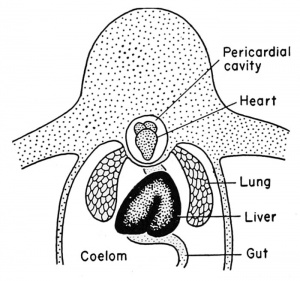
Just posterior to the pharynx the coelomic space extends into the heart mesenchyme as the pericardial cavity, to be described below.
During the tail bud stage, for instance, one could inject a colored dye into the coelom at the yolk level
and this would be carried anteriorly
to fill the forming pericardial cavity.
The frog develops lung respiration
by the time of metamorphosis but it
does not develop a diaphragm to
separate the coelom further into a
pleural and peritoneal cavity as in
the case of higher vertebrates. These
cavities remain continuous, in the
frog, as the pleuro-peritoneal cavity,
but the pericardial cavity becomes
cut off entirely from the embryonic coelom. This is accomplished by
the aid of the developing ductus Cuvieri which helps to form the
septum trans versum at about the time of metamorphosis.
The Heart
The heart rudiment develops early from splanchnic mesenchymal cells which grow ventrally toward the mid-line below the pharynx. There they aggregate to form paired spaces (anterior coelomic spaces) at about the tail bud stage. This seems to occur under the inductive influence of the archenteric floor. In the meantime there appear scattered and possibly endodermal cells dorsal to the point of junction of the converging mesodermal masses and ventral to the pharynx. These are known as endothelial cells which become endocardial cells.
It will be remembered that the mesoderm of the ventral lip of the blastopore is at first continuous with the floor endoderm of the gut by way of the yolk. It may be suggested that the separation of the heart endothelial cells may be a later (hence more anterior) phase of the same separation of endoderm and mesoderm. It is purely an academic consideration whether the endocardium is endodermal or mesodermal in origin, for it arises from original endoderm in a manner similar to most mesoderm. The converging sheets of mesoderm then fuse above and below, enclosing these endothelial cells. The wall of the inner tube thus formed becomes greatly thickened during this convergence and is known as the myocardium. It will begin to give rise to the striated, syncytial, and involuntary muscles of the heart by the 5 mm. body length stage of development. The enclosed endothelial cells will form the lining of the heart known as the endocardium. The cavity surrounding the myocardium is the pericardial cavity which itself is enclosed by an outer (splanchnic) pericardium. The pericardial portion of the embryonic coelom is separated from the peritoneal cavity by growth of the common cardinal veins, the liver, and peritoneal folds, all of which comprise the septum transversum. This process is completed during metamorphosis.
As in the gut region there will develop a double-layered suspensory membrane of the heart known as the dorsal mesocardium. This remains attached to the heart at the anterior and posterior extremities of that organ, but ruptures at the level of the elongating and expanding heart itself. There is also a temporary ventral mesocardium,
which arises before the dorsal mesocardium. It soon ruptures through to provide a single continuous enclosing pericardial cavity completely surrounding the heart except for the previously described dorsal mesentery.
The mesocardium then disappears both dorsally and ventrally at the level of the heart, and this is accomplished in part by the rapid growth and expansion of the heart itself. The heart arises by the bilateral downgrowth of mesenchyme which becomes a single short tube. This tube finally lengthens and becomes divided to form the coiled, folded, and three-chambered heart of the adult.
The early differentiated heart consists of a simple tube which becomes divided into three chambers. The fused pair of vitelline veins becomes the meatus venosus which empties into the first division of the heart, the sinus venosus. Then follows (more anteriorly) the thinwalled atrium, the thick-walled ventricle, and finally the bulbus arteriosus (bulbus aortae) which leads to the ventral aorta or truncus arteriosus.
The development of the heart is accomplished within the limited space provided by the surrounding body tissues and organs, so that this organ becomes a reversed "S" coiled tube. With the embryo facing to the right, and looking at it from the right side, the shape of the heart would be something like this:
The posterior part of the original tube becomes folded dorsally and anteriorly to the more anterior part of the original tube. Conversely, the anterior part (which is destined to become the muscular ventricle) is folded ventrally and posteriorly to the other parts of the heart. There is thus a reversal in the original anterior-posterior relations of the heart in respect to the embryo. The extremities of the heart, those parts which are held in place by the dorsal mesocardium, are the posterior sinus venosus and the anterior truncus or bulbus arteriosus. Anteriorly the bulbus arteriosus gives rise to three pairs of arteries (aortic arches) to each of the three pairs of external gills. Later a fourth pair is added. In the frog there are no aortic (arterial) arches in either the mandibular or hyoid (first or second visceral) arches.
Before the development of the heart has progressed this far (i.e., the 4 mm. stage) there appears a pair of blood vessels in the ventrolateral splanchnic mesoderm known as the vitelline veins. These are associated closely with the yolk mass, hence the name "vitelline" from the vitellus (yolk). They probably are derived, as is the heart, by a separation of yolk endoderm and ventral mesoderm. They grow anteriorly by the merging of blood islands or lacunae until the veins merge, anterior to the yolk and liver mass, and become the fused vitelline veins. This point of fusion of the vitelline veins is called the sinus venosus from the very beginning. It represents the most posterior limit of the heart. The part of the heart into which the sinus venosus empties is the atrium or the undivided embryonic auricles. The atrium and subsequent auricles remain thin-walled and become very elastic. Shortly (i.e., at the 7 mm. body length stage) there grows down from the roof of the atrial chamber a longitudinal sheet of endocardium which is known as the interatrial septum. This divides the atrium into a right and left auricle, and the sinus venosus now leads into the right auricle. Eventually the left auricle will receive blood from the pulmonary veins.
The ventricle or anterior portion of the original heart tube becomes very thick-walled and muscular and is never divided longitudinally into two as it is among mammals. There is a transverse division groove between the atrium and the ventricle at the folds in the inverted "S" coil just described. The ventricle is therefore folded caudally and ventrally to the atrium and leads into the truncus arteriosus which is suspended to the dorsal body wall by the permanent dorsal mesocardium at this level. The truncus arteriosus becomes partially subdivided by a vertical and longitudinal septum (the spiral valve) and gives rise to the paired ventral aortae which are connected with the various aortic arches.
The Circulatory System
The blood vessels arise from the fusion or confluence of blood islands which develop first in the splanchnic mesoderm and later in scattered mesenchyme. These are isolated groups of yolk-laden cells which form endothelial pockets containing blood-forming cells. These lacunae ("litde lakes") merge to form the blood vessels, and eventually the embryo acquires a complete and closed blood vascular system, continuous with the heart. This occurs even before there is any rhythmic pulsation of the blood or the heart. The blood islands give rise to the early embryonic blood, but this blood is later derived from the spleen and bone marrow and possibly the liver.
The Arterial System
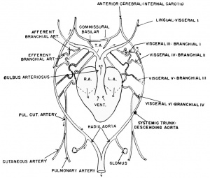
The dorsal aorta arises in splanchnic mesoderm dorsal to the enteron. Above the pharynx it is double and the vessels are known as the supra-branchial aortae or dorsal aortae. Aortic arches develop within the mesenchyme of the visceral arches (III to VI). They arise first as blood islands which merge to form blood vessels. The afferent branchial portion of each arch joins the ventral aortae, while the efferent branchial portion joins the dorsal aortae. This junction does not occur in either the mandibular or the hyoid arches, but it does occur in all the others. A closed circuit is established, therefore, between the ventral heart and the dorsal aortae by way of the viscerally located aortic arches.
The third, fourth, and fifth visceral arches (branchial arches I, II, and III) now give rise to secondary blood vessels or capillary loops. These connect dorsally (efferent branchial artery) and ventrally (afferent branchial artery) with the corresponding aortic arch. The intermediate section of each new vessel then grows out into the developing external gill. That portion arising from the ventral part of the aortic arch, and therefore nearest the heart, is then known as the afferent branchial loop. The returning portion from the gill which is connected with the dorsal part of the aortic arch is the efferent branchial loop. These loops coil through the filaments of the developing external gills, but the original portion of the aortic arch in each of the visceral arches remains for a brief time. The blood may therefore flow through either of two courses (i.e., the arch or the branchial loop), during the early functioning of the external gills. Usually no external gill grows outward from the sixth visceral arch and as a result an external branchial vessel never develops. The aortic arches tend to disappear or become relatively non-functional, as the external gills handle the major volume of the blood flow in this region.
As the external gills degenerate there develops a short-circuited connection between the afferent (ventral) and the efferent (also ventral) ends of the branchial vessels. This probably is aided by the remnant of the original aortic arches. This short circuit supplies the newly forming internal gills. All of the blood from the ventral aorta then must pass through the filaments of the internal gills until the lung circulation develops. These gills degenerate at the time of metamorphosis.
| Blood vascular system of the developing frog embryo. |
|---|

Early embryo, from the right side. |
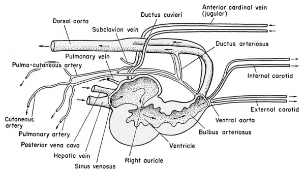
Late frog embryo, from the right side. |
The paired dorsal aortae extend into the head and the aortic arches of the first pair of branchial arches (visceral arch III), after the loss of branchial respiration, remain to carry blood into the head from the heart. These become the large anterior or internal carotids, originally the anterior extensions of the dorsal aortae. They proceed to the base of the skull and give rise to the palatine (to the roof of the mouth), the cerebral (to the brain), and the ophthalmic (to the eyes) arteries. The original paired ventral aortae proceed into the head as the smaller external carotids and join the blood islands and sinuses of the floor of the mouth and the tongue as the lingual arteries. The carotid glands, derived from the third visceral arch and having the function of filtering carotid blood, are located at the bifurcation of the internal and the external carotids. This is at the original ventral origin of the first branchial arch.
The aortic arches which pass through the second branchial (fourth
visceral) arch become greatly enlarged and retain their connection
with the dorsal aortae on both sides, to become the main systemic
trunk. That portion of the dorsal aorta anterior to the second branchial arch then degenerates, so that all of the arterial blood of the
first branchial arch must pass anteriorly and all of the blood of the
second branchial arch must pass posteriorly. The systemic trunk on each side gives rise to the laryngeal artery ( to the larynx and muscles
of the hyoid apparatus) and the oesophageal arteries (to the shoulder
and brachial arteries of the limb). The paired systemic arteries join
within the body to form the single dorsal aorta, sometimes called the
descending aorta, which in turn gives rise to many branches. These
include the renals to the glomi of the pronephros and glomeruli of
the mesonephros; the large coeliac artery to the stomach, liver, pancreas, and intestine; the gonadals; the lumbars; and finally the iliacs
to the hind legs.
The aortic arch which develops into the third branchial arch (fifth visceral) disappears. Those in the fourth branchial arches, however (sixth visceral), develop connections with blood islands in the skin and the lungs to become the pulmo-cutaneous arteries. In the frog, the skin represents about 60 per cent of the necessary respiratory surface. Some time after metamorphosis the connection of this fourth pair of branchial aortic arches severs connections with the dorsal aorta (now known as the systemic trunk), leaving only a vestigial strand of tissue known as the ductus Botalli or ductus arteriosus. In this manner the systemic arteries to the viscera and hmbs are separated from the respiratory arteries to the skin and lungs.
The arterial circulation of the frog is not entirely efficient for the simple reason that each truncus arteriosus is divided only partially by two septa into three channels, and some of the recently aerated blood from the lungs will be sent again to the lungs. One of the channels leads to the carotid arches; a second joins the systemic arches; a third leads from the right side of the heart, carrying venous blood to the pulmo-cutaneous arches. The largest exit is to the systemic trunk, and, until the time of metamorphosis, the two main anterior arteries must pass through the gill circulation where the blood can adequately exchange its carbon dioxide for oxygen. The systemic trunks give rise to the pharyngeal arteries, which supply the mandibular arches, and to the subclavians, which supply the forelimbs, and then they join to form the single dorsal aorta. This large vessel then gives off several thoracic and lumbar arteries to the back, a large coeliac artery and smaller mesenteric arteries to the viscera, iliac arteries to the limbs, and a caudal artery to the tail.
The Venous System
The vitelline veins are the first blood vessels to develop and they appear as blood islands ventro-lateral to the yolk mass. The fused anterior vitelline veins comprise the meatus venosus, later to become the hepatic vein emptying into the sinus venosus. The right posterior vitelline vein shortly disappears and the left one remains as the hepatic portal vein, bringing blood from the viscera to the liver. Shortly after hatching, a hepatic vein arises from the liver substance to pass directly to the sinus venosus via the path of the original anterior vitelline vein. After the development of the posterior vena cava the two main liver lobes direct their separate hepatic veins into the sinus venosus.
The paired common cardinals grow obliquely in a dorso-lateral direction from the sinus venosus toward the body wall and then divide to send branches anteriorly and posteriorly. The paired outgrowths of the sinus venosus are known as the ducts of Cuvieri or Cuvierian sinuses. The anteriorly growing extension of the common cardinal vein is the anterior cardinal vein. This receives blood from the tongue, hyoid, thyroid, parathyroid, and the floor of the mouth by a branch known as the external (inferior) jugular vein. It also receives blood from the brain, shoulder, and forelimb by way of another branch, the internal (superior) jugular vein. A third contributing vessel is the subclavian vein which brings blood from the brachial vessel of the forelimb and the cutaneous vein of the skin. All of these vessels contribute to the original anterior cardinal vein and at their point of junction are known as the anterior or superior vena cava. Since the posterior cardinal veins fuse mesially and become separated from the common cardinal, the anterior vena cava becomes the only remnant of the embryonic ductus Cuvieri.
The posterior cardinal veins are directed posteriorly from the paired common cardinals (ductus Cuvieri). Each of these forms a sinus around the temporary pronephric tubules and then grows posteriorly as the single median cardinal vein between the mesonephroi and into the tail. During its course it receives vessels from the dorsal body wall.
The posterior cardinals go through considerable change during the pre-metamorphic development of the frog. There is degeneration of the entire left cardinal and most of the right cardinal veins. The sections posterior to the pronephric sinuses tend to fuse mesially to form the single posterior vena cava (post-caval vein or inferior vena cava) which empties directly into the sinus venosus, having moved posteriorly from the ductus Cuvieri. The paired hepatic veins, from the liver lobes, are now incorporated into the enlarged posterior vena cava as it enters the sinus venosus. These changes leave the ductus Cuvieri connected with only the anterior or superior vena cava, which may now be called the pre-caval vein.
Posteriorly the vessel produced by fusion of the posterior cardinal
veins, now considered as the median cardinal vein, is enlarged as sinuses which shortly become organized into a vessel. This vessel connects with the hepatic vein through a new junction anterior to the
liver. The median vein remains as the posterior vena cava to receive
blood from the tail, which degenerates at metamorphosis, and from
the kidneys (several short renal veins). After metamorphosis there is
no remnant of this vessel posterior to the kidneys, since the tail disappears. Thus the post-caval vein is composed of several elements: a
hepatic vein derived from the original left vitelline vein; a short portion of the right posterior cardinal vein; the median channel derived
from the fused right and left posterior cardinal veins; and a short
new section just posterior to the hepatic section, which grows posteriorly.
The lateral pair of vessels on the outer margin of the mesonephros brings blood from the hindlimbs (sciatic and femoral) by way of the common iliac, and from the dorsal body wall (lumbar). These are known as the renal portal veins and terminate in the mesonephroi.
The pulmonary veins arise at about the 6 mm. body length stage as posterior dorsal outgrowths of the sinus venosus to the lung buds. These veins eventually fuse and open directly into the left auricle, bringing blood from the lungs after metamorphosis.
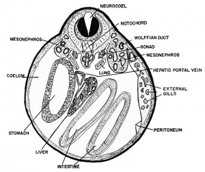
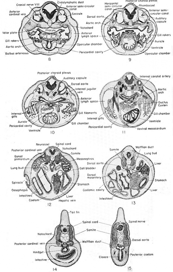
Serial transverse sections of the 11 mm. frog larva.
The single abdominal vein arises as a pair of veins growing posteriorly from the sinus venosus along the ventral body wall to the level of the cloaca, making junction with the femoral veins of the hind legs. These paired vessels then fuse, anterior to the cloaca, into a single median ventral abdominal vein. Tt then loses its connection with the sinus venosus and establishes a new connection with the hepatic portal vein, originally the right vitelline vein, which enters the liver lobes.
The Lymphatic System
The lymphatic system of the frog is made up of very large lymph spaces in various parts of the body, without well-defined connecting lymph vessels such as occur in mammals. However, all of the four lymph hearts are protected by valves so that the lymph always passes from a lymph heart to a lymph or blood vessel, and never in the reverse direction. There are no muscular coats to the lymph spaces, but they are lined with flat endothelial cells and connective tissue. The connective tissue forms septa which divide the lymph spaces and attach them to the underlying muscle.
Between the third and fourth somites between the peritoneum and
the integument, at the time of hatching (6 mm. body length), there
are developed crude anterior lymph hearts, surrounded by muscle
fibers. These are connected by an ill-defined pair of vessels just beneath the skin, the subcutaneous lymph spaces, one of which extends
anteriorly and the other posteriorly, each to merge with the venous
plexuses. The anterior paired lymph vessels send extensions ventrally
to the branchial region considerably after the time of hatching. Paired
thoracic ducts develop from the anterior lymph hearts (at the 26 mm.
stage) just between the posterior cardinal vein and the dorsal aorta.
The posterior vessels give rise to both dorsal and ventral vessels in the
larval tail. After metamorphosis posterior lymph hearts appear in
association with the intersegmental veins near the hindlimbs. As previously described, there is flow of lymph from the peritoneal cavity
into the venous sinuses of the kidney by way of the ciliated peritoneal
funnels of that organ.
The Spleen
This hematopoietic organ arises in the vicinity of the stomach at about the 10 mm. body length stage as a cluster of cells within the dorsal mesentery. By the 15 mm. stage the spleen is seen as a definite projection from the mesentery, covered by coelomic epithelium. Apparently some wandering cells from the intestinal epithelium invade the spleen at this time. By the 30 mm. stage these cells become reticular and are vascularized and can be recognized as splenic cells, and the organ as the ovoid spleen of the adult.
Summary of Embryonic Development of the 11 mm Frog Tadpole - Mesodermal Derivatives
- Mesenchyme — loose, primitive mesoderm found scattered throughout tadpole. There are condensations in head region where cartilage-forming centers are developing, later to give rise to neurocranium except dura and pia mater, which come from neural crest. Connective and blood vascular tissues are also to be derived from mesenchyme. Around mesodermal notochord are sclerotomal (mesenchyme) cells which will form axial skeleton.
- Epimere — most dorsal mesodermal masses appearing as metameric somites, the most anterior of which are being transformed. About 12 somites in trunk and 32 in tail remain.
- Dermatome — distinct band of mesenchymatous cells lying dorso-laterally just beneath ectoderm, to form dermis (cutis).
- Myotome — central portion of somite which is being organized into muscle bundles, divided in tail region into dorsal and ventral bundles.
- Sclerotome — thin layer of mesenchyme cells surrounding nerve cord and notochord from which will develop axial skeleton.
- Mesomere — intermediate mesodermal mass from which urogenital system is derived.
- Pronephros — primary, embryonic kidney made up of a few coiled tubules and ciliated nephrostomes.
- Pronephric ducts — lateral to dorsal aorta and dorsal to posterior cardinals, these ducts lead from pronephros posteriorly to join each other just before they fuse with cloaca.
- Mesonephros — large mass of nephrogenic tubules, without nephrostomes, forming permanent frog kidney.
- Gonads — cell masses hanging into coelom from dorsal peritoneum between mesonephros and gut, in vicinity of lung buds.
- Hypomere — lateral plate mesoderm, ventral to mesomere, and consisting of somatic and splanchnic layers with intermediate coelomic cavity.
- Pericardial cavity — surrounds heart, not yet separated from true peritoneal cavity.
- Peritoneal cavity — body cavity, surrounding viscera.
- Mesenteries — double-layered dorsal mesentery supports viscera. A remnant of ventral mesentery (gastrohepatic omentum) is found between stomach and liver.
- Spleen — accumulation of cells lateral to mesenteric artery in dorsal mesentery.
- Circulatory System — differentiated as heart, arteries, veins, lymphatics, and contained corpuscles.
- Heart — already a three-chambered structure as in the adult.
- Sinus venosus — junction of post-caval and common cardinals, leading to right atrium.
- Atria — thin-walled, paired heart chambers, at least partially separated from each other by interatrial septum. Right atrium receives systemic veins while left atrium receives pulmonary vein from lung buds.
- Ventricle — thick-walled, muscular, and generally ventral to atria. Leads into truncus and bulbus arteriosus.
- Bulbus arteriosus — tubular extension of early embryonic heart which leads directly into aortic arches.
- Truncus arteriosus — short, ventrally directed vessel leading into bulbus arteriosus. (Syn., ventral aorta.)
- Arteries — major arteries only.
- Afferent branchial arteries.
- Lingual (external carotid) — extensions of ventral aorta into lower jaw.
- First branchial within third visceral arch.
- Second branchial — forked portion of first branchial found within fourth visceral arch.
- Third branchial — temporarily found within fifth visceral arch; degenerates.
- Fourth branchial — within sixth visceral arch.
- Efferent branchial arteries.
- Anterior cerebral (internal carotid) — gives rise to basilar artery and commissural artery passing beneath infundibulum to other side of head (see diagram). Extensions of first branchial artery from within first visceral arch.
- Second branchial — within fourth visceral arch, enlarges and grows posteriorly as descending aorta to join corresponding vessel of other side to form systemic trunk. Junction about level of liver.
- Third branchial — within fifth visceral arch; degenerates.
- Fourth branchial — within sixth visceral arch, loses its connection (ductus arteriosus) with descending aorta and grows posteriorly to form pulmonary and cutaneous arteries (see diagram).
- Dorsal aorta — single large artery formed by junction of two descending aortae (radices aortae) and extending posteriorly into tail, giving off various smaller arteries en route.
- Intersegmental arteries — paired and metameric arteries which grow from dorsal aorta dorso-laterally between myotomes.
- Glomi — short branches from radices aortae which grow toward pronephric chambers. Really undeveloped glomeruli.
- Mesenteric artery — single large vessel growing ventrad from dorsal aorta to supply viscera. Later becomes coeliac artery.
- Caudal artery — extension of dorsal aorta into tail.
- Afferent branchial arteries.
- Veins — major veins only. Generally walls are thinner than arterial walls.
- Cardinal system.
- Anterior cardinals (superior jugulars) — irregular in cross section, found in head, closely associated with internal carotid arteries.
- Inferior jugulars — bring blood from lower jaw, accompanying external carotid arteries, and join common cardinals.
- Posterior cardinals — ventro-lateral to dorsal aorta, dorsal to nephrogenic tissue. Carry blood to common cardinals.
- Subcardinals — smaller veins, ventral to mesonephric tissue, and bringing blood from caudal vein. Become renal portals.
- Common cardinals — ducts of Cuvier which receive blood from anterior and posterior cardinal vessels, and pronephric sinus, and convey it to sinus venosus at its postero-lateral margin.
- Post-caval vein — posterior vena cava or inferior vena cava. This vessel has grown posteriorly from hepatic vein and incorporates mesonephric level of right posterior cardinal vein. It therefore passes through liver into sinus venosus ventral to entrance of right common cardinal vein (see diagram).
- Hepatic system — derivatives of original vitelline veins.
- Hepatic vein — fused vitelline veins carrying blood from liver sinuses to the sinus venosus. Short and difficult to distinguish from post-caval vein to which it gives rise.
- Portal vein — common vessel receiving blood from pancreas, stomach, intestine, etc., representing original vitelline veins.
- Lymphatic system — not well developed at this stage.
- Cardinal system.
- Chondrocranium — cartilage centers may be found, within which bone will develop in formation of the embryonic skull.
- Neurocranlum — skeleton around brain and primary sense organs. Anterior to posterior.
- Labial (supra-ostral) cartilages — paired, short, within upper lip and lateral to mouth. (May be ectodermal)
- Cornua trabecularum (trabecular horns) — paired cartilage bars which accompany olfactory tubes. (May be ectodermal)
- Ethmoid (internasal) plate — fused trabeculae within median line, anterior to ascending process of pterygoquadrate.
- Pterygoquadrate (palatoquadrate) — derived from maxillary portion of first visceral arch and joins posterior end of trabecula by its posterior ascending process. At its anterior end joins anterior ascending process. (May be ectodermal)
- Basicranial fontanelle — skeletal cavity for pituitary gland.
- Trabeculae — paired cartilage bars on either side of basicranial fontanelle, converging slightly anteriorly.
- Basilar plate — single, fused cartilage mass immediately anterior to notochord. Joins parachordals and trabeculae.
- Parachordals — lateral to notochord, at its anterior end.
- Splanchnocranium — derived from the visceral arches. Anterior to posterior.
- Mental (infra-rostral) cartilages — paired, but meet in mid-line of lower lip.
- Meckelian cartilages — laterally projecting flanges to which muscles will be attached joining pterygoquadrate. (May be ectodermal)
- Ceratohyals — large cartilages arising within hyomandibular arches, posterior to Meckelian cartilages. (May be ectodermal)
- Basibranchial (copula) — single cartilage between ceratohyals. (May be ectodermal)
- Hypobranchials — paired, posterior to ceratohyals, and derived from third visceral arches. Articulates also with basibranchial. (May be ectodermal)
- Ceratobranchials — three or possibly four slender processes within third to sixth visceral arches, extending from hypobranchial on each side. (May be ectodermal)
| Historic Disclaimer - information about historic embryology pages |
|---|
| Pages where the terms "Historic" (textbooks, papers, people, recommendations) appear on this site, and sections within pages where this disclaimer appears, indicate that the content and scientific understanding are specific to the time of publication. This means that while some scientific descriptions are still accurate, the terminology and interpretation of the developmental mechanisms reflect the understanding at the time of original publication and those of the preceding periods, these terms, interpretations and recommendations may not reflect our current scientific understanding. (More? Embryology History | Historic Embryology Papers) |
Reference
Rugh R. Book - The Frog Its Reproduction and Development. (1951) The Blakiston Company.
Cite this page: Hill, M.A. (2024, April 28) Embryology Book - The Frog Its Reproduction and Development 13. Retrieved from https://embryology.med.unsw.edu.au/embryology/index.php/Book_-_The_Frog_Its_Reproduction_and_Development_13
- © Dr Mark Hill 2024, UNSW Embryology ISBN: 978 0 7334 2609 4 - UNSW CRICOS Provider Code No. 00098G

