Book - Human Embryology and Morphology 19
| Embryology - 13 Mar 2026 |
|---|
| Google Translate - select your language from the list shown below (this will open a new external page) |
|
العربية | català | 中文 | 中國傳統的 | français | Deutsche | עִברִית | हिंदी | bahasa Indonesia | italiano | 日本語 | 한국어 | မြန်မာ | Pilipino | Polskie | português | ਪੰਜਾਬੀ ਦੇ | Română | русский | Español | Swahili | Svensk | ไทย | Türkçe | اردو | ייִדיש | Tiếng Việt These external translations are automated and may not be accurate. (More? About Translations) |
Keith A. Human Embryology and Morphology. (1902) London: Edward Arnold.
| Historic Disclaimer - information about historic embryology pages |
|---|
 |
Chapter XIX. The Body Wall, Ribs and Sternum
Bilateral Symmetry of the Body
From a developmental point of view the body is made up of two symmetrical halves; each half of the blastoderm, taking the medullary groove as the line of division, contributes equally to the formation of the body. Each produces a half of the nervous system, each a half of the vascular, muscular and alimentary systems, so that each individual is in reality made up of two identical halves, right and left.
The Ventral Line of the Body
The tw.o halves are united along the ventral line from the mouth to the anus (see Fig. 227). In this line are developed the symphysis of the lower jaw, the body of the hyoid bone (copula), the white line of the neck and angle of the thyroid cartilage, the sternum, the supra-umbilical part of the linea alba, umbilicus, infra-umbilical part of the linea alba, symphysis pubis, the septum of the penis, and of the scrotum and perineal raphe. The ventral line is continued forwards on the face between the parts derived from the mesial nasal processes (Chap. 1.).
The idea was at one time prevalent that the whole of this line was formed by the fusion of one somatopleure with the other ; the median ventral line was the suture formed by the union. Such is not the case. The blastoderm, which lies at first like a cap on the yolk-sac (Fig. 75), is produced or folded anteriorly to form the fore-gut and the part of the body above the umbilicus ; it is produced posteriorly to form the hind-gut and the part of the body below the umbilicus (Fig. 75). The blastoderm grows out from the umbilicus to form the embryo in much the same way as a soap bubble is blown from the bowl of a pipe. In an embryo, at the commencement of the 3rd week the greater part of the ventral line is occupied by the umbilicus (Fig. 228). At that time the umbilicus is 3 mm. long, the entire ventral line being about 4 mm. At the 6th week the ventral line measures 15 mm., the umbilicus retains its former size, about 3 mm.

|

|
| Fig. 227. Diagram of the Structures formed in the Median Ventral Line of the Body. | Fig. 228. The Median Ventral Line in an embryo of three weeks, to contrast with the Corresponding Line in the Adult. |
At first the somatopleure shows no trace of segmentation. The paraxial masses of mesoblast become segmented early and form the muscle plates (Fig. 126). From each muscle plate of the primitive segments a process grows down into the somatopleure. The somatopleure thus becomes segmented secondarily, the process of segmentation spreading from the dorsal to the ventral side of the plate, but along the median ventral line of the body wall a band of the primitive mesoblastic tissue remains unchanged and undifferentiated. In the ventral band between the left somatopleure and the right are formed the sternum and the linea alba (Fig. 227). In lower vertebrates, in fishes, and to a less marked extent in amphibians and reptiles, the somatopleure becomes segmented from end to end of the trunk, each muscle segment being distinctly recognizable in the adult.

Fig. 229. Scheme of the Manner in which the Somatopleure is segmented.
Formation of Ribs
Ribs, like all true skeletal bones, pass through three stages :
- They are represented by a membranous basis in the fibrous tissue (septa) between the muscular segments of the somatopleure (Fig. 229).
- The fibrous basis of the rib becomes cartilaginous.
- Ossification of the cartilage begins in the 7th week, but the process of ossification leaves the ventral parts of the costal segments untouched ; they form the costal cartilages ; in lower forms they may become ossified and form sternal ribs.
In lower vertebrates, such as reptiles, each rib articulates with the neural arch of a vertebra by two heads, dorsal and ventral (Fig. 123).
In man and mammals only the first and the lowest two ribs retain the vertebral attachment. The heads of the upper ribs (2nd to 10th) migrate forwards and gain attachments to the disc and vertebra in front of their own segments. The dorsal head of the rib in man is represented by the tuberosity (see also p. 152).
The Sternum
In man and anthropoids the sternum has become flat and highly modified with the alterations in the shape of the thorax (Fig. 208). With the adaptation to the upright posture the thorax becomes flattened from back to front ; its transverse diameter is as great, or greater, than the anteroposterior. The sternum also becomes wider and shorter. To understand its true nature it is necessary to note the characters of the sternum of a pronograde mammal, such as the dog or ape (Fig. 230). In such the sternum is typically made up of seven segments :
- A modified anterior segment, the pre-sternum.
- Five narrow, cylindrical segments or sternebrae, forming the body of the sternum.
- The ensiform process, a hind segment, complex in nature and ending in the middle ventral line. The ensiform process usually bifurcates and probably belongs to several segments.

|

|
| Fig. 230. The Form of Sternum in a Pronograde (quadrupedal) Mammal. | Fig. 231. The Form of Sternum in a Mammal adapted to the orthograde (upright) Posture. The Points of Ossification are also shown. |
The chief changes in the human sternum are :
- Each segment has become flat and wide ;
- The segments of the body fuse together during the years of adolescence, the fusion beginning behind and passing forwards ;
- The 4th sternebra of the body is usually vestigial; is probably made up of two or more fused segments.
In low primates 8 or 9 pairs of ribs may reach the sternum, six or more sternebrae being then present. In man the number has been reduced to seven pairs, the sternal ends of the seventh pair lying in front of the base of the ensiform process. It is not uncommon to find the 8 th rib reaching the sternum, especially on the right side; it is rare to find the 7th pair fail to reach the sternum. The more frequent presence of an 8 th sternal rib on the right side is due to right-handedness (Cunningham) or the pressure of the underlying liver requiring support (Tredgold).
Development of the Sternum

The sternum is developed in the mesoblast of the ventral median line between the first 8 or 9 dorsal segments (Paterson). It is developed in two parts — a right from the right somatopleure, a left from the left somatopleure. These two halves, the right and left fibrous sternal bars, fuse gradually in the middle line, the process of fusion commencing at the presternum and spreading back (Fig. 232). The bifurcated end of the ensiform process represents the posterior extremities of the sternal bars.
The sternum is described here as a structure arising independently in the median ventral line. This, however, is not the commonly accepted view. Euge's researches led him to the conclusion that the segments of the sternal bars were produced as buds from the ventral ends of the ribs, to which they correspond, and his conclusions are supported by the evidence of comparative anatomy.
In its development the sternum passes through three stages — fibrous, cartilaginous and bony.
- Fibrous Stage — At the 6th week (Fig. 232) the costal cartilages are already chondrified. The mesoblast on each side of the median line, in which they end, has become condensed, and forms the membranous basis of the two sternal bars (Paterson). The bars begin to fuse together in front.
- Cartilaginous Stage — The mesoblast of each sternal bar begins to chondrify in the intervals between the ends of the costal cartilages. The process of chondrification and fusion proceed apace, and by the commencement of the third month the segments of each side have united to form the cartilaginous sternal bars (Paterson). At the same time, the two bars gradually unite, the cellular tissue between them becoming chondrified. Thus a single cartilaginous plate formed out of the two cartilaginous bars is formed. Fibrous joints are subsequently formed between the presternum and mesosternum and between the mesosternum and ensiform process. A fibrous and synovial joint is also developed at the union of the costal cartilages with the sternum, except in the case of the first pair.
- Ossification — A centre appears for each sternebra ; those for the third and fourth of the mesosternum are usually double, oue being placed on each side. The centres for the 4th segment are more frequently absent than present (Paterson). The centre for the presternum (there may be two or even more) appears about the 4th month ; the centres behind appear in order ; that for the 4th sternebra of the mesosternum appearing about the time of birth, that for the ensiform after birth. The process of fusion of segments begins behind about puberty; the segments of the mesosternum are united together by the 30 th year. Occasionally a median foramen may be seen in the sternum ; it is due to imperfect union of the sternal bars.
The Presternum is complex in nature. It probably represents more than the simple sternebra between the first and second pairs of ribs. Part of it may belong to the segment in front, the last cervical. When the last (7th) cervical rib is fully developed it reaches the presternum. It may also contain another element. In more primitive types of vertebrates, in fact in all below the higher mammals, a part of the coracoid element of the shoulder girdle, the epi-coracoid (pre-coracoid), is situated at the cephalic end of the sternum (Fig. 244). The supra-sternal bones, not infrequently seen on the upper border of the human manubrium sterni (Fig. 231), appear to represent the epi-coracoids. These bones are probably always present, but their presence is difficult to detect because they are commonly more or less completely fused with the presternum (Paterson). Occasionally the first segment of the meso-sternum joins the manubrium instead of the body of the sternum — a union frequently seen in some anthropoids.
The presternum is the first part of the sternum found in ascending the scale of vertebrate animals, and is developed as a median ventral support for the shoulder girdle.
Chapter Figures
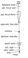
Fig. 227. Diagram of the Structures formed in the Median Ventral Line of the Body.
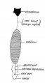
Fig. 228. The Median Ventral Line in an embryo of three weeks, to contrast with the Corresponding Line in the Adult.
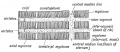
Fig. 229. Scheme of the Manner in which the Somatopleure is segmented.
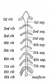
Fig. 230. The Form of Sternum in a Pronograde (quadrupedal) Mammal.
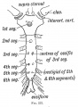
Fig. 231. The Form of Sternum in a Mammal adapted to the orthograde (upright) Posture. The Points of Ossification are also shown.
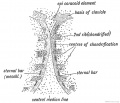
Fig. 232. The Sternal Bars in a human embryo of six weeks (after Paterson).
| Historic Disclaimer - information about historic embryology pages |
|---|
|
Human Embryology and Morphology (1902): Development or the Face | The Nasal Cavities and Olfactory Structures | Development of the Pharynx and Neck | Development of the Organ of Hearing | Development and Morphology of the Teeth | The Skin and its Appendages | The Development of the Ovum of the Foetus from the Ovum of the Mother | The Manner in which a Connection is Established between the Foetus and Uterus | The Uro-genital System | Formation of the Pubo-femoral Region, Pelvic Floor and Fascia | The Spinal Column and Back | The Segmentation of the Body | The Cranium | Development of the Structures concerned in the Sense of Sight | The Brain and Spinal Cord | Development of the Circulatory System | The Respiratory System | The Organs of Digestion | The Body Wall, Ribs, and Sternum | The Limbs | Figures | Embryology History
Reference
Keith A. Human Embryology and Morphology. (1902) London: Edward Arnold.
Cite this page: Hill, M.A. (2026, March 13) Embryology Book - Human Embryology and Morphology 19. Retrieved from https://embryology.med.unsw.edu.au/embryology/index.php/Book_-_Human_Embryology_and_Morphology_19
- © Dr Mark Hill 2026, UNSW Embryology ISBN: 978 0 7334 2609 4 - UNSW CRICOS Provider Code No. 00098G