Book - Human Embryology and Morphology 18
| Embryology - 27 Apr 2024 |
|---|
| Google Translate - select your language from the list shown below (this will open a new external page) |
|
العربية | català | 中文 | 中國傳統的 | français | Deutsche | עִברִית | हिंदी | bahasa Indonesia | italiano | 日本語 | 한국어 | မြန်မာ | Pilipino | Polskie | português | ਪੰਜਾਬੀ ਦੇ | Română | русский | Español | Swahili | Svensk | ไทย | Türkçe | اردو | ייִדיש | Tiếng Việt These external translations are automated and may not be accurate. (More? About Translations) |
Keith A. Human Embryology and Morphology. (1902) London: Edward Arnold.
| Historic Disclaimer - information about historic embryology pages |
|---|
| Pages where the terms "Historic" (textbooks, papers, people, recommendations) appear on this site, and sections within pages where this disclaimer appears, indicate that the content and scientific understanding are specific to the time of publication. This means that while some scientific descriptions are still accurate, the terminology and interpretation of the developmental mechanisms reflect the understanding at the time of original publication and those of the preceding periods, these terms, interpretations and recommendations may not reflect our current scientific understanding. (More? Embryology History | Historic Embryology Papers) |
Chapter XVIII. The Organs of Digestion
Development of the Liver
The human liver, in its development, repeats the forms met with in ascending the animal scale. In Amphioxus the liver is merely a caecal diverticulum of the digestive canal ; in amphibians the liver is a compound tubular gland — the hepatic cells being arranged in cylinders round the bile ducts. In birds and mammals the tubular arrangement is lost and the lobular form substituted.
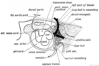
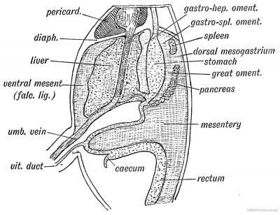
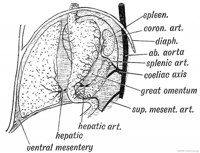
Fig. 212. The Mesentery of the Fore-gut and its Contents, -viewed from the left side (schematic).
To understand the development of the liver, the condition of parts at the commencement of the third week must be studied.
At that time, the anterior wall of the yolk-sac and that part of the fore-gut which becomes the stomach, lie in the septum transversum (Figs. 211, 212). When the liver bud grows out, it springs from the junction of the fore-gut and yolk-sac (Fig. 221); and spreads into the ventral mesentery and septum transversum. The part of the gut from which it springs afterwards becomes the second stage of the duodenum It is at first a hollow diverticulum of the gut hypoblast (Fig. 218). The diverticulum is surrounded in the mesogastrium by a mass of mesoblastic cells which form the vessels, capsule and connective tissue of the liver. It divides almost at once into right and left solid processes of hypoblastic cells, to form the right and left lobes of the liver (Fig. 218.)
The hepatic buds are developed just behind the sinus venosus and between the vitelline veins which are also situated in the ventral mesentery (Figs. 202 and 212). The veins are broken up by the ingrowth ; from them starts an invasion of venous capillaries, which, with the mesoblast of the septum transversum, penetrates the liver buds and breaks the solid processes of hypoblast into reticulating cylinders. Secondary processes start from the primary hepatic reticulating cylinders and form smaller and smaller meshes of hepatic cells. By the third month the hepatic lobules, with the intra- and sub-lobular arrangement of portal and hepatic veins, have appeared. The bile ducts probably represent the lumina of the original tubular hepatic buds. The umbilical veins also are cut by the hepatic invasion (Figs. 188 and 190).
The liver develops rapidly; in the 2nd and 3rd months it occupies more than half the abdominal space. At the commencement of the 2nd month, the yolk-sac becomes gradually smaller and, owing to the development of the duodenum and jejunum, it gradually retreats from the abdomen and region of the diaphragm and liver. The second and third stages of the duodenum and the jejunum are developed behind the ventral mesentery. As the liver grows it gradually frees itself from the diaphragmatic part of the ventral mesentery. The right lobe develops more rapidly than the left and comes in contact with the inferior vena cava. The stomach also frees itself from the basis of the diaphragm and comes to lie with the liver in the mesentery. The common bile duct is developed out of the hollow stalk of the hepatic bud. So is the common hepatic duct, while the right and left hepatic ducts are formed from the stalks of the processes which form the right and left lobes of the liver. The gall bladder arises as a diverticulum of the common bile duct.
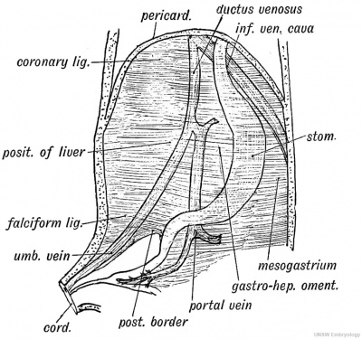
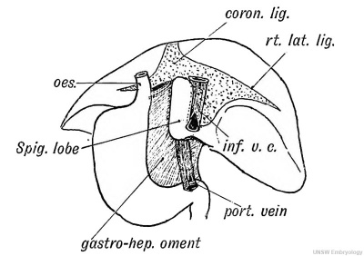
Fig. 213. The origin of the Peritoneal Ligaments connected with the Liver.
A. Diagram of the foetal relationship of the ventral mesentery to veins and the stomach, the liver being removed. B. The liver viewed from behind to show its relationship to the gastro-hepatic omentum, part of the ventral mesentery.
The Ligaments of the Liver
When the liver grows out from the septum transversum it withdraws within the ventral mesentery. Out of the ventral mesentery are formed all the ligaments of the liver (Fig. 213 A). These are the following: 1. The Gastro-hepatic Omentum is that part of the ventral mesentery which passes from (1) the oesophagus, (2) lesser curvature or ventral border of stomach, and (3) first stage of duodenum to (1) the diaphragm, (2) the posterior part of the longitudinal fissure of the liver, the ductus venosus lying within its hepatic attachment, and (3) the transverse fissure of the liver (Fig. 213 B). The portal and umbilical veins lie in the ventral mesentery (Fig. 213^4); the hepatic artery passes by it to the liver. The right or free border of the gastro-hepatic omentum, with the falciform ligament containing the remnant of the umbilical vein, represents the posterior border of the primitive ventral mesentery (Fig. 213 A).
2. The Falciform Ligament, containing the umbilical vein, also represents part of the ventral mesentery (Fig. 213 A). At an early stage the umbilical veins reached the sinus venosus by passing through the septum transversum. The development of the liver led to its being thrust out within the falciform ligament.
3. The coronary, the right and left lateral ligaments, and the attachments to the vena cava and diaphragm (Fig. 213 5) are developed as the liver emerges from the septum transversum, and separates itself from the diaphragm.
Fig. 214. Diagram of a mammalian Liver viewed from behind and below.
Morphology of the Liver
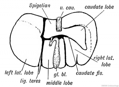
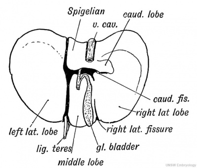
The liver of orthograde animals (man, anthropoids) differs widely in form and lobulation from that of mammals generally, but Professor Arthur Thomson has shown that traces of the fissures and lobes of the typical mammalian liver can be seen in the human organ. The liver of a dog or dog-like ape consists of three main lobes — right, middle and left, and two accessory lobes- — the caudate and Spigelian (Fig. 214). In man the right and middle lobes have fused, but traces of the fissure which separates them (the right lateral fissure) are frequently to be seen in the liver of the newly-born child (Fig. 215). The caudate lobe has been reduced in man to a vestige, but in the third month foetus it is of considerable size (Fig. 215). It projects from the liver at the upper boundary of the foramen of Winslow; in many animals it rivals the right lobe in size. The caudate fissure separates the caudate from the right lobe, and a trace of this fissure is very frequently to be seen in the human liver (Fig. 215).
Fig. 215. The lower surface of the Liver of a human foetus during the 3rd month, showing Vestiges of Fissures and Lobes of the typical mammalian Liver.
After the second month the growth of the right lobe is more rapid than that of the left. At birth the left lobe still touches the spleen, a relationship more frequently retained in women than in men. Eiedel's lobe is a linguiform prolongation of the right lobe below the 10th right costal cartilage caused by compression.
Gall Bladder.— It is developed from the common stalk (common bile duct) as a diverticulum in the second month (Fig. 218). It may represent a modified lobule of the liver. In some animals it is absent. The cystic duct represents the stalk of the bud.
The Oesophagus and Stomach. — The Oesophagus in a third week's foetus is the junctional piece of fore-gut that unites the pharynx to the scarcely differentiated stomach (Fig. 202, p. 244). It is extremely short, but with the development of the lungs and recession of the diaphragm it becomes elongated. In the foetus the mucous membrane is thrown into four longitudinal folds which end abruptly at the orifice of the stomach in a circular epithelial ridge which sometimes persists and acts as a valve at the cardiac orifice of the stomach. These four ridges can be seen in adult apes. At one stage of development the cervical part of the oesophagus is filled with epithelium and thus closed.
The Stomach
The stomach is developed out of that part of the fore-gut which lies between the oesophagus and pharynx in front and the yolk sac, duodenum and liver bud behind (Fig. 187, p. 229). At first it is contained in that part of the primitive mesentery to which the name of septum transversum has been given, and is situated below the lower cervical and upper dorsal region of the spine (Fig. 204). In the 3rd week the gastric part of the fore-gut shows a dorsal bulging — the greater curvature. As the liver and gut are developed, the stomach separates itself from the septum transversum and comes to be suspended from the dorsal wall of the coelom by the dorsal mesogastrium (Fig. 213 A). The gastrohepatic omentum is part of the ventral mesogastrium. The oesophageal end of the stomach is fixed in the septum transversum (diaphragm) ; the outgrowth of the liver bud fixes its pyloric end in the ventral mesogastrium. Three changes quickly ensue, the one being partly dependent on the other : (1) Its dorsal margin grows rapidly so as to produce the greater curvature ; this growth particularly affects the left side of the cardiac end of the stomach and thrusts the attachment of the dorsal mesogastrium towards the right side at the cardiac end of the stomach. The greater part of the fundus or cardiac sac of the stomach is produced from the left side.
(2) The dorsal mesogastrium being too limited in extent to allow for the growth of the greater curvature, the stomach rotates so that its left surface, with the left vagus nerve, comes to be anterior ; the other posterior.
(3) To allow this rotation to take place and probably for some other unknown reason, part of the dorsal mesogastrium, which is attached to the great curvature or dorsal border of the stomach, undergoes a rapid growth and forms the great omentum (Fig. 2 1 4).
The stomach loses its original vertical position only to a slight degree ; even in the adult the pyloric orifice lies not far from the middle line ; the oesophageal slightly to the left of it. The glands of the stomach begin to form during the third month. They appear as tubular invagination of the gastric hypoblast. The peptic cells are formed by the 7 th month.
The Spleen
The Spleen appears in the dorsal mesogastrium above the cardiac end of the stomach (Fig. 216) and grows out on the left surface of the mesogastrium (Fig. 217). It appears at the end of the second month by a collection of lymphoid follicles in the mesoblast of the mesogastrium. The tail of the pancreas (Fig. 216) reaches its point of origin. The splenic artery is one of the vessels of the inesogastrium (Fig. 219), its branches end in the developing tissues of the spleen. The splenic blood spaces are probably formed from the veins which in the developing spleen are lined by a layer of columnar cells. The trabecular and muscular tissue and the capsule are derived from the mesoblast of the dorsal mesogastrium.
Fig. 216. The Relationship of the Spleen, Pancreas, and Liver to the Mesogastrium in the Embryo.
The gastro-splenic omentum is that part of the dorsal mesogastrium which unites the spleen to the stomach (Figs. 216 and 217). It becomes elongated and stretched as the stomach rotates.
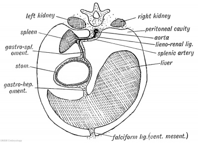
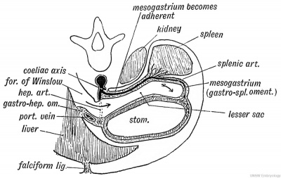
Fig. 217. A diagrammatic transverse Section of the Mesogastrium viewed from behind.
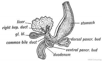
The spleen comes to lie against the posterior (right) surface of the cardiac end of the stomach. The dorsal part of the mesogastrium between the roof of the coelom and the spleen becomes the lienorenal ligament. The rotation of the stomach also leads to the spleen being thrust towards the left side ; the upper or renal surface of the spleen becomes applied to the peritoneum covering the anterior surface of the left kidney (Fig. 217). The part of the mesogastrium between the spleen and oesophagus adheres to the diaphragm and forms the suspensory ligament of the spleen. The Pancreas. — The Pancreas appears during the 4th week as two, or perhaps three, processes of hypoblast from that part of the gut which afterwards becomes the second stage of the duodenum (Fig. 218). Of the two buds, one is a minor process which springs from the ventral aspect of the duodenum in common with the hepatic diverticulum. This ventral bud only forms the lower part of the head of the pancreas. The greater part is formed from a process which springs from the dorsal border of the duodenum, nearer the stomach than the ventral process, and grows into the dorsal mesogastrium above the stomach until it reaches the spleen (Fig. 219). It unites in the mesogastrium with the ventral bud. The ducts of both processes may persist, the duct of the dorsal bud (duct of Santorini) opening half an inch above the opening of the bile duct ; the duct of the ventral bud (Wirsung's) opens with the common bile duct. The terminal part of the duct of Santorini commonly becomes obliterated, and even if it persists the secretion from the dorsal bud reaches the duodenum mostly through the duct of the ventral bud — the duct of Wirsung (Fig. 218). A third pancreatic bud has been observed in the human embryo. It arises from the ventral aspect of the gut, and corresponds to a third bud observed in the development of the pancreas in lower vertebrates.
Fig. 218. The Pancreatic and Hepatic Processes of a 4th week human embryo. (After Kollmann.)
The developing pancreatic processes are at first hollow, like the primary liver process, but the secondary processes are solid and cylindrical. They divide and re-divide, acquire lumina, and form an acino-tubular gland. The capsular and connective tissue of the pancreas are derived from the mesoblast of the dorsal mesentery.
Relationship of the Pancreas to the Peritoneum and Vessels. 1. In the Embryo. — The pancreas develops between the layers of the dorsal mesogastrium (Fig. 216); it is completely surrounded by peritoneum, and it lies with its tail directed forwards against the spleen and its head on the dorsal bend of the duodenum. It is parallel to the great curvature (dorsal border) of the stomach. This is the relationship during the 5th and 6th weeks. The dorsal mesogastrium is then attached in the middle line, in front of the aorta. The coeliac axis (Fig. 219) is the artery of the mesogastrium and of the structures which it contains. It supplies the fore-gut and its derivatives, between the septum transversum in front and yolk sac behind. The coronary artery passes direct to the cardiac end of the stomach ; the splenic is a short vessel ending on the cardiac dilatation of the stomach and supplying the spleen ; the hepatic passes on the right side of the pancreas to the duodenum and pyloric end of the stomach, and ends in the liver by passing through the ventral mesentery.
Fig. 219. The Arrangement of Vessels in the Dorsal week (diagrammatic).
2. In the Adult. — The development of the great omentum and the rotation of the stomach to the left lead to the pancreas being pressed against the posterior wall of the abdomen on the left side. That part of the dorsal mesogastrium which lies between the stomach and pancreas becomes elongated enormously to form the great omentum, and hence the two anterior layers of the great omentum are attached to the great curvature of the stomach and to the gastro-splenic omentum (Pig. 219). The two posterior layers of the omentum end on the lower (formerly ventral) border of the pancreas. The duodenal loop, with the head of the pancreas in its concavity, is also pressed against the posterior abdominal wall. During all the changes which take place in the position of the pancreas and spleen, owing to the rotation of the stomach and intestine, one structure remains firm, and that is the coeliac axis. The part of the mesogastrium in which the spleen and tail of the pancreas are situated becomes greatly drawn out. Both structures, instead of being situated near the middle line dorsal to the stomach, come to occupy a situation in front of the left kidney, the pancreas thus coming to lie across, instead of along, the abdominal cavity. The mesogastrium is ballooned out towards the left side to form the lesser sac of the peritoneum, and as the splenic artery lies in the mesogastrium it also is drawn towards the left, circumventing the lesser sac of the peritoneum (Fig. 220).
Fig. 220. Diagram to show the Formation of the Lesser Sac of the Peritoneum from the Dorsal Mesogastrium.
In the 5 th week the pancreas lies between the layers of the dorsal mesogastrium ; thus right and left surfaces are covered by peritoneum. The left surface, which becomes anterior, retains its covering, but the right becomes applied to the posterior abdominal wall in front of the aorta, crura of the diaphragm and left kidney (Fig. 220). Its peritoneal covering gradually disappears, and thus in the adult the pancreas comes to appear as if it lay behind and outside the cavity of the peritoneum. The complete application and fixation of the pancreas to the posterior abdominal wall only occurs in animals adapted to the upright posture.
The part of the dorsal mesogastrium between the pancreas and aorta (Fig. 219) is also applied to the posterior abdominal wall, and forms the posterior wall of the lesser sac.
The Lesser Sac is really the bursa of the stomach. In its anterior wall are situated (Fig 220): (1) The gastro-hepatic omentum or ventral mesentery, which is at first vertical and median ; (2) The stomach ; (3) The gastro-splenic omentum, a part of the dorsal mesentery ; (4) The two anterior layers of the great omentum, also parts of the dorsal mesentery.
In its posterior wall are situated: (1) The lieno-renal ligament (dorsal mesentery) ; (2) The dorsal mesentery of pancreas.
The Primary Divisions of the Alimentary Canal
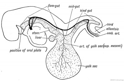
At the commencement of the 3rd week the embryo appears to be situated as a cap on the yolk sac (Fig. 75, p. 96). As the chorion and amnion represent the extra-embryonic part of the somatopleure, so the yolk sac represents the extra-embryonic part of the splanchnopleure (Fig. 72, p. 93). At the end of the 3rd week the alimentary tract shows three divisions (Fig. 221): 1. A short anterior diverticulum — the fore-gut; it becomes the pharynx, oesophagus, stomach, and that part of the duodenum situated in front of the opening of the common bile duct ; 2. The hind-gut — a short posterior diverticulum ; it forms the rectum, sigmoid and descending colon ; its terminal part forms the cloaca from which the allantois grows out ;
3. The mid-gut or roof of the yolk sac, from which is formed that part of the gut between the opening of the common bile duct in front to the splenic flexure of the colon behind. Only the fore-gut has a ventral mesentery; the mid-gut and hind-gut (excepting the cloaca) are suspended by a dorsal mesentery only.
Fig. 221. The Form of the Alimentary Canal in a human embryo of the 3rd week.
The Yolk Sac and Meckel's Diverticulum. — The fore and hind gut may be regarded as diverticula of the yolk sac. The yolk sac reaches its maximum size in the 4th week. In the 3rd week the umbilicus extends along the whole length of the abdomen, from the septum transversum to the allantois. The neck of the yolk sac completely fills it. The vitelline arteries, which afterwards form the superior mesenteric, end on its walls, the vitelline veins commence on them (Fig. 221).
In the fifth week the form of the alimentary tract is that shown in Fig. 222. The condition then differs from that shown in the 3rd week in the following points :
1. The production of the mid -gut as a U-shaped loop from the roof of the yolk sac ;
2. The formation of a long neck to the yolk sac — the vitellointestinal canal ; if it persists it forms a Meckel's diverticulum ;
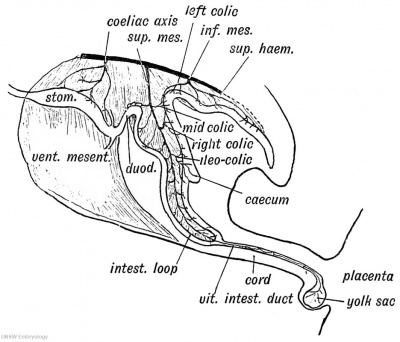
3. The yolk sac, by the constriction of the umbilical orifice and formation of the cord, comes to lie on the placenta (Fig. 222). The neck of the yolk sac, the vitello-intestinal duct, lies within the umbilical cord. By the sixth week the sac is in a state of retrogression. The U-shaped intestinal loop, which is formed from the mid-gut, is at first really extra-abdominal, being situated within a funnel-shaped cavity formed by the somatopleure at the umbilicus. The vitelline artery, afterwards the superior mesenteric, is the artery of the TJ-shaped loop ; it terminates at the vitellointestinal canal — the elongated neck of the yolk sac (Fig. 222).
Fig. 222. The Form of the Alimentary Canal during the 5th week.
Persistence of Certain Embryonic Structures
Many of the features seen in the human embryo at the stage of development reached during the fifth or sixth weeks may persist.
1. The most common structure to remain is the intestinal end of the neck of the yolk-sac — Meckel's diverticulum. It occurs in 2 per cent, of subjects, and commonly forms a fingerglove-like sac on the free border of the ileum about four feet above the ileo-caecal valve. Hence we know that this part of the ileum forms the apex of the U-shaped loop of intestine.
2. The neck of the yolk sac, instead of closing, may remain open at the umbilicus and form a faecal fistula at birth.
3. The artery of the yolk-sac, the terminal part of the superior mesenteric, may persist as a fibrous band which stretches from the mesentery at the situation of Meckel's diverticulum to the umbilicus. Over it, the gut may become strangulated.
4. The U-shaped loop, instead of retreating within the abdomen at the end of the second month, may remain within the umbilical funnel of somatopleure. This gives rise to a congenital umbilical hernia. Such hernias occur in all degrees ; they may contain a piece of intestine, or almost the whole of the abdominal contents. In such cases the somatopleure, or belly wall, which forms the covering of the hernia, is commonly thin and transparent.
Derivatives of the Hind-Gut
The Rectum and cloaca are formed out of the posterior end of the hind-gut. The manner in which they become separated from the allantois and open in the proctodaeurn at the anal depression has been described (page 118).
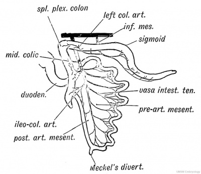
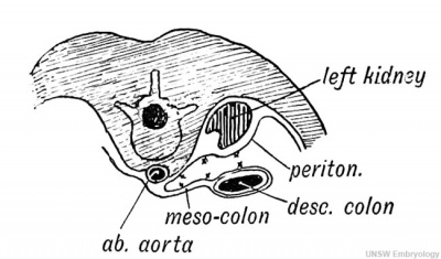
The Descending Colon and Sigmoid Flexure are also formed out of the hind-gut. The artery of the hind-gut is the inferior mesenteric (Fig. 222). Hence it supplies the rectum, sigmoid, and descending colon. In the fifth week the hind-gut is suspended from the front of the aorta and spine by the dorsal mesentery of the hind-gut. This becomes transformed into the meso-rectum, meso-sigmoid and descending meso-colon. The angle between the hind-gut and U-shaped loop becomes the splenic flexure (Fig. 222). At the commencement of the third month the U-shaped loop has become twisted round on the axis of the superior mesenteric artery (Fig. 223^4), so that the part of the hind-gut which forms the splenic flexure is turned forwards and to the left until it touches the spleen (Fig. 226). It carries its artery, the left colic, with it. At this time the anterior limb ■of the U-loop grows rapidly, and is produced into toils of small intestine— the jejunum and ileum — which press the descending meso-colon against the kidney and the parietal peritoneum covering the left kidney (Fig. 223 B). The left surface and layer ■of the meso-colon adheres to the pre-renal layer of the peritoneum, both layers subsequently being absorbed. Thus the descending meso-colon, originally situated in the middle line, comes to be attached in the left lumbar region.
Fig. 223 A. The mesentery of the hind-gut. The Position assumed by the Colon after the rotation of the Gut has taken place.
Fig. 223 B. Diagram to show how the descending Meso-colon becomes applied to the parietal Peritoneum of the left Lumbar Region.
The Intersigmoid Fossa
The sigmoid flexure, after the rotation of the gut, forms a loop, with its convexity directed towards the liver. The meso-sigmoid is originally attached in the middle line, but the pressure of the developing loop of small bowel pushes the meso-sigmoid against the posterior abdominal wall and left iliac fossa. The extent to which it adheres varies; it may become completely adherent like the descending meso-colon, or only partially. When the sigmoid is lifted up a recess or fossa may be apparent beneath the meso-sigmoid, to the outer side of the left common iliac artery, which is due to a failure of adhesion between the meso-sigmoid and parietal peritoneum. It occurs opposite the convexity of the sigmoid loop.
Development of the Colon and Caecum
In the sixth week a process or diverticulum is seen to grow out from the free border of the posterior limb of the U-shaped loop (Fig. 225 A). The diverticulum forms the caecum and appendix. It continues to grow outwards and downwards. The part of the posterior loop above the caecal diverticulum becomes increased in diameter and forms the ascending and transverse parts of the colon. As the superior mesenteric (vitelline) artery descends in the loop, it gives off three branches to the posterior limb — the middle colic, right colic and ileo-colic arteries (Figs. 222 and 223 A). The mesentery of the U-shaped loop may be divided into two parts, the fate of the two parts being different : 1. The mesentery of the anterior limb in front of the superior mesenteric artery — the pre-arterial part. This forms the greater part of the mesentery of small bowel.
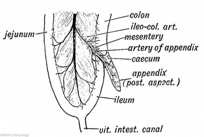
Fig. 224. Diagram of the Apex of the Caecum at the time of birth and the Diverticula which may be produced in the Fundus of the Caecum afterwards.
2. The mesentery of the posterior limb, behind the artery — the post-arterial part. It forms the mesentery of the ascending and transverse colon and also the lower part of the mesentery of the . small bowel.
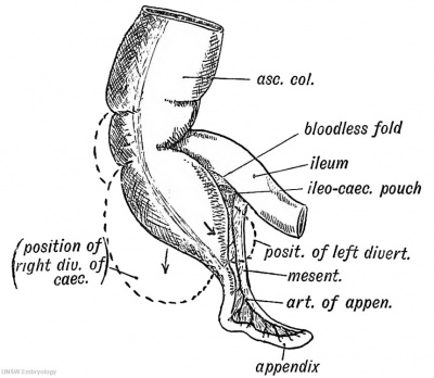
Fig. 225 A. The Appendix and Peritoneal Folds at the end of the 2nd month of foetal life. The Intestinal Loop is viewed from the left side.
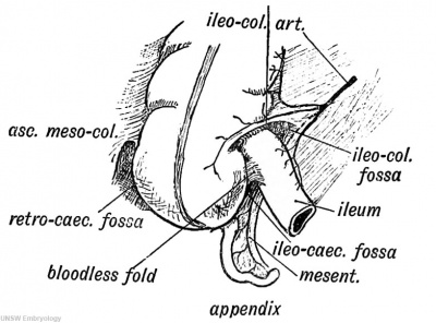
Fig. 225 B. Peritoneal Fossae in the lleo-caecal Region.
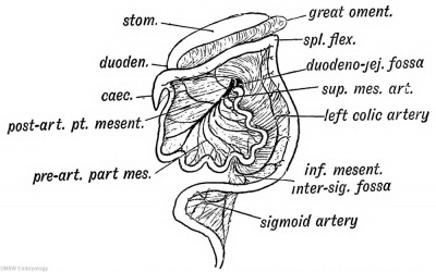
At the seventh week the great growth of the anterior limb, to form the coils of the jejunum and ileum, causes the U-shaped loop to rotate so that the splenic flexure of the colon comes against the spleen. This brings the transverse meso-colon, containing the middle colic artery, against the part of the mesogastrium which forms the great omentum (Fig. 226). These two layers adhere. The rotation places that part of the loop mesentery, which forms the ascending meso-colon, against the duodenum, and at the same time the duodenal loop is pressed into its permanent position in front of the right kidney and inferior vena cava. The caecum thus comes to be situated in front of the right kidney, near the gall-bladder, and there it remains until about the time of birth, when both the caecum and ascending colon undergo a gradual migration towards the right iliac fossa. The cause of this migration is not known, but it occurs only in animals adapted to the upright posture. Thus the attachment of the ascending meso-colon is formed by a secondary adhesion to the parietal peritoneum during the migration of the colon and caecum. The appendix, during . the migration, may be caught behind the colon ; it is then lodged and fixed in the ascending meso-colon.
Fig. 226. To show the Rotation of the Intestinal Loop and Formation of the Duodenojejunal Fossa.
The Appendix
At first and until the third month, the caecal diverticulum is of the same calibre throughout, but from the third month onwards, the appendix remains small while the caecum grows, keeping pace in diameter with the colon. At birth the appendix is still the tapered apex of the caecal diverticulum (Fig. 224), but during childhood, an outer, or an inner, sacculation, or both together, arise in the fundus of the caecum and thrust the appendix backwards and to the left into an asymmetrical position.
Although a distinctly marked appendix is only seen in man, the anthropoids, lemur, and opossum, still a corresponding lymphoid structure is present generally in mammals. The appendix is a lymphoid diverticulum of the caecal apex (E. J. Berry). It must be regarded as a lymphoid structure, and although it can be dispensed with, is not therefore to be regarded as vestigial in nature any more than is the tonsil. The caecum is largest, as is also the bowel, in herbivorous animals.
Ileo-caecal Fossae. — When the caecal diverticulum grows out from the hinder limb of the U-shaped loop it carries with it three folds (see Fig. 224):
- The ileo-colic fold, a process from the right side of the mesentery containing the anterior caecal artery ;
- The bloodless fold, a process from the coat of the ileum ;
- The mesentery of the appendix, a process from the left side of the mesentery, containing the artery to the appendix (Fig. 225 A).
These three folds give rise to three fossae (Fig. 225 5):
- The ileo-colic, between the termination of ileum and ileocolic fold ;
- The ileo-caecal, between the bloodless fold and mesentery of the appendix ;
- The retro-caecal, between the mesentery of the appendix and commencement of the ascending meso-colon.
The Duodenum
The part of the duodenum above the entrance of the common bile-duct is formed from fore-gut, the part behind from the mid-gut. The liver and pancreatic buds arise from its ventral border at the junction of these two parts. At first it is entirely covered by peritoneum and suspended in the mesentery, in which it forms a minor loop (Figs. 219, 222). In its concavity rests the head of the pancreas.
In the 7th week the U-shaped loop of intestine rotates so as to bring the ascending colon, with its mesentery, against the duodenal loop, which, with the head of the pancreas, is thus pressed against the right kidney and inferior vena cava (Fig. 226). The peritoneum covering the right aspect of the head of the pancreas and duodenal loop adheres to the parietal peritoneum covering the kidney, and disappears. The transverse meso-colon gains an attachment to the front of the duodenum.
The Duodeno-jejunal Fossa is formed as the U-shaped loop of bowel rotates, dragging the transverse meso-colon after it (Fig. 226). The inferior mesenteric vein passes forwards along the dorsal mesentery to the splenic vein, which is also then in the dorsal mesentery (Fig. 187, p. 229). The mesentery of the hind-gut is stretched as the ascending colon migrates to the right ; the inferior mesenteric vein checks it and gives rise to a fold, which bounds the fossa (Fig. 226).
The Mesentery of the small gut is formed out of the primitive mesentery of the U-shaped intestinal loop, chiefly from that part of it (the pre-arterial) which lies between the superior mesenteric artery and the anterior limb of the loop (Fig. 223 A). After the rotation, the aspect of the mesentery, which was directed towards the right, becomes left and anterior. During the rotation of the gut the superior mesenteric artery comes to lie in front of the third stage of the duodenum. At first the mesentery is attached in front of the spine only at the origin of the superior mesenteric artery. Its oblique attachment to the posterior abdominal wall, from the duodenum to the right iliac fossa, is a secondary adhesion, formed after the rotation of the gut, and this extensive attachment is found only in animals adapted to the upright posture.
| Historic Disclaimer - information about historic embryology pages |
|---|
| Pages where the terms "Historic" (textbooks, papers, people, recommendations) appear on this site, and sections within pages where this disclaimer appears, indicate that the content and scientific understanding are specific to the time of publication. This means that while some scientific descriptions are still accurate, the terminology and interpretation of the developmental mechanisms reflect the understanding at the time of original publication and those of the preceding periods, these terms, interpretations and recommendations may not reflect our current scientific understanding. (More? Embryology History | Historic Embryology Papers) |
Human Embryology and Morphology (1902): Development or the Face | The Nasal Cavities and Olfactory Structures | Development of the Pharynx and Neck | Development of the Organ of Hearing | Development and Morphology of the Teeth | The Skin and its Appendages | The Development of the Ovum of the Foetus from the Ovum of the Mother | The Manner in which a Connection is Established between the Foetus and Uterus | The Uro-genital System | Formation of the Pubo-femoral Region, Pelvic Floor and Fascia | The Spinal Column and Back | The Segmentation of the Body | The Cranium | Development of the Structures concerned in the Sense of Sight | The Brain and Spinal Cord | Development of the Circulatory System | The Respiratory System | The Organs of Digestion | The Body Wall, Ribs, and Sternum | The Limbs | Figures | Embryology History
Reference
Keith A. Human Embryology and Morphology. (1902) London: Edward Arnold.
Cite this page: Hill, M.A. (2024, April 27) Embryology Book - Human Embryology and Morphology 18. Retrieved from https://embryology.med.unsw.edu.au/embryology/index.php/Book_-_Human_Embryology_and_Morphology_18
- © Dr Mark Hill 2024, UNSW Embryology ISBN: 978 0 7334 2609 4 - UNSW CRICOS Provider Code No. 00098G

