Book - Human Embryology and Morphology 16
| Embryology - 1 May 2024 |
|---|
| Google Translate - select your language from the list shown below (this will open a new external page) |
|
العربية | català | 中文 | 中國傳統的 | français | Deutsche | עִברִית | हिंदी | bahasa Indonesia | italiano | 日本語 | 한국어 | မြန်မာ | Pilipino | Polskie | português | ਪੰਜਾਬੀ ਦੇ | Română | русский | Español | Swahili | Svensk | ไทย | Türkçe | اردو | ייִדיש | Tiếng Việt These external translations are automated and may not be accurate. (More? About Translations) |
Keith A. Human Embryology and Morphology. (1902) London: Edward Arnold.
| Historic Disclaimer - information about historic embryology pages |
|---|
| Pages where the terms "Historic" (textbooks, papers, people, recommendations) appear on this site, and sections within pages where this disclaimer appears, indicate that the content and scientific understanding are specific to the time of publication. This means that while some scientific descriptions are still accurate, the terminology and interpretation of the developmental mechanisms reflect the understanding at the time of original publication and those of the preceding periods, these terms, interpretations and recommendations may not reflect our current scientific understanding. (More? Embryology History | Historic Embryology Papers) |
Chapter XVI. Development of the Circulatory System
Veins
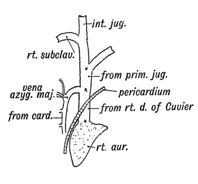
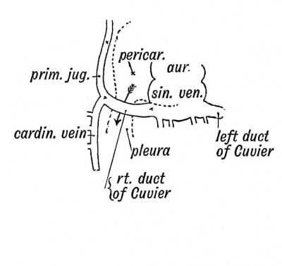
(1) The Superior Vena Cava arises from the following foetal vessels (Figs. 181 and 182): (a) The part above the entrance of the vena azygos is the terminal part of the right primitive jugular or anterior cardinal vein ; (b) The part below the entrance of the vena azygos major arises from the right duct of Cuvier. The condition of these venous trunks, the anterior and posterior cardinal veins and Ducts of Cuvier, in a human embryo of the 3rd week is shown in Fig. 182. The condition shown is retained permanently in lower vertebrates (Fishes, etc.).
|
|
| Fig. 181. The Superior Vena Cava of the Adult. | Fig. 182. The Embryonic Venous Trunks out of which the Superior Vena Cava is formed. |
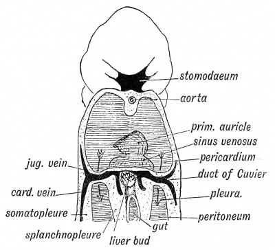
The anterior Cardinal or primitive Jugular Vein, which drains the anterior half of the body on each side with the posterior cardinal vein, which drains the posterior half of the body, receive a tributary (segmental vein) from each body segment. The cardinal veins lie in the mesoblast on the dorsal side of the coelom at the junction of the splanchnopleure and somatopleure (Fig. 183). By their union they form on each side the Duct of Cuvier which conveys the blood to the sinus venosus — a contractile chamber opening into the primitive auricle. The sinus venosus remains as a separate chamber of the heart in lower vertebrates, but in the course of mammalian development it becomes partly merged in the right auricle of the heart.
Fig. 183. Diagram to show the maimer in which the Ducts of Cuvier encircle the Coelom at the junction of the Pericardial and Pleural Parts (Iter venosum). (After His.).
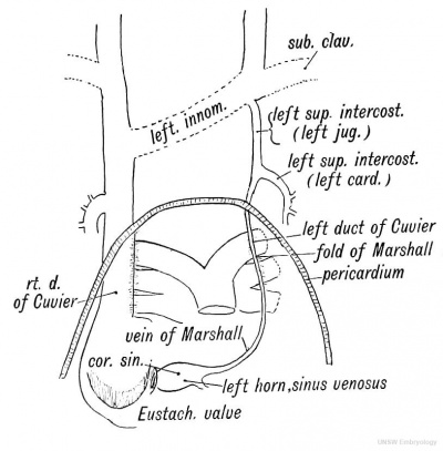
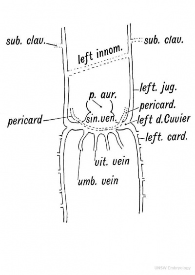
It is important to notice how each duct of Cuvier reaches the sinus venosus (see Fig. 183). They pass from the dorsal to the ventral surface of the body in the somatopleure and thus encircle the coelom. The right and left ducts of Cuvier lead to a constriction of the coelom, the fold in which each descends being known as the lateral or venous meso-cardium (Fig. 205, p. 251). "Ultimately, by the end of the 4th week, the part of the coelom lying in front of the] ducts of Cuvier is cut off from the rest; the part so cut off forms the pericardium (Fig. 201). Thus the ducts of Cuvier are instrumental in separating the pericardial from the pleural cavity. If the primitive pleuropericardial communication (iter venosum of Lockwood) persists between them, it occurs as a foramen in the pericardium behind the part of the superior vena cava derived from the duct of Cuvier. 2. The Vestigial Fold and Oblique Vein of Marshall. — In the human embryo during the 3rd week and for some weeks afterwards there is a right and left duct of Cuvier and corresponding cardinal veins (Fig. 185). A left superior vena cava is present and may persist. The vestigial fold and oblique vein of Marshall (Fig. 184) are all that usually remain of the left superior vena cava. The right superior vena cava within the pericardium passes in front of the right pulmonary vessels, and is bound to them by a mesentery or fold of serous pericardium ; the left has a similar relationship (Fig. 184); when it disappears this fold remains in front of the left pulmonary vessels as the vestigial fold. The intra-pericardial part of the left vena cava or duct of Cuvier becomes the oblique vein (Fig. 184): it turns round the left auricle to terminate in the left horn of the sinus venosus (coronary sinus). The extra-pericardial part of the left duct of Cuvier joins the superior intercostal vein (Fig. 184). Both right and left superior venae cavae persist in some lower mammals.
Fig. 184. The Remnants of the Left Superior Vena Cava, derived from the Structures shown in Fig. 185.
Fig. 185. Diagram of the Sinus Venosus and Duets of Cuvier of the human embryo about the 3rd week.
The left superior intercostal vein represents the following embryonic vessels (see Fig. 184): (a) Anterior part of the left posterior cardinal vein ; (b) The extra-pericardial part of the left duct of Cuvier ; (c) The terminal part of the left primitive jugular vein.
3. The Left Innominate Vein opens up as a channel of communication between the two primitive jugular veins, the left superior vena cava undergoing a simultaneous process of atrophy (Fig. 184).
4. The Subclavian Veins are developed in the 4th week with the outgrowth of the fore-limb buds ; they open into the primitive jugulars (Fig. 184).
5. The Primitive Jugular Veins escape from the cranial cavity in front of the ear. A trace of the opening may occasionally be detected at the root of the zygoma behind the post-glenoid spine (A. Cheatle). The petro-squamous sinus represents the intracranial part of this vein (p. 56). As the internal jugular vein opens up, the primitive jugular vein within the skull becomes atrophied. The temporo-maxillary vein and the external jugular vein probably represent the extra-cranial part of the primitive jugular vein. It must be remembered that the caudalward migration of the heart affects the primitive relationship of the veins in the neck. They are drawn backwards with it.
Veins formed from the Posterior Cardinals of the Embryo. — 1. From the branches of the Cardinal Veins. Each posterior cardinal vein receives on its own side — (a) A branch from each body segment from the lower cervical to the last caudal. These become the intercostal, lumbar and sacral veins. (5) The segmental veins of the intermediate cell mass (Fig. 85), which become the suprarenal, renal, spermatic, ovarian, uterine and vesical veins. (c) When the hind-limb buds grow out their veins join that part of the cardinal veins which become the common iliac. The lateral sacral, ilio-lumbar and ascending lumbar veins are anastomotic channels formed between the segmental veins.
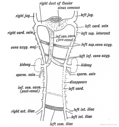
From the right cardinal vein are formed (1) the vena azygos major ; (2) the post-renal part of the inferior vena cava (Fig. 186).
The part above the entrance of the right renal becomes the vena azygos major ; the part below, the inferior vena cava. Hence it is that the origin of the vena azygos can commonly be traced to the renal vein. The ascending lumbar vein, which also ends in the vena azygos major, is, as already mentioned, a new anastomotic channel.
From the left cardinal arise (Fig. 186) — (1) Part of the left superior intercostal vein ; (2) Left superior azygos vein ; (3) Left inferior azygos, which commences in the left renal vein, and also receives the left ascending lumbar. The post-renal part of the left cardinal disappears in higher mammals; occasionally it persists in man and very frequently in the rabbit.
Fig. 186. The Remnants of the Posterior Cardinal Veins in the Adult. The new channels are shaded. (After Hoohstetter.)
The greater part of the left common iliac vein arises, like the left innominate, as a communicating channel between the posterior cardinals. It is formed as the post -renal part of the left cardinal becomes obliterated (Fig. 186).
The Inferior Vena Cava
The post-renal part is formed from the right cardinal vein (Fig. 186). The pre-renal part, with the mesial part of the left renal vein, is quite a new formation which opens up a short circuit to the heart for the blood of the lower half of the body. The formation of this new channel leads to the retrogression of the thoracic stages of the cardinal veins and their formation into the azygos veins. The exact date of the origin of the inferior vena cava in the human foetus is not known ; the newchannel is said to grow out from the ductus venosus of the liver, and growing downwards opens up a connection with the right and left cardinal veins at the entrance of the renal vessels (Fig. 188). Thus in the formation of the inferior vena cava are included three elements — (1) the terminal or upper part of the ductus venosus ; (2) the new channel ; (3) the inferior or postrenal part of the right cardinal.
Occasionally it happens that pressure on the hepatic part of the vena cava, from tumours, cirrhosis of the liver, etc., leads to the azygos veins, the early foetal channels, being again opened up.
The Portal Vein.
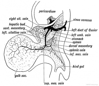
The Portal Vein is formed out of the two vitelline veins — the first of all the veins to be developed. They end in the posterior chamber of the tubular heart of the embryo — the sinus venosus. The vitelline veins, right and left, arise from ramifications on the yolk sac and pass in the splanchnopleure to the sinus venosus (Fig. 187). The nutriment within the yolk sac is thus carried to the heart and distributed by the heart to the tissues of the embryo and yolk sac. The commencement of the left vitelline or omphalo-meseraic vein disappears. The right (Fig. 187) forms the superior mesenteric vein. It commences on the yolk sac, of which a remnant (the neck) may remain as Meckel's diverticulum.
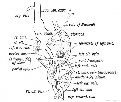
The terminal parts of the two vitelline veins are joined by three transverse communications, the upper two of which are included in the portal vein. The uppermost of the three afterwards lies in the transverse fissure of the liver (Fig. 188). The middle communication lies on the dorsal aspect of the duodenum (Fig. 188). The splenic vein and inferior mesenteric pass in the dorsal mesentery (Fig. 187) and join this transverse communication which may be named the supra-duodenal junction. The third or lowest of the junctional trunks is situated on the ventral aspect of the duodenum (Fig. 188). Two parts may be recognised in the portal vein of the adult : (1) the part which lies behind the pancreas and duodenum ; (2) the part in the gastro-hepatic omentum and transverse fissure of the liver.
Fig. 187. The Left Vitelline Vein of an Embryo of the 4th week.
The retro-duodenal part is formed out of the left vitelline vein and the supra-duodenal junction (Fig. 188) ; the omental stage is formed out of the right vein and the uppermost of the junctional trunks (Fig. 188). The part of the portal vein within the transverse fissure of the liver represents the third or uppermost junction between the left and right vitelline veins.
The Hepatic Veins are formed out of the terminal parts of the vitelline and umbilical veins. These veins end at first in the sinus venosus (Fig. 187). The liver is developed between and around the vitelline and umbilical veins, near their termination in the sinus venosus. The veins are broken up and a fine intra-hepatic venous network takes their place. Thus it comes about that the vitelline veins are transformed into the veins of the portal and hepatic circulation. All the foetal and umbilical blood is at first poured through the liver.
Fig. 188. Diagram showing the Formation of the Ductus Venosus, and the fate of the Umbilical and Vitelline veins. The arrows show the parts of the Vitelline Veins which become the Portal Vein.
The Ductus Venosus is a new channel formed between the uppermost of the anastomoses between the right and left vitelline veins and the sinus venosus whereby the greater part of the umbilical blood is short-circuited to the heart without passing through the liver. It appears after the liver bud has broken up the vitelline venous trunks (Fig. 188). After birth, when a short circuit is no longer required between the foetal circulation and heart, it becomes reduced to a fibrous cord. It occupies the posterior part of the longitudinal fissure of the liver and lies within the hepatic attachment of the gastro-hepatic omentum.
The pre-renal part of the inferior vena cava is developed as an outgrowing channel from the ductus venosus (Fig. 189).
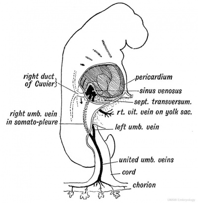
The Umbilical Veins. — The umbilical vein at birth consists of two parts: (1) A part within the umbilical cord; (2) another within the body, enclosed in the falciform ligament and anterior half of the longitudinal fissure of the liver. It joins there the ductus venosus and portal vein (Fig. 189). The condition of the umbilical veins in a human embryo of three weeks is shown in Fig. 190. They are formed after the vitelline veins but before the ducts of Cuvier, which afterwards terminate in the sinus with the umbilical veins. The veins lie in the somatopleure and drain the blood of the chorion, a derivative of the somatopleure. It passes from the chorion to the body wall in the umbilical cord, which is also formed from the somatopleure as well as allantois. In front the vein of each side joins the sinus venosus with the duct of Cuvier. There is a right and left vein, but in the cord they have fused into one. Within the body the right umbilical vein completely disappears during early embryonic life. The outgrowth of the liver-bud breaks up not only the vitelline veins but also the umbilical at their junction with the sinus venosus (Figs. 185 and 188). Thus the umbilical blood as well as the vitelline comes to be poured into the liver. The left umbilical vein within the body remains permanently ; when its terminal part is broken up by the outgrowth of the liver it becomes united with the uppermost of the transverse communications between the right and left vitelline veins, which, as already mentioned, form the part of the portal vein within the transverse fissure of the liver. The left umbilical vein thus comes into communication with the ductus venosus (see Figs. 188 and 189).
Fig. 189. Diagram of the remnants of the Umbilical Vein in the Adult-viewed from behind.
Fig. 190. Diagram of the Right Umbilical Vein in an embryo of 3 weeks, before the outgrowth of the Liver Bud. (Modified from His.)
Development of the Heart
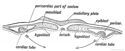
The Heart. — In the 2nd week, while the embryo is still in the blastoderm stage, a contractile tube appears in the splanchnopleure on each side (Fig. 191). Each tube receives blood from the yolk sac by a vitelline vein ; each pumps its blood into an aortic stem which terminates as the artery of the yolk sac.
Fig. 191. Transverse section of the Blastoderm showing the Right and Left Cardiac Tubes situated in the Splanchnopieure.
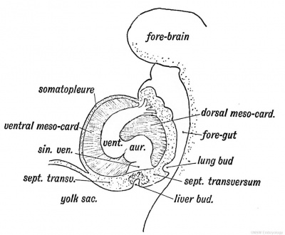
These two tubes become fused together to form a simple tubular heart by the 3rd week. The cardiac tubes unite as the splanchnopleures come together (see Figs. 191 and 192). The lining membrane of the cardiac tubes is probably furnished by an invagination of the hypoblast (Fig. 191). The heart is at first suspended by a mesentery, which stretches from the fore-gut (Fig. 192) to the ventral median line of the body wall and separates the anterior end of the coelom into right and left halves. The mesentery is formed out of the fused splanchnopleures (Fig. 192). The part of the cardiac mesentery between the heart and the fore-gut forms the dorsal mesocardium, the part between the heart and the ventral wall, the ventral mesocardium. The anterior end of the coelom which is occupied by the heart becomes the pericardium. The pericardium is situate beneath the primitive pharynx.
Fig. 192. Transverse section at a more advanced stage showiDg the union of the Splanchnopleures to form the Mesocardia and the fusion of the Right and Left Cardiac Tubes.
Demarcation into Chambers
In the third week the heart undergoes three important changes : (1) The dorsal and ventral mesocardia disappear and the heart, all but the anterior and posterior extremities, is left free in the anterior end of the coelom. This can best be understood by a reference to Fig. 193. The heart is there seen suspended between the fore-gut and ventral wall. The tubular heart is formed behind by the union of the right and left vitelline veins from the yolk sac and ends in front as the first or mandibular aortic arches. The posterior end of the mesocardium, both the ventral and dorsal parts, persists — the part which encloses the sinus venosus and this forms the primitive basis of the diaphragm or septum transversum (Fig. 202, p. 244).
Fig. 193. Lateral view of the Heart and Pericardium to show the Attachments of the Dorsal and Ventral Mesocardia (schematic).
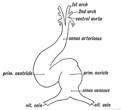
(2) The tubular heart shows demarcations into four parts (Fig. 194): (a) Sinus venosus; (b) Primitive auricle; (c) Primitive ventricle ; (d) Conus or bulbus arteriosus. In this condition (third week) the human heart is exactly like that of a fish, viz., a tubular four-chambered heart which pumps blood into the branchial or aortic arches.
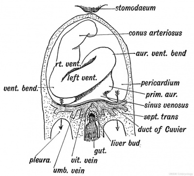
(3) The disappearance of the mesocardium allows the heart to become twisted and bent. Two chief bends are formed which materially help to give the heart its adult shape (Fig. 195):
(a) The Ventricular Bend. — The ventricular part of the tube is bent into a V-shaped piece, the apex of the V-shaped loop beingturned towards the right.
(b) The Auriculo-ventricular Bend. — The ventricular part is bent in front of the auricular so that the auricle becomes dorsal to the ventricle (Fig. 195).
Fig. 194. The Primitive Divisions of the Heart.
The Sinus Venosus. — The sinus venosus, the first chamber of the foetal heart, is formed by the union of the vitelline veins ; the umbilical veins and ducts of Cuvier come subsequently to open in it (Fig. 185). The sinus is imbedded in the persistent posterior part of the mesocardium (Fig. 202).
Through the ventral part of the mesocardiurn the ducts of Cuvier reach the sinus from the somatopleure (Fig. 183). In fishes and in the human embryo the sinus pumps the blood into the primitive auricle, and its orifice into the auricle is protected by two valves, right and left (Fig. 197).
Fig. 195. Showing the two chief Bends which occur in the Heart during the 3rd week.
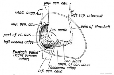
Fate of the Sinus Venosus (Fig. 196). — The sinus venosus shifts towards the right side of the primitive auricle and ultimately forms part of the right auricle and the coronary sinus. The part which it forms of the right auricle is indicated by the entrance of the following vessels which primarily terminate in the sinus : (1) The superior vena cava (the right duct of Cuvier); (2) The inferior vena cava, which also opens into the sinus ; (3) The oblique vein of Marshall (left duct of Cuvier), which opens into the left horn of the sinus venosus. The left horn of the sinus becomes the coronary sinus. A groove, the sulcus terminalis, which is marked on the interior of the right auricle by a crest, runs down on the anterior wall of the right auricle from the superior to the inferior vena cava, and indicates the junction of the primitive auricle with the sinus venosus.
Fig. 196. Showing the Structures formed from the Sinus Venosus.
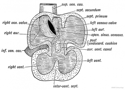
Fig. 197. Section of the Heart of a 5th week human foetus showing the Right and Left Venous Valves which guard the entrance of the Sinus Venosus into the Primitive Auricle. (After His.)
The Valves of the Sinus Venosus
Eight and left lateral valves (venous valves) guard the entrance of the sinus to the primitive auricle and prevent the regurgitation of blood when the auricle contracts (Fig. 197). The right valve becomes reduced, and ultimately forms the Eustachian valve, and the Thebesian which is always connected with the Eustachian (Fig. 196). The part of the Eustachian valve prolonged to the annulus ovalis is a new formation. What becomes of the left valve is not certain ; it may disappear completely, but more probably it amalgamates with, and forms part of the septum primum (see Figs. 196, 197, and 198). The Eustachian and Thebesian valves also help to indicate the part of the right auricle, which is formed from the sinus venosus, for they bound the right side of the entrance of the sinus to the auricle.
The Cardiac Septa
Every modification of the heart, from its simple tubular form in fishes to its complete division into separate pulmonary and systemic pumps, can be seen in the vertebrate series. In amphibians the primitive auricle becomes divided into right and left chambers, but the ventricle is undivided. In crocodilia an incomplete ventricular septum, with a complete auricular, may be seen. In birds and mammals the auricular and ventricular septa are complete, and the sinus venosus becomes part of the right auricle. Occasionally the septa are incompletely formed in man, conditions found in the lower vertebrates being thus produced.
The division of the simple tubular heart into right and left halves is rendered possible by four changes which take place in its shape :
(1) The ventricle, instead of being a bent tube, becomes dilated and bag-like.
(2) The opening of the sinus venosus in the primitive auricle migrates towards its right side, and thus opens in that part of the primitive auricle which becomes the right (Fig. 197).
(3) The auricle sends out an appendix on each side; these grow forwards and nearly surround the bulbus arteriosus.
(4) The communication between the common auricle and ventricle is drawn out into a tube — the auriculo-ventricular canal (Fig. 198).
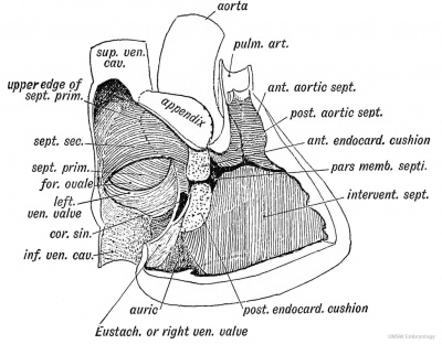
The Division of the Heart is effected by five Elements or Septa {Fig. 198). These are:
- The endo-cardial cushions.
- The Inter- ventricular septum.
- The Aortic septa.
- The septum primum of the auricle.
- The septum secundum of the auricle.
(1) The auricular or auriculo-ventricular canal is divided by anterior and posterior endocardial cushions, which grow out and meet, dividing the auricular canal into the right and left auriculoventricular orifices.
(2) The inter-ventricular septum rises from the floor of the common ventricle and grows upwards towards the aortic orifice and endocardial cushions (Figs. 197 and 198).
(3) In the aortic bulb (Fig. 198) appear anterior and posterior aortic ridges, arranged spirally. They fuse together ; their lower extremities unite with the inter-ventricular septum. The conus or bulbus arteriosus is thus divided into an anterior or pulmonary part connected with the right ventricle, and a posterior or aortic part connected with the left ventricle.
Fig. 198. Diagram of the Septa of the Heart viewed on the right side.
Pars Membranacea Septi.— The three septa just described meet about the 6 th week, and are fused together by a thin membrane — the pars membranacea septi (Fig. 198). It is seen under cover of the septal segment of the tricuspid valve and is the last part of the septum to form ; occasionally it fails to form, and a communication exists between the ventricles. This condition is commonly associated with a partial failure in the formation of the pulmonary artery. Such a condition may give rise to cyanosis ; the subjects of it commonly die before 15 of pulmonary tuberculosis. The walls of the right and left ventricle in such cases are of equal thickness, for both act as common systemic and pulmonary pumps.
(4) The septum primum of the auricle springs from the posterior wall of the primitive auricle, and falls like the blade of a guillotine on the endocardial cushions with which it fuses (Figs. 197 and 198). Its upper border separates from the auricular wall. It forms the septum ovale. As it descends it involves and fuses with the left venous valve.
(5) The septum secundum grows down from the roof and posterior wall of the primitive auricle to the right of the septum primum. It forms the annulus ovalis and the auricular septum above it (Fig. 198).
The foramen ovale (Fig. 198) is an interval left between the two auricular septa ; the Eustachian valve becomes connected with the annulus (septum secundum). In 75 per cent, of people, following Fawcett's statistics, the foramen becomes closed in the first year after birth. In 2 5 per cent, it remains partially open, but even when an opening remains the blood could pass from the right to the left auricle only when the pressure within the right auricle is greater than within the left. The valvular action of the septum ovale prevents the passage of blood from the left to the right auricle. The foramen ovale is an adaptation to the foetal form of circulation.
Thus the primitive auricle is divided into right and left chambers by two septa — septum 'primum and secundum ; the bulbus arteriosus by the anterior and posterior aortic septa ; the auriculo-ventricular canal by the anterior and posterior endocardial cushions; 'and, lastly, the primitive ventricle into right and left by the inter-ventricular septum.
Tricuspid and Mitral Valves
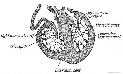
It is to be remembered that the interior of the ventricular cavities during the second month is filled with a muscular sponge work, like the ventricle of the frog's heart (Fig. 199). Out of this muscular network are formed : (1) Chordae tendineae ; (2) Musculi papillares ; (3) The moderator band ; (4) Trabeculae and columnae carneae; (5) Musculi pectinati (in the auricular appendages).
Fig. 199. Section of the Ventricles of the Foetal Heart, showing the Muscular Sponge Work within their Cavities. (After His.)
The endocardium lining the auricular canal (see Figs. 197 and 199) becomes folded or protruded within the ventricles by a shortening of the auricular canal. The funnel-shaped fold or inflection of endocardium is the basis of the auriculo-ventricular valves. They are formed at the same time as the septa of the heart. Originally funnel-shaped, the left or mitral comes to show two cusps — septal and left, the right or tricuspid three segments — septal, anterior and posterior. The funnel shape is frequently partially retained. The cardiac tissue on the outer aspect of the valves is opened out into a sponge-work continuous with that of the ventricle. Hence the attachment of the chordae tendinae on the outer surfaces and edges of the valves.
The Semilunar Valves
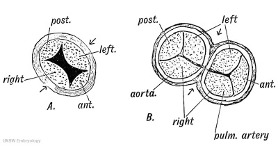
The bulbus arteriosus is divided by the aortic septa into an anterior and right part — the pulmonary aorta, and a posterior and left — the ascending aorta. Just before the division takes place four cushions of the endocardium arise at the junction of venticle and aortic bulb (Fig. 200 A). The four cushions — anterior, posterior and two lateral — become cupped above and valvular. When the aortic septa are formed, each lateral cushion is divided in two, thus forming the two posterolateral semilunar valves of the pulmonary aorta and the two antero-lateral of the aorta. The anterior and posterior cushions remain as the anterior pulmonary semilunar valve and the posterior aortic valve (Kg. 200 5).
Fig 200. The origin of the semilunar valves.
A. The four Endocardial Cushions in the Aortic Bulb.
B. The division of the Lateral Cushions to form two Aortic and two Pulmonary Semilunar Valves.
Remnants of the Foetal Circulation in the Adult
The nature of these remnants has been already described ; they need be only enumerated here. They are : 1. The Obliterated Hypogastric Arteries ; 2. The Umbilicus ; 3. The Eound Ligament of the Liver ; 4. The Fibrous Eemnant of the Ductus Venosus ; 5. The Eustachian Valve ; 6. The Foramen Ovale ; 7. The Fibrous Eemnant of the Ductus Arteriosus.
The Coelom, Splanchnocele or Cavity of the Body Wall
We have already seen that in the primitive blastoderm (Fig. 191) a cleft is formed between the somatopleure and splanchnopleure. It is highly probable that the mesothelium which lines the coelom is a derivative of the hypoblast, and that the coelom was originally a series of segmental diverticula derived from inflections of the hypoblast ; yet, from a clinical point of view, it is better to regard it as part of the lymphatic system, and really a very extensive lymph space. It allows the heart, lungs and abdominal viscera to undergo movements with a minimum of friction.
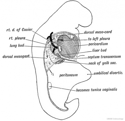
Fig. 201. The Form of the Coelom in a 3rd week Embryo as viewed from the right side.
As may be seen from Fig. 72, page 93, the coelom at first extends beyond the embryo, between the layers of the somatopleure, which go to form the foetal membranes. With the union of the right and left layers of the somatopleure at the umbilicus part of the blastodermic coelom is shut within the body of the embryo— the intra-embryonic part of the coelom. It is out of this part that the pericardium, pleurae, peritoneum and tunioae vaginales are formed. The coelom commences in front below the pharynx and ends behind on the hind gut (Pig. 201). This cavity is originally divided into a right and left half by the dorsal and ventral mesentery, formed by the united splanchnopleures (Pig. 192). The ventral mesentery disappears in the anterior and posterior parts, so that the right and left halves of the coelom communicate ; an intermediate part persists and helps to form the diaphragm (Fig. 202).
The Divisions of the Coelom. — Out of the anterior part of the coelom (Fig. 201) is formed the pericardium. It lies beneath and behind the primitive pharynx (Fig. 202). The pericardial part of the coelom by the third week (Fig. 201) is expanded and thrust downwards and backwards, and communicates by a narrow neck or isthmus (iter venosum) with the pleuro-peritoneal part on each side of the mesentery. The duct of Cuvier encircles the neck or isthmus on each side, and causes the constriction (Pig. 183).
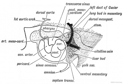
Fig. 202 Diagram to show the manner in which the heart is fixed within the pericardium by the Arterial and Venous Mesocardia in a human embryo of 3 weeks.
This condition is permanent in many fishes. With the origin of the lungs the part of the coelom which forms the isthmus undergoes a great expansion by the ingrowth of the lung buds (Fig. 201). Thus out of the isthmus of the right and left sides are gradually formed the pleural cavities. The pleurae expand until, instead of lying as minute passages above and behind the pericardium, they come to completely cover it. The abdominal part of the coelom forms the peritoneal cavity ; a small part on each side is shut off in the scrotum, the tunica vaginalis.
The Pericardium
In Fig. 202 the relationship of the heart to the pericardium is shown during the 3rd week of foetal life. The heart is tubular ; the dorsal and ventral mesocardia (Fig. 193) have already disappeared except at two points, where the heart still remains attached. These two points are (Fig. 202): (1) In front where the bulbus arteriosus passes out under the pharynx to divide into right and left ventral aortae from which the right and left visceral aortic arches arise. The mesocardium which binds it here may be named the arterial mesocardium.
(2) Behind the sinus venosus is imbedded in the mesentery or posterior mesocardium, through which the great veins reach it. The mesocardium which binds it behind may be named the venous mesocardium.
Fig. 203. View of the Interior of the Pericardium showing the attachments of the heart to its dorsal aspect by the Arterial or Venous Mesocardia.
In Fig. 203 is shown the fixation of the heart within the pericardium of the adult. The arterial and venous mesocardia can be recognised, only somewhat altered in position and form.
(1) The bulbus arteriosus becomes the ascending aorta and pulmonary aorta, and these are attached together to the fibrous pericardium by the arterial mesocardium (Fig. 203).
(2) The sinus venosus, which becomes part of the right auricle, and its vessels are still attached by the venous mesocardium. The heart has become so doubled on itself that the venous mesocardium comes almost in contact with the arterial inesocardium, the narrow space which separates them being the transverse sinus (Fig. 203). By comparing Figs. 3 93, 202 and 203 it will be seen that the transverse sinus is formed by the breaking down of the dorsal mesocardium of the heart.
The venous mesocardium becomes much more extensive by the ingrowth of the pulmonary veins. These grow in from the lungs, and pierce the pericardium to reach the left auricle. They reach the auricle through the mesentery or venous mesocardium of the sinus venosus (Fig. 202). The ingrowth of the left pulmonary veins causes a prolongation of the venous mesocardium to the left side; when the heart is removed the venous mesocardium is seen to be F-shaped in section. The oblique sinus lies in the concavity of the venous mesocardium (Fig. 203).
Ectopia Cordis. — Occasionally children are born with their hearts exposed on the surface of the chest. In extreme cases, only the dorsal wall of the pericardium is present, and it is flush and continuous with the skin of the chest. In such cases there has been either a failure on the part of the right and left soinatopleures to unite in the ventral line, or, more probably, they did unite, but an early cystic condition led to a rupture of the pericardium and overlying layer of the somatopleure. In such cases the sternum is partially absent, or if present it is cleft, the right and left halves being widely parted. The pathology of the condition is probably similar to that of ectopia vesicae.
The Dorsal Aortae
In the 3rd week there are still two dorsal aortae (Fig. 205). The blood passes from the two ventral aortic stems through the aortic visceral arches to end in a dorsal aorta on each side (Fig. 202). These run backwards side by side, and at first end in the yolk sac, but with the formation of the allantois they pass out on the body stalk to the membranes and placenta. The umbilical arteries are thus the direct continuations of the dorsal aortae (Young and Robinson) ; the middle sacral artery is an inter-segmental vessel of later formation (Fig. 221, p. 273).
About the end of the 3rd week the dorsal aortae fuse together from about the region of the 4th dorsal segment to the 4th lumbar. In front of and behind these points they remain separate.
The fate of the two dorsal aortae in front of the 4th dorsal vertebra has been already dealt with (p. 35 and Figs. 28 and 29). It remains to consider what becomes of them in the posterior extremity of the body. Following the researches of Young and Eobinson, the dorsal aortae beyond the 4th lumbar segment, where they remain apart, form :
(1) The common iliac arteries;
(2) The internal iliac — in part at least ;
(3) The hypogastric arteries — intra-abdominal parts of umbilical ;
(4) The umbilical arteries — in the umbilical cord.
The authors just mentioned have described in the embryos of several mammals a system of arterial arches on each side of the hind gut resembling the aortic arches on each side of the fore gut (see Fig. 221, p. 273).
Red Corpuscles of the Blood
The first red corpuscles are nucleated, capable of division, and are formed in mesoblastic cells of the splanchnopleure of the yolk sac and in the somatopleure of the amnion and chorion. The nucleated red corpuscles gradually become the biconcave, non-nucleated disc, but even at birth nucleated red corpuscles are still present. In the early half of foetal life the liver plays a leading part in their formation ; in the latter half the spleen, red marrow and connective tissues become the chief sites of their formation. After birth they are formed almost entirely in the red marrow.
White blood corpuscles are produced at a later date than the red blood corpuscles. They also are produced by cells of the mesoblast.
Lymphatic Vessels
Lymphatics are developed in the chick by the end of the 3rd day. It is some time before the lymphatic system opens into the blood system by the formation of a thoracic duct.
Certain important changes take place in the formation of the human lymphatic system about the 5th month. Lymphatic glands then begin to form in the connective tissue throughout the body. Peyer's patches begin to appear in the intestine. Until then the connective tissue of the body contains a jelly-like ground substance corresponding to Wharton's jelly in the umbilical cord. At the 5th month the mucoid substance in the connective tissue begins to be absorbed (Berry Hart).
Until the fifth month the marrow of bones is composed of embryonic mesoblastic tissue (primary marrow). After the fifth month this becomes invaded by lymphoid tissue (white marrow) which in certain bones (vertebrae, etc.) becomes transformed further into red marrow. (Hamar.) .
Chapter Figures
- Circulatory System: Fig. 181. Adult Superior Vena Cava | Fig. 182. Embryonic Venous Trunks forming Superior Vena Cava | Fig. 183. Ducts of Cuvier | Fig. 184. Left Superior Vena Cava Remnants | Fig. 185. Sinus Venosus and Ducts of Cuvier 3rd week | Fig. 186. Posterior Cardinal Veins Remnants | Fig. 187. Left Vitelline Vein 4th week | Fig. 188. Diagram showing the Formation of the Ductus Venosus, and the fate of the Umbilical and Vitelline veins. The arrows show the parts of the Vitelline Veins which become the Portal Vein. | Fig. 189. Umbilical Vein Remnants | Fig.190. Right Umbilical Vein 3 weeks | Fig.191. Right and Left Cardiac Tubes | Fig.192. Fusion of Right and Left Cardiac Tubes | Fig.193. Dorsal and Ventral Mesocardia | Fig.194. Primitive Heart Divisions | Fig.195. Heart Bends 3rd week | Fig.196. Sinus Venosus | Fig.197. Heart 5th week | Fig.198. Heart Septa | Fig.199. Fetal Heart Ventricles | Fig 200. Semilunar valves | Fig 201. Embryo Coelom 3rd week | Fig. 202 Heart Arterial and Venous Mesocardia week 3 | Figures
| Historic Disclaimer - information about historic embryology pages |
|---|
| Pages where the terms "Historic" (textbooks, papers, people, recommendations) appear on this site, and sections within pages where this disclaimer appears, indicate that the content and scientific understanding are specific to the time of publication. This means that while some scientific descriptions are still accurate, the terminology and interpretation of the developmental mechanisms reflect the understanding at the time of original publication and those of the preceding periods, these terms, interpretations and recommendations may not reflect our current scientific understanding. (More? Embryology History | Historic Embryology Papers) |
Human Embryology and Morphology (1902): Development or the Face | The Nasal Cavities and Olfactory Structures | Development of the Pharynx and Neck | Development of the Organ of Hearing | Development and Morphology of the Teeth | The Skin and its Appendages | The Development of the Ovum of the Foetus from the Ovum of the Mother | The Manner in which a Connection is Established between the Foetus and Uterus | The Uro-genital System | Formation of the Pubo-femoral Region, Pelvic Floor and Fascia | The Spinal Column and Back | The Segmentation of the Body | The Cranium | Development of the Structures concerned in the Sense of Sight | The Brain and Spinal Cord | Development of the Circulatory System | The Respiratory System | The Organs of Digestion | The Body Wall, Ribs, and Sternum | The Limbs | Figures | Embryology History
Reference
Keith A. Human Embryology and Morphology. (1902) London: Edward Arnold.
Cite this page: Hill, M.A. (2024, May 1) Embryology Book - Human Embryology and Morphology 16. Retrieved from https://embryology.med.unsw.edu.au/embryology/index.php/Book_-_Human_Embryology_and_Morphology_16
- © Dr Mark Hill 2024, UNSW Embryology ISBN: 978 0 7334 2609 4 - UNSW CRICOS Provider Code No. 00098G

