Book - Human Embryology and Morphology 12
| Embryology - 27 Apr 2024 |
|---|
| Google Translate - select your language from the list shown below (this will open a new external page) |
|
العربية | català | 中文 | 中國傳統的 | français | Deutsche | עִברִית | हिंदी | bahasa Indonesia | italiano | 日本語 | 한국어 | မြန်မာ | Pilipino | Polskie | português | ਪੰਜਾਬੀ ਦੇ | Română | русский | Español | Swahili | Svensk | ไทย | Türkçe | اردو | ייִדיש | Tiếng Việt These external translations are automated and may not be accurate. (More? About Translations) |
Keith A. Human Embryology and Morphology. (1902) London: Edward Arnold.
| Historic Disclaimer - information about historic embryology pages |
|---|
| Pages where the terms "Historic" (textbooks, papers, people, recommendations) appear on this site, and sections within pages where this disclaimer appears, indicate that the content and scientific understanding are specific to the time of publication. This means that while some scientific descriptions are still accurate, the terminology and interpretation of the developmental mechanisms reflect the understanding at the time of original publication and those of the preceding periods, these terms, interpretations and recommendations may not reflect our current scientific understanding. (More? Embryology History | Historic Embryology Papers) |
Chapter XII. The Segmentation of the Body
Segmentation of the Body. — The human body or trunk consists of 33 or 34 segments. Each segment is fundamentally of the same type, but the resemblance is obscured owing to extensive modifications which they undergo to form the cervical, dorsal (thoracic), lumbar (abdominal), sacral (pelvic) and caudal regions of the body. The outgrowth of the limbs also renders it difficult to recognise in the adult the simple system of segments which can be seen in the embryo at the end of the third week (Fig. 233, p. 289).
Until lately the segmentation of the human body was a matter of only speculative importance, but recent advances in our knowledge of the distribution of nerves, has shown that it has a direct bearing on diagnosis and treatment.
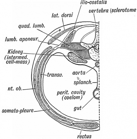
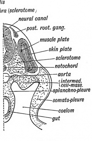
Constitution of a Typical Segment (11th Dorsal). — It is better to study the development of one typical body segment and from that the student will be able to note for himself the modifications which have taken place in the more highly differentiated segments of the body. By the end of the third week,, the process of segmentation, which began in the occipital region a few days previously, has spread backwards and separated the 18th body segment (11th dorsal) from the one in front and behind. As already explained, the process of segmentation affects only the paraxial block of mesoblast which lies on each side of the neural canal and notochord. In Figs. 125 and 126 a segment is represented in the adult and in the embryonic condition.
The following elements make up the 11th dorsal segment: (1) Its skeletal basis; (2) Muscular element; (3) Eenal element; (4) Vessels; (5) Nerves; (6) Neural segment. Although the
epiblast and hypoblast are never segmented, yet a definite area
of each is associated with every body segment. The origin of each element will be taken separately.
|
|
| Fig. 125. A transverse section showing the Elements of the 1st Lumbar Segment in the Adult. | Fig. 126. A corresponding section of an Embryo about the end of the 3rd week (diagrammatic). |
I. The skeleton of the 11th dorsal segment is represented by the adjacent halves of the 11-12 dorsal vertebrae and the disc between them, for, as already pointed out, the vertebrae are intersegmental in origin (Fig. 126). The transverse processes, the spinous process and 11th and 12 th ribs are also formed in the septa in front of and behind the 11th segment. The septum in the rectus muscle a little below the umbilicus represents' the inter-segmental septum corresponding to the 11th rib. Sometimes another septum occurs in the rectus, midway between the pubes and umbilicus, marking the lower limit of the 11th segment. The linea alba separates the segments of the two sides.
In the linea alba or ventral median line of the thoracic region, the sternum is developed. The inter-segmental septa are well marked in the thoracic region ; the ribs and their cartilages are developed in them. In the neck the septa are almost lost ; the intermediate tendon of the omo-hyoid and the septa occasionally found in the sterno-hyoid and -thyroid, complexus and trachelomastoid muscles are the only representatives of them in the cervical region.
IT. The Muscles of the 11th Dorsal Septum. — All the muscles of this segment are developed from the muscle plate of the primitive segment (see Figs. 125 and 126). There is a cavity, which probably arises as a diverticulum of the coelom, in each primitive segment (Fig. 6 9, p. 90). The cells of the mesoblast on the inner side of the segmental cavity become columnar and form the muscle plate (Fig. 126). Each segment has its own muscle plate. The cells of each plate increase rapidly in number ; they spread into the somatopleure, and form the muscles of the body-wall and limbs. Each cell becomes elongated and directed across its segment from septum to septum. The intercostal muscles retain this arrangement, but in the abdominal region the fibres fuse with those of neighbouring segments to form muscular sheets — the external oblique, internal oblique, transversalis and rectus. In fishes the embryonic segmental arrangement of the musculature persists. The manner in which the final groups of muscles are derived from the muscle plates is not known, but in the typical segment with which we are at present dealing it will be seen that the musculature falls into two groups (see Fig. 125): (1) axial (acting on the spine — the erector spinae, etc.), and (2) ventro-lateral or body-wall muscles (intercostals, rectus, oblique muscles, etc.). The musculature of the limbs is derived from the ventro-lateral group.
Many of the ventro-lateral muscles (trapezius, rhomboids, and latissimus dorsi), migrate dorsalwards over the axial muscles and take origin from the spines of the vertebrae (Fig. 125).
Each muscle fibre is a cell derived from the endothelial cells which make up the muscle plate. The protoplasm of each cell is converted into a living contractile substance (myosin), which reacts to nerve stimuli.
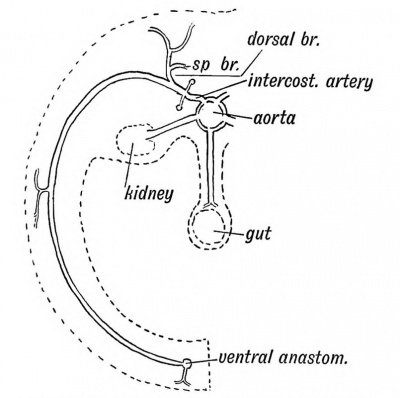
III. The Arteries of the 11th Segment (Fig. 127).— The 11th intercostal is the artery of the segment. It gives off a dorsal branch to supply the axial muscles, the spinal column, spinal cord and membranes and skin. The segmental artery joins at its termination with a ventral longitudinal vessel, the deep epigastric. The primitive arrangement in vertebrates appears to have been a dorsal and ventral longitudinal vessel, with the segmental artery passing between them. The vertebral, ascending cervical, deep ■cervical, ascending lumbar and lateral sacral arteries are examples of the anastomoses that may arise between segmental arteries.
Fig. 127. The distribution of a typical Segmental Artery.
Segmental arteries also arise from the aorta to supply the structures formed from the intermediate cell mass (the kidney, testis, ovary, etc., Figs. 125 and 127). As a rule only one renal segmental artery persists, but frequently accessory renals are seen. These are persistent embryonic vessels of the several segments from which the mesoblast of the kidney arose. The splanchnopleure shows no trace of segmentation ; hence its vessels (coeliac axis and mesenteric) are not segmental in origin.
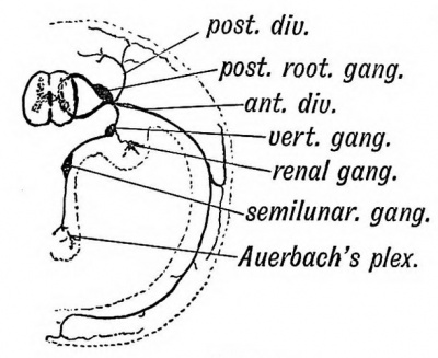
IV. The Nerve Elements of the 11th Segment (Fig. 128). — (1) Although the spinal cord of the human embryo shows no certain sign during development of being definitely divided into segments corresponding to those of the body, yet from what we know of its condition in embryos of other animals and from clinical evidence there can be little doubt that such a segmentation does take place, and that it possesses segments corresponding to those of the body. A group of cells in each segment, afterwards those of the anterior horn, sends out processes to all the muscles of the primitive body segment in which it is situated. The anterior root of a spinal nerve is thus formed. Other motor cells send out processes which reach viscera through the white rami communicantes and sympathetic system (Fig. 128).
Fig. 128. Diagram of the Nerve System of the 11th Dorsal Segment.
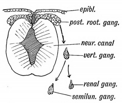
Fig. 129. A diagram showing the derivation of the Parts of the Nerve System of the 11th Segment in the Embryo.
(2) Besides the anterior horn cells, two other nerve groups become connected with each segment. At the point where the medullary plates are cut off from the epiblast to form the neural canal, a crest, the neural crest, grows out on each side (Fig. 129) composed of the cells which formed the junctional line between medullary plates and epiblast. A group of these neuroblasts grows into each segment and forms the posterior root ganglion. Each neuroblast within the ganglion sends out a process which bifurcates, one branch or fibre growing into the cord and ending in the posterior column and cells of the posterior horn, the other passing to the skin, muscles, etc., of the segment. The posterior nerve root is thus formed by the ingrowing processes of the cells of the posterior root ganglion, and thus the body segment in which the outgrowing processes are distributed is brought into sensory communication with the central nervous system. The anterior and posterior roots unite to form a spinal or segmental nerve. Like the artery it divides into a posterior division for the epaxial part of the segment and an anterior for the ventro-lateral part (Fig. 128) (3) A third group of cells, the sympathetic, is also connected with each segment. The origin of these cells is not yet certain, but most of the evidence goes to show that the cells are derived, with the posterior root ganglion, from the neural crest and that a group enters each segment. Professor Paterson's research on their origin led him to the conclusion that the nerve cells of the sympathetic arose from the mesoblast. The sympathetic group of nerve cells (Pigs. 128 and 129) is broken up into —
(a) The prevertebral ganglion situated on the vertebra (in the gangliated chain), ventral to the exit of the spinal nerve ; (i) A group to the intermediate cell mass (renal ganglion) ;
(c) Another to the splanchnopleure (in the semilunar ganglia) ;
(d) To the viscera (cells of Auerbach's plexus, etc.).
Groups (c) and (d) show no trace of segmentation in their arrangement, but, clinically, evidence is to be found that every viscus or part of a viscus is connected with certain segments of the spinal cord. The cells of the sympathetic ganglia throw out axis-cylinder processes, which pass to the spinal cord by a white ramus communicans and posterior root, and act as sensory pathways from the viscera. The distal end of the axis-cylinder process ends in a viscus. In this manner certain segments of the spinal cord are brought into touch with the viscera. The vasomotor supply of each body segment passes to it from the sympathetic ganglia by a grey ramus communicans.
It will thus be seen that all the parts of a segment — -body wall (somatopleure), kidney (intermediate cell mass), and a part of the abdominal or thoracic viscera (splanchnopleure) are connected by nerves to a corresponding segment of the spinal cord. In diseased conditions of any part of the body segment the corresponding spinal segment of the cord is disturbed, the disturbance being reflected from the cord to the segment. The nervous mechanism of the whole segment is affected. Thus, for instance, a stone in the pelvis of the kidney (which is supplied from the 10th, 11th, and 12th dorsal segments) is frequently accompanied by pain referred along the 11th and 12 th intercostal nerves. The skin supplied by these nerves may become hyper-aesthetic.
| Historic Disclaimer - information about historic embryology pages |
|---|
| Pages where the terms "Historic" (textbooks, papers, people, recommendations) appear on this site, and sections within pages where this disclaimer appears, indicate that the content and scientific understanding are specific to the time of publication. This means that while some scientific descriptions are still accurate, the terminology and interpretation of the developmental mechanisms reflect the understanding at the time of original publication and those of the preceding periods, these terms, interpretations and recommendations may not reflect our current scientific understanding. (More? Embryology History | Historic Embryology Papers) |
Human Embryology and Morphology (1902): Development or the Face | The Nasal Cavities and Olfactory Structures | Development of the Pharynx and Neck | Development of the Organ of Hearing | Development and Morphology of the Teeth | The Skin and its Appendages | The Development of the Ovum of the Foetus from the Ovum of the Mother | The Manner in which a Connection is Established between the Foetus and Uterus | The Uro-genital System | Formation of the Pubo-femoral Region, Pelvic Floor and Fascia | The Spinal Column and Back | The Segmentation of the Body | The Cranium | Development of the Structures concerned in the Sense of Sight | The Brain and Spinal Cord | Development of the Circulatory System | The Respiratory System | The Organs of Digestion | The Body Wall, Ribs, and Sternum | The Limbs | Figures | Embryology History
Reference
Keith A. Human Embryology and Morphology. (1902) London: Edward Arnold.
Cite this page: Hill, M.A. (2024, April 27) Embryology Book - Human Embryology and Morphology 12. Retrieved from https://embryology.med.unsw.edu.au/embryology/index.php/Book_-_Human_Embryology_and_Morphology_12
- © Dr Mark Hill 2024, UNSW Embryology ISBN: 978 0 7334 2609 4 - UNSW CRICOS Provider Code No. 00098G

