Book - Human Embryology and Morphology 10
| Embryology - 28 Apr 2024 |
|---|
| Google Translate - select your language from the list shown below (this will open a new external page) |
|
العربية | català | 中文 | 中國傳統的 | français | Deutsche | עִברִית | हिंदी | bahasa Indonesia | italiano | 日本語 | 한국어 | မြန်မာ | Pilipino | Polskie | português | ਪੰਜਾਬੀ ਦੇ | Română | русский | Español | Swahili | Svensk | ไทย | Türkçe | اردو | ייִדיש | Tiếng Việt These external translations are automated and may not be accurate. (More? About Translations) |
Keith A. Human Embryology and Morphology. (1902) London: Edward Arnold.
| Historic Disclaimer - information about historic embryology pages |
|---|
| Pages where the terms "Historic" (textbooks, papers, people, recommendations) appear on this site, and sections within pages where this disclaimer appears, indicate that the content and scientific understanding are specific to the time of publication. This means that while some scientific descriptions are still accurate, the terminology and interpretation of the developmental mechanisms reflect the understanding at the time of original publication and those of the preceding periods, these terms, interpretations and recommendations may not reflect our current scientific understanding. (More? Embryology History | Historic Embryology Papers) |
Chapter X. Formation of the Pubo-Femoral Region, Pelvic Floor and Fascia
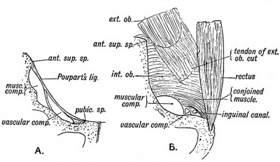
Inguinal and femoral hernia occur so rarely amongst mammals generally that they may be considered human peculiarities. Their frequency in man is due to certain structural changes in his pubo-fenioral region, changes which have resulted mainly from his adaptation to upright progression. His susceptibility to hernia is due to : — (1) The unique form of Poupart's ligament in man. It is scarcely developed in any other animal (Fig. 107). In the orang, for instance, also an upright primate, the external oblique has no attachment to the crest of the ilium, and takes no part in forming the outer part of Poupart's ligament (Fig. 107), but its tendon terminates over and strengthens the region of the inguinal canal. This is the usual termination in the mammalia.
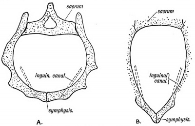
Fig. 106. A. The form of Pelvis and Inguinal Canal in Man. B. The corresponding forms in the Lower Primates.
(2) The internal oblique and transversalis (conjoined parts) in the orang, and in all primates except man, arise from the firm tubular sheath of the ilio-psoas, also from the extensive anterior border of the ilium, and arching over the spermatic cord end in a long insertion on the ileo-pectineal line. They act as a powerful compressor or sphincter of the inguinal canal, and thus prevent hernia (Fig. 107.6).
(3) The human manner of walking, and the great head of the human child at birth require a wide pelvis. All mammals adapted to the prone posture have a narrow pelvis, and hence a narrow anterior abdominal wall (Figs. 106.4 and B) through which the inguinal canal passes very obliquely. The course of the canal is more direct in man, and therefore offers a greater facility to the escape of the abdominal contents.
Fig. 107. Poupart's Ligament and the Crural Passage of Man. B. Poupart's Ligament, Crural Passage, and sphincter-like Conioined Muscle of the Orang.
(4) The size of the space between the edge of the pelvis and Poupart's ligament (the crural passage) is very much greater in man than in any other animal (Figs. 107-4 and £). In him, the most internal part of the passage is left unfilled, and this unfilled space forms the femoral or crural canal through which femoral hernia may escape. The crural passage is relatively larger in women than in men, owing to the greater size of the female pelvis, and hence femoral hernia are much more common in women than in men. Some hint as to the method of treatment of hernia in man may be obtained from a consideration of the arrangement of structures which prevent them in other animals.
The Pelvic Floor
The Coccyx. — It is necessary to consider the coccyx here because the changes which it has undergone in the evolution of the human body are intimately connected with the formation of the pelvic floor.
The coccyx in man is commonly composed of four vertebrae, more or less vestigial in nature, which represent the basal caudal vertebrae of tailed mammals. Evidence of their vestigial or retrograde nature is to be found in :
- Only their centra are developed — with the exception of the first, which shows partial formation of transverse processes and neural arches (superior cornua) ;
- Delay in the appearance of the centres of ossification. These, instead of beginning in the 7th week as in a typical vertebra, commence after birth. The centre for the 1st coccygeal vertebra appears in the 1st year, that for the 4th vertebra about the 25th year; the 2nd and 3rd at intermediate periods.
- Late in life, between the 40th and 60th year, the vertebrae fuse together, and then unite with the sacrum.
The number of coccygeal vertebrae varies ; four is the normal number, but there may be three or five. In the young foetus (2nd month) there are commonly 5, 6 or 7 (Eosenberg). The first coccygeal vertebra may join the sacrum, making 6 sacral vertebrae.
The evidence of the former existence of a true tail in the ancestral human stock consists of:
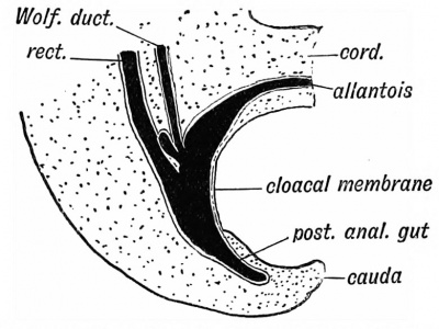
- In the second month the coccygeal region of the spine protrudes (Fig. 108).
- Vestiges of the extensor and flexor muscles of the tail are frequently found (10°/ o of bodies) on the dorsal and ventral aspects of the sacrum and coccyx.
- True tails, consisting of external prolongations of the coccyx, commonly fibrous, rarely containing vertebrae, occasionally occur.
- The post-anal pit, always to be seen in the newly-born child, marks the point at which the coccyx disappears below the surface. In man the coccyx forms part of the perineal floor. Instead of projecting far beyond the gut, as in tailed mammals, it terminates 1\ inches above the commencement of the anal canal.
Fig. 108. The caudal end of the body in a human embryo of the 3rd week.
The Pelvic Floor is peculiarly extensive in man, an adaptation to his upright posture. The floor is formed by the following structures :
- The levator ani and its sheath (recto- vesical and anal fasciae) on each side.
- The coccyx and coccygeus muscles.
- The constrictor urethrae and its sheath (the triangular ligament).
- The pyriformis and its sheath may also be included. Development of the Pelvic Floor.— The pelvic floor has been evolved in man by a transformation of the tail and the caudal muscles.
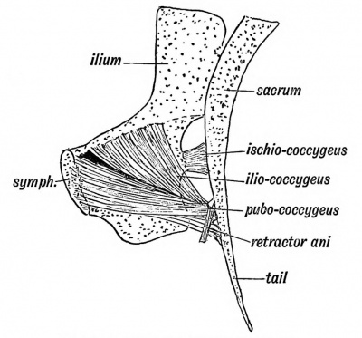
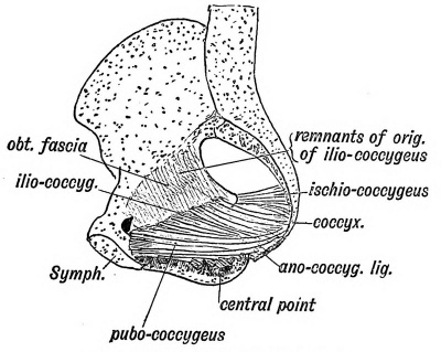
The arrangement of tail muscles in a four-footed mammal, such as the monkey or dog, is shown in Fig. 109, and the modification of this form in man in Fig. 110. In mammals two muscles, the pubo-coccygeus and ischio-coccygeus (Fig. 109), act as depressors of the tail, which in four-footed animals plays the part of a perineal shutter. They are attached to the small V-shaped chevron bones on the under surface of the basal caudal vertebrae. Another muscle, the ischio- or spino-coccygeus, acts as a lateral flexor of the tail (Fig. 109). It is attached to the transverse processes of the caudal vertebrae, and rises from the dorsal border of the ischium. In man the pubo-coccygeus and iliococcygeus unite into one sheet and form the levator ani. The shrinkage of the tail leaves the muscle partly stranded on the ano-coccygeal ligament. Other fibres of the pubo-coccygeus loose their primary insertion to the coccyx and become attached to the prostrate, central point of the perineum and to the anal canal. Both muscles, especially the ilio-coccygeus, retain in part their primitive attachment to the coccyx (cauda). The spino-coccygeus, or coccygeus muscle, is partly fibrous in man, its outer laminae forming the small sacro-sciatic ligament ; its inner laminae remain muscular and form the coccygeus. In man, too, the origin of the ilio-coccygeus has sunk from the pelvic brim of the ischium on to the obturator fascia (P. Thompson) ; traces of the primitive origin from the pelvic brim can often be detected. The white line, a structure peculiar to man, marks the new point of origin of the levator ani from the obturator fascia. Further details of changes undergone by the pelvic muscles and fasciae may be found in papers by Dr. P. Thompson in the Journal of Anatomy and Physiology, vol. xxxv.
Fig. 109. The pelvic-caudal Muscles of a Monkey.
Fig. 110. The corresponding Muscles of Man.
On the dorsal and ventral aspects of the sacrum and coccyx, fibrous or muscular vestiges of the anterior and posterior sacrococcygeal muscles (elevators and flexors of the tail) are commonly to be found in man.
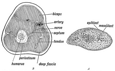
The Pelvic Fascia and Fasciae in General. — It has been customary to regard fasciae as separate structures forming distinct sheets with devious and complex courses. It is possible by dissection to prepare and display them according to accepted descriptions, but the structures so displayed are artificial and not the true structures with which the surgeon or physician has to deal with in actual practice. Embryology is the best guide to their nature. Take the development of the fasciae seen on making a section of the upper arm, for example. When the limb bud has appeared, which it begins to do about the end of the 3rd week of development, a section through it reveals a uniform composition of more or less rounded mesoblastic cells with a covering of epiblast (Fig. Ill A). Very soon the central cells near the axis of the bud are densely grouped and form the basis of the humerus. Others arrange themselves to form the biceps, triceps and muscles of the arm ; others form the walls of vessels and the sheaths of nerves.
Fig. 111. A. Diagrammatic section of the Arm Bud of an embryo at the commencement of the 4th week. B. Corresponding section of the Adult Arm.
After these various groups of cells have become differentiated, there are numerous cells left over which form a basis in which the specialized cells and groups of cells are packed and ensheathed. The undifferentiated mesoblast forms the connective tissue or fascial system of the part. From the manner of its origin it is evident that the connective tissue system — the fasciae and septa — must form a continuous formation of sheaths, each being in continuity with that of every surrounding structure. The sheaths of the biceps, triceps and brachialis anticus, the periosteum of the humerus, the deep fascia, internal and external intermuscular septa, the sheaths of the vessels and nerves of the arm, represent the mesoblastic tissue which was left over after the structures which they enclose were differentiated, and are, from the manner of their origin, necessarily in continuity.
They can only be artificially separated from each other. It is more accurate and easier to describe fasciae, then, not as separate structures, but as adjuncts of the structures which they surround or ensheath.
The Pelvic Fascia, which strengthens the pelvic floor, is composed of the sheaths of four muscles :
- The Levator Ani;
- The Obturator Internus ;
- The Pyriformis ;
- The Constrictor Urethrae and deep Transversus Perinei.
The fibrous capsules of the following viscera also form part of it:
- Prostate and Vesiculae Seminales in the male ;
- Vagina and Uterus in the Female ;
- The Bladder ;
- The Eectum. Under the title of pelvic fascia these eight elements are combined.
I. The Obturator Fascia is the sheath on the inner or pelvic aspect of the obturator interims ; the sheath on the outer side of the muscle is formed by the periosteum and obturator membrane. The obturator fascia is attached at the circumference of the muscle. There it becomes continuous with the periosteum of the os innominatum. The part above the white line (supra-linear) is intra-pelvic ; the part below (infra-linear) is perineal and situated on the outer wall of the ischio-rectal fossa.
II. The Recto-vesical and Anal Fasciae. — The levatores ani form a muscular floor for the pelvis, stretching from the white line of one side to the white line of the other. The sheath on their under surface — on the inner wall of the ischio-rectal fossa — forms the anal fascia. On the upper surface, their sheath forms the greater part of the recto-vesical fascia. The pelvic viscera rest on the upper surface of the levatores ani and the capsules of the viscera are continuous with the sheath on the upper surface of the muscles. The combined visceral capsules and upper sheath of the levatores ani form the recto-vesical fascia.
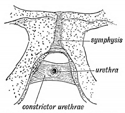
III. The Triangular Ligament is the sheath of the constrictor urethrae muscle (Fig. 112). The inferior transverse fibres of the constrictor form really a separate muscle — the deep transverse perineal. The apex of the prostate rests on the muscle, its capsule being continuous with the posterior layer of the muscle sheath — the deep layer of the triangular ligament.
IV. The inner sheath of the pyriformis forms the pyriform fascia. As the muscle arises between the sacral foramina, the sacral plexus lies within the sheath, the iliac vessels on its inner aspect. The coccygeus is continuous with the levator ani and its sheath forms part of the recto- vesical fascia.
Fig. 112. The Constrictor Urethrae Muscle.
The Cervical Fascia. — From what has been said of the pelvic fascia, the nature and arrangement of the cervical fascia will be readily understood. It is composed of (1) the sheaths of the cervical muscles (sterno-mastoid, etc.) ; (2) of the sheaths of vessels (carotid sheath, etc.) ; (3) the sheaths of nerves (axillary sheath, etc.) ; (4) the fascial capsules of viscera, such as the thyroid body, salivary glands, and pharynx.
| Historic Disclaimer - information about historic embryology pages |
|---|
| Pages where the terms "Historic" (textbooks, papers, people, recommendations) appear on this site, and sections within pages where this disclaimer appears, indicate that the content and scientific understanding are specific to the time of publication. This means that while some scientific descriptions are still accurate, the terminology and interpretation of the developmental mechanisms reflect the understanding at the time of original publication and those of the preceding periods, these terms, interpretations and recommendations may not reflect our current scientific understanding. (More? Embryology History | Historic Embryology Papers) |
Human Embryology and Morphology (1902): Development or the Face | The Nasal Cavities and Olfactory Structures | Development of the Pharynx and Neck | Development of the Organ of Hearing | Development and Morphology of the Teeth | The Skin and its Appendages | The Development of the Ovum of the Foetus from the Ovum of the Mother | The Manner in which a Connection is Established between the Foetus and Uterus | The Uro-genital System | Formation of the Pubo-femoral Region, Pelvic Floor and Fascia | The Spinal Column and Back | The Segmentation of the Body | The Cranium | Development of the Structures concerned in the Sense of Sight | The Brain and Spinal Cord | Development of the Circulatory System | The Respiratory System | The Organs of Digestion | The Body Wall, Ribs, and Sternum | The Limbs | Figures | Embryology History
Reference
Keith A. Human Embryology and Morphology. (1902) London: Edward Arnold.
Cite this page: Hill, M.A. (2024, April 28) Embryology Book - Human Embryology and Morphology 10. Retrieved from https://embryology.med.unsw.edu.au/embryology/index.php/Book_-_Human_Embryology_and_Morphology_10
- © Dr Mark Hill 2024, UNSW Embryology ISBN: 978 0 7334 2609 4 - UNSW CRICOS Provider Code No. 00098G

