Book - Developmental Anatomy 1924-4
| Embryology - 27 Feb 2026 |
|---|
| Google Translate - select your language from the list shown below (this will open a new external page) |
|
العربية | català | 中文 | 中國傳統的 | français | Deutsche | עִברִית | हिंदी | bahasa Indonesia | italiano | 日本語 | 한국어 | မြန်မာ | Pilipino | Polskie | português | ਪੰਜਾਬੀ ਦੇ | Română | русский | Español | Swahili | Svensk | ไทย | Türkçe | اردو | ייִדיש | Tiếng Việt These external translations are automated and may not be accurate. (More? About Translations) |
Arey LB. Developmental Anatomy. (1924) W.B. Saunders Company, Philadelphia.
| Historic Disclaimer - information about historic embryology pages |
|---|
| Pages where the terms "Historic" (textbooks, papers, people, recommendations) appear on this site, and sections within pages where this disclaimer appears, indicate that the content and scientific understanding are specific to the time of publication. This means that while some scientific descriptions are still accurate, the terminology and interpretation of the developmental mechanisms reflect the understanding at the time of original publication and those of the preceding periods, these terms, interpretations and recommendations may not reflect our current scientific understanding. (More? Embryology History | Historic Embryology Papers) |
Chapter IV Age, Body Form and Growth Changes
Age Size And Weight Of Embryos
The age of a human embryo can not be determined with certainty, because too little is known of the time relations existing between ovulation and menstruation, and between ovulation, coitus, and fertilization (p. 27). This lack of a reliable basis makes any computation approximate, although the errors thus introduced are significant only in young specimens.
From numerous clinical observations it is certain that ovulation does not immediately precede menstruation, as was long held, but on the contrary follows it (p. 24). Experience proves that most pregnancies date from a coitus within a week or ten days after the menses cease. Hence, it is a])proximately correct to compute the age of an embryo from the tenth Jay after the onset of the last menstruation.
Careful studies on embryos which were accompanied by adequate data as to menstruation, coitus, and clinical history have led to the establishment of certain age-norms. By comparing a given specimen with such standards its age can be determined with reasonable accuracy. It is simplest to make these comparisons on the basis of size, although young embryos vary sufficiently so that structure must be taken into account as well. Embryos are measured in two ways. Commonest is the crown-rump length (designated CR), or sitting height; this is the measure from vertex to breech. The second is the crown-heel length (CH). or standing height.
The following table, based on data by Mall and Scammon, lists the size and weight of human embryos corresponding to definite ages : Ratio of increase .
Crown-rump length .
Crown-heel length
to weight at
(.CR), or sitting .
(CH). or standing .
Weight in .
beginning of .
Age .
height (mm.).
height (mm.) .
grams .
month .
Three weeks .
0.5 .
0.5 .
Four weeks .
2.5 .
2-5 .
.
8000.00 .
Five weeks .
5-5 .
5-5 .
. 004 .
.
Six weeks .
I I . 0 .
11.0 .
.
.
Seven weeks .
17.0 .
19 . 0 .
.
.
Second lunar month .
25.0 .
30. 0 .
2 .
499 . 00 .
Third lunar month .
68.0 .
98 . 0 .
24 .
I I . 00 .
Fourth lunar month .
I 2 I . 0 .
180.0 .
120 .
4.00 .
Fifth lunar month .
167 . 0 .
250.0 .
330 .
1-75 .
Sixth lunar month .
210.0 .
3130 .
600 .
0.82 .
Seventh lunar month ,.
. 245 . 0 .
370.0 .
1000 .
0.67 .
Eighth lunar month .
284.0 .
425 0 .
1600 .
0.60 .
Ninth lunar month .
316.0 .
470.0 .
2400 .
0.50 .
Tenth lunar month .
345 â– 0 .
500. 0 .
3200 .
0.33 .
68 .
For estimating the age of an embryo when its size is known, or the reverse, the following rules are useful ;
Standing height (in cm.) X 0.2 = Age {in months)
Sitting height (in cm.) X 0.3 = ,-lg^ {in months) (For embryos less than 10 cm. long, add one month to the result)
Age (in months) ^ 0.2 = Standing height (in cm.)
Age {in months) -u 0.3 = Sitting height {in cm.) (For embryos of the first 3 months, subtract 4 cm. from the result)
Of practical interest is the determination of the date of delivery of a pregnant woman. Most labors occur ten lunar months, or 280 days, from the first day of the last menstrual period. The month and day of this date are easily found by counting back three months from the first day of the last period, and then adding one week. As some women menstruate once or more after becoming pregnant this computation is not infallible.
For comparison and reference, the gestation periods of a few representative mammals are appended ;
Opossum 13 days Pig Mouse 20 days Sheep Rat 21 days Cow Rabbit 30 days Horse Cat 8 W - eeks Rhinoceros Dog, guinea pig 9 weeks Elephant . .
The early history of the human ovum, including implantation and the development of membranes for its protection and nutrition, has been described on previous pages. The present section will deal with the appearance of the embryo and fetus at successive stages of uterine existence.
Period of the Embryo
Embryos of the Second Week
The youngest known embryo is the Miller specimen. It is somewhat like the diagram represented in Fig. 40 A. The central embryonic anlage is solid, without amnion cavity or yolk sac; it measures oI mm. in length. The extra-embryonic mesoderm is unsplit by a coelom. The chorion has both syncytial and Langhans layers, but true mesodermal villi are absent; its internal cavity' measures 0.44 mm.
The Bryce-Teacher ovum (Fig. 40 B) differs from the foregoing specimen chiefly by possessing an amniotic cavity^ and y'olk sac.
A well-defined extra-embryonic coelom divides the mesoderm of Peter - s specimen into somatic and splanchnic layers, and there is also the beginning of true villi (Figs. 40 C and 46). The ectodermal embryonic disc measures 0.19 mm.; it is thickened and separated from the entoderm by a layer of mesoderm (Fig. 41). Strands of mesoderm, known as the magma reticulare, bridge the extra-embryonic body cavity, which is 0.9 X 1.6 mm. in diameter (Fig. 41).
17 weeks 21 weeks 41 weeks 48 weeks
18 months 20 months .
These ova all belong to the latter part of the second week. The yolk sac is smaller than the amnion and the villi are mostly unbranched. The embryo is merely a plate combined from the three germ layers. Neither primitive streak nor allantois has appeared. Even in the oldest, a broad zone of mesoderm connects embryo to chorion.
Embryos of the Third Week
The Mateer ovum is shown as Fig. 32 A. It possesses a distinct primitive groove and allantois. The embryonic disc is 0.9 mm. in length.
Fig. 57. Dorsal view of a human embryo of 1.54 mm. (Spee). X 23.
A head process with its contained notochordal canal features the advance illustrated by the Ingalls embryo (Fig. 32 B). There is also the beginning of a neural groove. The chorionic vesicle has an internal diameter of 7 mm.
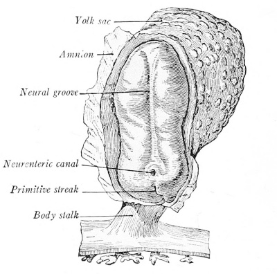
Spec’s specimen has progressed still further (Fig. 57). The embryonic disc measures 1.54 mm. and is slightly constricted from the yolk sac. The primitive streak is confined to the caudal end of the embryonic disc, the neural folds are well-marked, and a neurenteric canal opens as a pore into the primitive intestinal cavity. In longitudinal section it is evident that the floor of the head process has disappeared, leaving its roof as the notochordal plate (Figs. 40 D and 43). The fore-gut is forming and there are indications of a future heart anlage.
In this group as a whole, the continued extension of the extra-embryonic coelom has separated the embryo from the chorion except in the region of the body stalk, which constitutes a bridge that contains the allantois. The yolk sac is now larger than the amnion. The chorionic villi branch freely and there is evidence of blood-vessel formation in the wall of the yolk sac (Fig. 43), and, usually, in the body stalk and chorion.
Embryos of the Fourth Week
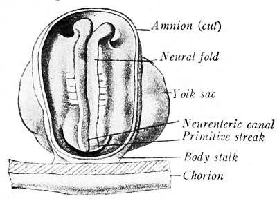
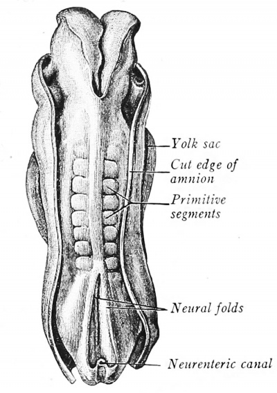
Embryos of this period are early characterized by the presence of high neural folds (Fig. 58) whose edges soon unite along part of their extent to form a tube which is the anlage of the brain and spinal cord (Figs. 59 and 245). The expansive brain portion is already recognizable. The mesoderm of each side of the midplane becomes arranged in blocks, the primitive (mesodermal) segments, visible externally.
In the embryo shown in Fig. 245 there are 14 pairs. The primitive streak is now insignificant (Figs. 44 and 58).

|

|
| Fig. 58. Kromer human embryo of 1.8 mm., in dorsal view (after Keibel and Elze). X 20. | Fig. 59. Human embryo of 2.11 mm. in dorsal view (Eternod). X 35. |
Growth at the head and tail regions appears to constrict the embryo from the yolk (Figs. 58 and 245). In a longitudinal section of an embryo at the middle of this period (Fig. 44), both fore- and hind-gut are evident and the heart is conspicuous. A system of blood vessels is established connecting with the heart (Figs. 180 and 181). The embryo is now cylindrical, its body wall encloses two more or less complete tubes (neural and enteric) with the axial notochord between. During this period there is an increase in length from 0.5 to 2.5 mm.
Embryos of the Fifth Week
Specimens corresponding to Figs. 60 and 61 stand at the turn between the fourth and fifth weeks, whereas one like Fig. 62 is more representative of this period. The progressive separation of embryo from yolk sac is evident. The primitive segments have increased until the 2.6 mm. specimen (Fig. 61) has 35 of the definitive 38 pairs. The convex curvature of the back is characteristic. External swellings indicate the three primary brain vesicles and the head becomes hexed at a right angle in the mid-brain region. On each side of the future neck appear branchial arches, separated by grooves. The hrst pair of arches bifurcates into maxillary and mandibular processes that will form the upper and lower jaws; between them is a depression, the oral fossa or stomodeum, where the mouth will be. The heart is large and flexed. The body ends in a blunt tail, and, toward the end of the period, bud-like outgrowths indicate the anlages of the upper and lower limbs. An idea of the extent of internal organization may be gained by examining Figs. 91, 183 and 184.
Embryos of Six to Eight Weeks
These embryos range between 5.5 and 25 mm. and show marked changes. Their external form comes to resemble more the adult condition, and, after the second month, the developing young is designated a fetus. This external metamorphosis may be followed by referring to the illustrations of embryos of 7 mm. (Fig. 63), 9 mm. (Fig. 227), 12 mm. (Fig. 64), 18 mm. (Fig. 65), and 23 mm. (Fig. 66; two months). It is due principally to the following factors: (1) Changes in the flexures of the body; the dorsal convexity is lost, the head becomes erect, and the body straight. (2) The face develops (also illustrated in Fig. 68). (3) The external structures of the eye, ear, and nose appear. (4) The prominent tail of the sixth week regresses and becomes inconspicuous, largely through concealment by the growing buttocks. (5) The umbilical cord encloses both yolk stalk and body stalk and constitutes the sole attachment, limited to the region of the umbilicus. (6) The heart, which formed the chief ventral prominence in earlier embryos, now shares this distinction with the rapidly growing liver, and the two determine the ventral body shape until the eighth week when the gut dominates the belly cavity and the contour of the abdomen is more evenly rotund. (7) The appearance of a neck region, due chiefly to the settling of the heart caudad and the loss of the branchial arches. (8) The external genitalia appear in their - sexless - condition.
Fig. 62. Human embryo of 4.2 mm. (His). X 15.
Fig. 63. Human embryo of 7 mm. (Mall in Kollman). X 14. 1, u, HI, Branchial arches; u, lit., heart; L, liver; 0, otic vesicle; R, olfactory placode.
Fig. 64. Human embryo of 12 mm. (Prentiss). X 4 .
Period of the Fetus
During the third month the fetus definitely resembles a human being, but the head is still disproportionately large (Fig. 66); the umbilical herniation is reduced by the return of the intestine into the abdomen; the eyelids fuse, nail anlages form, and sex can now be distinguished readily. In the fourth month, the muscles become active and cause fetal movements; lanugo hair makes its appearance (Fig. 66). At five months, hair is present on the head. During the sixth month the eye brows and lashes grow and vernix caseosa forms; the body is lean but in better proportion. At seven months, the fetus looks like a dried-up, old person with red, wrinkled skin; the eyelids reopen. In the eighth month, the testes usually are in the scrotum ; infants of this age born prematurely may generally be reared. In the ninth month, the dull redness of the skin fades, wrinkles smooth out, the panniculus adiposus develops, the limbs become rounded, and nails extend to the finger tips. At ten months, the child is - at full term, - ready to cope with an extrauterine existence (Fig. 55).
Fig. 65. Human embryo of 18 mm. with its membranes. X 2. The chorion is opened and reflected; the upper half of the amnion has been cut away.
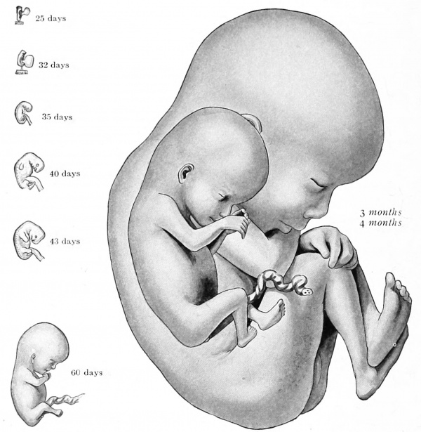
Fig. 66. Human embryos of three weeks to two months (His), and fetuses of three and four months (De Lee). Natural size.
The Establishment of External Form
Although the preceding section deals largely with the aquisition of fetal form, this topic requires supplementary treatment.
The Head and Neck
Since development in the cephalic region maintains its early advantage, the head and neck of an embryo are for a long time disproportionately large. In Fig. 63 the last cervical segment is midway on the body. The gradual adjustment of size relations may be traced in Fig. 6g .
Anomalies
Many grossly abnormal embryos are found at operation or spontaneous abortion. Various pathological conditions in the embryo commonly accompany those disturVjances which induce its stunting or death. Degenerative changes are common also in the fetal membranes, although the chorionic sac sometimes continues to grow quite normally after the embryo has died or disappeared. Dead, retained fetuses are usually resorbed, but they may mummify and persist indefinitely,
The head is composed of two portions almost from the start. One is neural in nature and includes the brain, eyes, and internal ears, and their supporting structures. The other is the facial, or visceral, part that contains the cephalic ends of the alimentary and respiratory tracts. The neural portion is much the larger in young embryos and this superiority is never lost completely, although the subsequent differentiation and growth of the nose, jaws, and pharynx reduces the early disparity.
Branchial Arches
The formation of the face and neck involves the history of the branchial arches. These are bar-like prominences, separated by grooves, which occur on the lateral surfaces of the neck (Figs. 6 1 to 63). They correspond to the gill-bearing arches of fishes that are separated by clefts through which respiratory water flows. In amniotes they never assume a respiratory function, but occur as transitory vestiges that are applied to various purposes, then disappear. The human embryo develops five such arches, separated by four ectodermal grooves; subjacent to these grooves the entoderm of the pharynx bulges correspondingly (Fig. 87). The thin plates thus formed by the union of ectoderm and entoderm sometimes rupture to make temporary openings, reminiscent of the gill-slit condition.
The last arch lies caudal to the fourth cleft and is poorly defined along its posterior margin. Toward the end of the sixth week, the first and second arches overlap the other three and obScure them. Fig. 63 shows the beginning of this process. Fig. 227 an advanced stage, and in Fig. 64 it is complete. The caudal arches sink into a triangular depression called the cervical sinus. When the posterior edge of the second arch fuses with the thoracic wall, the sinus and its contained arches are closed off. This cavity eventually degenerates.
Various muscles and bones form from the arches, and from the entodermal pouches certain glandular organs arise. The completion of this metamorphosis marks the appearance of a neck (Fig. 65) which is characteristic of amniotes alone.
Anomalies
Imperfect closure of the branchial clefts (usually the second) leads to the formation of cysts, diverticula, or even fistulae. Such structures may be derived either from an ectodermal groove or the complementary entodermal pouch.
The Face
Pig embryos show clearly how the face forms. In Fig. 369 the expansive jronto-nasal process represents much of the front of the head. The olfactory pits are present, and the first branchial arches have not only bifurcated into maxillary and mandibular processes but the mandibular segments have already united as the lower jaw. Laterally, the olfactory pits subdivide the fronto-nasal process into paired lateral and median nasal processes (Figs. 394 and 67 A). Soon, the median nasal processes fuse with each other and with the maxillary processes; this constitutes the upper jaw (Fig. 67 B). The lateral nasal processes likewise join the maxillary process, thereby obliterating the lacrimal groove, and forming the wings and margins of the nose and the adjacent cheek region. Meanwhile, the mesial portion of the original fronto-nasal process becomes the forehead and the septum and bridge of the nose.
The early development of the human face is essentially the same. These changes may be followed in Fig. 68. At first the nose is broad and fiat, with the nostrils set far apart and directed forward (Fig. 68 C). In the later fetal months the bridge is elevated and prolonged into the apex, and the nostrils look downward (Fig. 68 D). The line of fusion of the median nasal processes is evident in the adult as the philtrum . The chin is a median projection from the fused mandibular processes. During the formation of the jaws the originally broad mouth opening is reduced in its lateral extent. Epithelial ingrowths begin to separate the lips from the alveolar portions of the jaws at the fifth week (Fig. 79); at birth the inner edges of the lips bear numerous villosities. Progressive modelling of the face continues until the individual becomes fully grown.
Fig. 67. Development of the pig - s face (Prentiss). X 7. A, 12 mm.; B, 14 mm.
Anomalies
A common facial defect is hare lip. This is usually unilateral and on the left side. It may involve both lip and maxilla. Hare lip is attributed to the failure to fuse of the median nasal and maxillary processes (Kdlliker), or the lateral and median nasal processes (Albrecht).
Fig. 68. Stages in the development of the human face (adapted). A, Five weeks; B, six weeks; C, eight weeks; D, si.xteen weeks. The fronto-nasal process is indicated by parallel lines, the median nasal processes by circles, and the lateral nasal processes by dots.
The Sense Organs
The eye, ear, and nose will be considered in detail in Chapter XV. The external nose has just been described. The eye makes its appearance in the early weeks, and, by the second month, lids are present. For a time the eyes are placed laterally and far apart, but gradually this distance is reduced (Fig. 68). The external ear is developed around the first branchial groove by the appearance of small tubercles which form the auricle (Figs. 64, 65 and 31 1). The groove itself becomes the external auditory meatus.
The Trunk
In young embryos the trunk is like a cylinder, flattened by lateral compression (Fig. 63). Its external contour is determined by the modelling of the viscera within. During the fetal period, this visceral mass becomes more rounded and the muscles and skeleton of the trunk appear, d'he trunk then assumes an ovoid form, circular in section, and largest at the umbilicus (Fig. 66). From the third fetal month through early infancy there is relatively little change in the trunk proportions. When erect posture is assumed, the dominance of the thorax and abdomen is reduced and the lumbar region gains in prominence and relative length. The thorax of the newborn is rather conical, with its base below, due to the ribs lying more horizontal. In the adult the thorax is barrel-shaped, that is, broadest in its middle. The characteristic curves of the spinal column are absent at birth. They appear partly through the drag of body weight, ])artly through the pull of the muscles, and are not pronounced until the posture becomes erect.
Anomalies
The embryonic tail sometimes persists and develops beyond its ordinary size. Specimens as long as 8 cm. have been recorded in the newborn. Most are soft and ileshy, but a few have contained skeletal elements. Some tumors of the coccygeal region are attributed to the activity of residual primitive-streak tissue.
The Appendages
The limbs appear during the fifth week as lateral buds. In a 4 mm. embryo (Fig. 62) limb buds may be recognized, but due to the early expanse of the head-neck region they seem to be located far down the body. The distal ends flatten (Fig. 63) and a constriction divides this paddle-like portion from the proximal, rounded segment (Fig. 196). Later, a second constriction separates the cylindrical part into two further segments (Figs. 64 and 65), and the three divisions of arm, forearm, and hand, or thigh, leg, and foot are respectively formed. Radial ridges, separated by grooves, first foretell the formation of digits (Figs. 64 and 65). These elongate as the definitive fingers and toes, and rapidly project beyond the original plates; the latter by a slower rate of growth become confined as webs about the basal ends of the digits (Fig. 66). The thumb and great toe early separate widely from the index finger and second toe.
The limbs as a whole undergo several changes of position. At the very start they point caudad (Figs. ig6 and 64), but soon project outward at right angles to the body wall. Next, they are bent ventrad so that the thumb (radial) side of the arm and the great toe ( tibial) side of the leg are directed forward; the palmar and plantar surfaces face the body; the elbow turns outward and somewhat caudad, the knee outward and slightly cephalad (Fig. 65). Finally, both sets of limbs undergo a torsion of 90° about their long axes, but in opposite directions. As a result, the radial side of the arm is outward (when radius and ulna are parallel) and the palm faces ventrad; on the contrary, the tibal side of the leg is the inner side, while the sole faces dorsad. By following through these changes it will be seen that the radial and tibial sides of arm and leg are homologous, as are palm and sole, elbow and knee.
The upper limb buds arise first and they maintain a slight advance in differentiation. Not until the second year of childhood are the two equal in length.
Anomalies
The extremities may either fail to develop, or become mere stubs; the hands and feet may join the body like flippers. Rarely, the hands or feet are partially duplicated or reduced. The presence of extra digits is polydactyly; a fusion of digits constitutes syndactyly. More or less complete union of the legs occurs as sympodia.
The developmental period of man is divided by the incident of birth into prenatal and postnatal periods. At birth the infant is sufficiently advanced to be cared for outside its mother - s body, yet its development is far from complete. In its new environment differentiation and growth, especially marked by changes in form and proportion, continue until the beginning of the third decade ; only then is full size and mature structure attained.
The several divisions of the developmental period are listed as follows by Scammon, from whose account much of the material of the succeeding paragraphs is taken :
Divisions of the Develop.mental Period in Man
^Period of the ovum (Fertilization to end of second week)
Prenatal life - Period of the embryo (Second to eighth week)
\Period of the fetus (Second to tenth month)
Birth
Period of the newborn (Neonatal period; birth to end of second week)
1 Infancy (Second week until assumption of erect posture at 13 to 14 months) j 1 Early childhood (Milk-tooth period; first to sixth year)
j - idliood childhood (Sixth to ninth or tenth year)
†\ Later childhood (Prepubertal period; from 9 or 10 years to 12-15
Postnatal life ! I years in females and 13-16 years in males)
I Puberty
I (Fourteenth year in females: sixteenth year in males)
I Adolescence (From puberty to the last years of the second decade in females I and to the first years of the third decade in males) .
Changes in Form
If an adult maintained the chubby newborn shape his weight would be twice the actual amount. Fig. 69 shows the proportions of the body at various developmental periods, all drawn as of the same height. Note: the great decrease in the size of the head; the constancy of the trunk length; the early completion of the arms and the tardier growth of the legs; the upward shift of the umbilicus and symphysis pubis, and the downward trend of the midpoint of the body.
Fig. 69. Diagrams to illustrate the changing proportions of the body during prenatal and postnatal growth (Scammon after Stratz).
Certain of these facts may be tabulated in terms of per cent of the total body volume:
Growth in Relative Volume of the Parts of the Body .
In per cent of the total body volume .
Age .
Head and neck .
Trunk .
Arms .
Legs .
Second fetal month .
45 .
50 .
3 .
3 .
Sixth fetal month .
37 .
40 .
8 .
15 .
Birth .
27 .
49 .
9 .
15 .
Two years .
22 1 .
50.5 .
9 .
17-5 .
Six years .
15 .
51 .
9 .
25 .
^Maturity .
7 .
53 .
10 .
30 .
Increase in Surface Area
The relation of surface area to body mass or volume has a profound influence on metabolism. This relation changes greatly during the postnatal period. At birth, the surface area is about 2500 sq. cm. This is doubled in the first year, tripled by the middle of childhood, and increases rapidly before puberty. At maturity, the total gain is seven-fold. Since, however, the weight of the body has increased some twenty-fold in the same time it is obvious that there has been a relative loss. Thus, in the newborn there are over 800 sq. cm. per kilogram of body weight, whereas in the adult there are less than 300 sq. cm.
Growth in Weight
During prenatal life the weight of the body increases several billion times, whereas from birth to maturity the increment is only twenty-fold. In absolute mass, however, 95 per cent of the final weight is acquired after birth. The ratio of increase during each fetal month to the weight at the beginning of that month is shown in the table on p. 68.
Growth in Length
Growth in length and in weight have certain features in common, although the relative increase in length is obviously smaller since weight is a three dimensional phenomenon. The increase in the second fetal month is ten-fold but thereafter the relative rate of growth gradually declines. The data of prenatal growth are given in the table on p. 68. The total postnatal increment is 3.3 times. During the first six months after birth, length increases 30 per cent; in the first year, 50 per cent. Throughout the most of childhood the linear increase is very slow, but at the prepubertal period there is an acceleration; as with weight, this is begun and ended earlier in girls than in boys. Growth is complete at about 18 years in females and soon after 20 in males. The body is heaviest in proportion to its length during late fetal life and early infancy. From the middle of the first year until after puberty there is a decline in relative weight. Thereafter there is an increase in relative mass which may continue throughout life. During infancy and childhood girls are relatively lighter than boys, but after puberty the reverse is true.
Growth of Organ Systems
The skeleton grows rather slowly until the ninth and tenth fetal months, when it shows an acceleration. At birth, it constitutes from 13 to 20 per cent of the body weight. Postnatal growth apparently parallels that of the body as a whole and shows neither relative loss nor gain. The musculature likewise grows slowly at first, but forms about 2 5 per cent of the weight of the newborn and 40 to 45 per cent of the adult. The central nervous system, on the contrary, is relatively huge in the young embryo. It decreases from about 25 per cent in the second month to about 15 per cent at birth and 2 to 2.5 per cent in the adult. Incomplete data on the peripheral nervous system and skin indicate a considerable reduction in relative weight during the postnatal years. As a whole, the visceral group decreases slowly and steadily in relative weight after the first two embryonic months. In the second month they comprise about 15 per cent of the body weight, about 9 per cent at birth, and from 5 to 7 per cent in the adult.
Growth of the Organs
Although the general course of relative growth in the individual organs follows that of the visceral group, each has its characteristic curve. Each usually increases more or less rapidly to a maximum relative size and then decreases in relative size through the subsequent prenatal and postnatal periods.
During fetal life the curves of absolute growth are quite similar. The various organs have an initial period of slow increase, followed after the fifth month by a terminal phase of rapid growth. However, this uniformity disappears at birth, and most of the organs can be arranged in four main divisions. The splanchnic group includes the digestive, respiratory, and urinary organs, and the heart, thyroid, and spleen. The nervous group comprises the brain, cord, and eyeballs. The genital group excludes the ovary and uterus which have special curves. The lymphoid group includes all but the spleen. Fig. 70 shows these relations graphically from embryo to adult.
Fig. 70. Chart showing the course of growth in the various organ groups (after Scammon). Growth is calculated in per cent of adult weight.
Anomalies
Giants and dwarfs may be of monstrous size when born at full term, or the acceleration or slowing may be secondary at some later period. This abnormal size is sometimes unilateral or even confined to specific parts of the body.
| Historic Disclaimer - information about historic embryology pages |
|---|
| Pages where the terms "Historic" (textbooks, papers, people, recommendations) appear on this site, and sections within pages where this disclaimer appears, indicate that the content and scientific understanding are specific to the time of publication. This means that while some scientific descriptions are still accurate, the terminology and interpretation of the developmental mechanisms reflect the understanding at the time of original publication and those of the preceding periods, these terms, interpretations and recommendations may not reflect our current scientific understanding. (More? Embryology History | Historic Embryology Papers) |
Reference
Arey LB. Developmental Anatomy. (1924) W.B. Saunders Company, Philadelphia.
Cite this page: Hill, M.A. (2026, February 27) Embryology Book - Developmental Anatomy 1924-4. Retrieved from https://embryology.med.unsw.edu.au/embryology/index.php/Book_-_Developmental_Anatomy_1924-4
- © Dr Mark Hill 2026, UNSW Embryology ISBN: 978 0 7334 2609 4 - UNSW CRICOS Provider Code No. 00098G


