BGDA Practical 3 - Gastrulation

From Embryology
BGDsmall.jpg
Practical 3: Oogenesis and Ovulation | Gametogenesis | Fertilization | Early Cell Division | Week 1 | Implantation | Week 2 | Extraembryonic Spaces | Gastrulation | Notochord | Week 3

Introduction

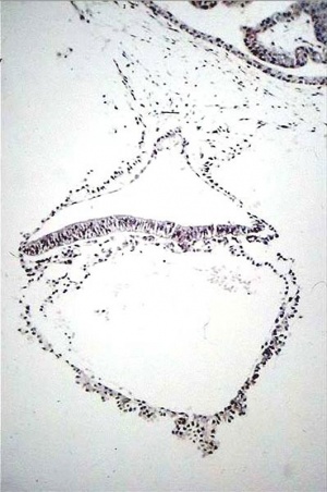
Human embryo day 18.jpg Gastrulation means "gut forming" and converts the inner cell mass which then formed the bilaminar embryo (epiblast, hypoblast) into the trilaminar embryo (ectoderm, mesoderm, endoderm).

The process involves the migration of cells from the epiblast layer through the primitive streak to form first the endoderm layer and then a second intermediate layer the mesoderm layer. Once all cells have left the epiblast layer it now becomes the ectoderm layer.

These three germ cell layers (ectoderm, mesoderm, endoderm) will form in a layer specific manner all the future tissues of the developing embryo.


Approximate cross-section of an 18 day human conceptus.

Identify the 3 layers of the trilaminar embryo: ectoderm (columnar cells), intraembryonic mesoderm (mesenchymal cells, endodermal cells (cuboidal single layer). Identify primitive groove with dense cluster of primitive streak cells below it.


(Image: Nishimura etal., 1977[1])


Stage 7

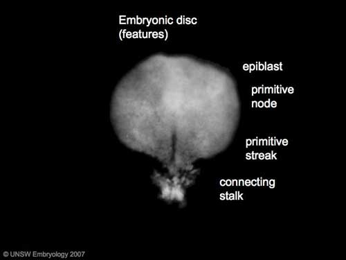
Stage7-sem2.jpg Stage7 features.jpg
Features: embryonic disc, primitive node, primative streak, primitive groove, yolk sac

Facts: Week 3, 15 - 17 days, 0.4 mm

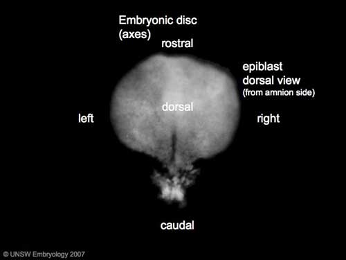
View 1: embryonic disc, showing the epiblast viewed from the amniotic (dorsal) side.

Events: Gastrulation is continuing as cells migrate from the epiblast, continuing to form mesoderm.

Mesoderm lies between the ectoderm and endoderm as a continuous sheet except at the buccopharyngeal and cloacal membranes. These membranes have ectoderm and endoderm only and will lie at the rostral (head) and caudal (tail) of the gastrointestinal tract.


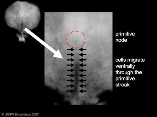
Gastrulation: Through the primitive streak cells migrate continuously through week 3 into week 4. Initial cells replace hypoblast as an epithelial layer the endoderm. Later migrating cells spread between the two epithelial layers to form mesoderm.
Stage7 primitive streak labelled.jpg Stage7 axes.jpg
Axes: embryonic disc is shown rostral (head) to top and caudal (tail) to bottom. Left and right are the lateral margins of the disc as shown.

Gastrulation

Carnegie Stage 7 and 8, gastrulation, migration of cells through the primitive streak to form endoderm and mesodermal layers of embryo.
Chicken-gastrulation3.jpg

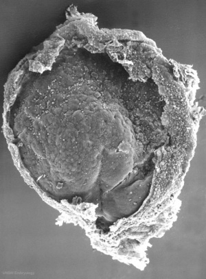
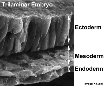
Trilaminar embryo.jpg Scanning electron micrograph showing the early forming 3 layers: ectoderm, mesoderm and endoderm. The embryonic disc has been broken to expose these early layers that form the entire embryo.
  1. Ectoderm - columnar epithelium
  2. Mesoderm - embryonic connective tissue (mesenchyme)
  3. Endoderm - cuboidal epithelium


  • The amniotic cavity would lie above the ectoderm layer.
  • The yolk sac would initially lie below the endoderm layer, later this would be the gastrointestinal tract within the embryo.
<html5media height="500" width="390">File:Mesoderm_001.mp4</html5media>

Click Here to play on mobile device

Mesoderm 001 icon.jpg

Now observe this brief animation of mesoderm development during week 3.

Mesoderm Development

  • This animation shows the migration of mesoderm throughout the embryonic disc during gastrulation.
  • The pink arrow show how mesodermal cells spread out between the ectoderm and endoderm layers, forming the third (middle) layer of the trilaminar embryo.
  • Axial process - the arrow running from the primitive node upward is the axial process which will later form the notochord.
  • 2 regions where no mesoderm is found, at the upper and lower ends of the gastrointestinal tract.
  1. cranial (top) - buccopharyngeal membrane
  2. caudal (bottom) - cloacal membrane.
  • Prechordal plate - lies above the buccopharyngeal membrane and is the cardiogenic mesoderm, that will form the heart.


Links: MP4 version | Gastrulation | Mesoderm | Week 3


References

  1. Nishimura H. Semba R. Tanimura T. and Tanaka O. Prenatal development of the human with special reference to craniofacial structures : An atlas. (1977) U.S. Dept. of Health, Education, and Welfare, Public Health Service, National Institutes of Health ; Washington. Bethesda, Md.


Interactive Component

Attempt the Quiz - Gastrulation 
BGDsmall.jpg

Here are a few simple Quiz questions that relate to Gastrulation from the lecture and practical.


1 Which of the following statements about gastrulation is most correct:

gastrulation is required to form the extra-embryonic coeloms
gastrulation is required to form the intra-embryonic coeloms
gastrulation occurs only at the primitive node
gastrulation occurs mainly during week 2 GA week 4 of development
gastrulation forms the trilaminar embryo

2 The primitive streak:

extends from the primitive node to the parachordal plate
is the primary inducer during organogenesis
is the site of migration of epiblast cells to form the mesoderm
persists at the parachordal plate
all of the above are correct

3 The migration of intraembryonic mesoderm occurs between the ectoderm and endoderm to give rise to:

the allantois
the somites
the buccopharyngeal membrane
the cloacal membrane
the neural crest


BGDsmall.jpg
Practical 3: Oogenesis and Ovulation | Gametogenesis | Fertilization | Early Cell Division | Week 1 | Implantation | Week 2 | Extraembryonic Spaces | Gastrulation | Notochord | Week 3