User:Z5001524
Introduction
During pregnancy, the maternal bloodstream does not only contain maternal cells but also fetal cells. Therefore, the fetus will expose its DNA in the maternal blood and they are generally known as cell-free fetal DNA (cffDNA) [1]. Studies have suggested that around 2-6% of the cells in maternal blood are from the fetus itself [2]. This proportion of fetal cells include nucleated red blood cells, fetal trophoblast cells, mesenchymal stem cells and leucocytes and they will circulate through the maternal blood stream freely. The understanding of their functions in maternal blood is still limited but this free moving fetal DNA facilitate the diagnosis of many birth defects and abnormalities with a lower risk of miscarriage [1].
Origin and microchimerism
The fetal cells which contain fetal DNA originate from trophoblast cells [3]. After implantation, the trophoblast cells will start to form the placenta. During this formation process, some trophoblast cells will undergo apoptosis and the DNA of the fetus will be fragmented. Eventually, they will be able to distribute and migrate into the maternal circulation through passing the intervillus space, which is filled with maternal blood [4]. This has explained the increase in the number of fetal cells in the maternal blood after implantation and presences of fetal cell can be detected starting from the 4 week of gestation [5]. However, the detection of fetal DNA in maternal blood in diagnostic tests is only reliable from 7 weeks of gestation onwards [6]. The level of fetal cells and its DNA in the maternal blood increase with gestational age and rise by approximately 64 fetal genomes per ml of maternal bloodstream between the first trimester and third trimester [7]. It will then reach its peak within last 8 weeks of gestation and decline after delivery [8].
More information on: Implantation| Placenta Development
Potential functions of fetal cells in the maternal blood
The functions of these cells that exist in maternal blood are still a mystery. However, it has been deduced that these cells would have contribute in tolerating the maternal immune system and alter the immune system cells from attacking the fetus.
Furthermore, the fetal cells in the bloodstream will also migrate to maternal organs such as lung and heart and the studies have found out the fetal cells and the placental cells have the ability to differentiate and repair the injuries in both the lungs and heart[9].
Fetal stem cells have also been deduced to have the functions of alleviating rheumatoid arthritis and as a preventative effect on breast cancer [8]. Although these cells are found in tumors, there are no specific findings that explain whether its effect in preventing or restricting the growth of tumors [10].
More information on: Rheumatoid arthritis| Breast cancer| Fetal cells differentiation in heart
Prenatal diagnosis uses
Problems of common diagnostic tests
Chorionic villus sampling and amniocentenses are the common diagnostic methods used to detect whether the fetus has Down Syndrome and other abnormalities. Although these methods have very false positive rates, the tests can only be conducted and indicate accurate results at a relatively late period. Besides, these two tests are invasive prenatal testings and both carry a chance of miscarriage or harming the fetus [11].
Uses of maternal blood in non-invasive prenatal testing
As the cffDNA has been discovered in maternal blood, this enables the testing the fetal genome from collecting maternal blood. This testing is regarded as a non-invasive prenatal testing (NIPT). The fetal blood group, sex and also chromosomal abnormalitiescan also be determined by this testing.
Some examples of cffDNA testing
Fetal rhesus D type --- The identification of the fetal blood status from maternal blood and to indicate the risk of having haemolytic disease.
Fetal sex --- The determination of Y chromosome sequences such as DYS or SRY. Male fetus with chances of having an X-linked disorder can be primarily tested with cffDNA testingto determine whether the fetus needs an invasive diagnostic test (PCR).
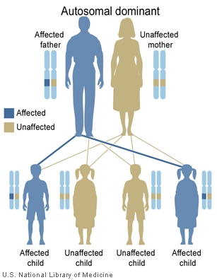
Single gene disorders --- The detection and elimination of the condition of the paternal allele inherited from a father with an autosomal dominant condition such as Huntington's disease.
Disadvantages of cffDNA testing
Since this test is used to screen disease nonspecifically for chromosomal aneuploidies, the pregnant women will be constantly informed about the findings, even those which are uncertain. This may lead to unnecessary worries. Besides, this risk also applies to ultrasound scanning and other microarray analyses. However, this problem can be solved when there is a better understanding on genetic diseases and, therefore, a specific and detailed examination will be established. http://www.rcog.org.uk/files/rcog-corp/SIP_15_04032014.pdf
References
- ↑ 1.0 1.1 <pubmed>10472878</pubmed>
- ↑ <pubmed>9504651</pubmed>
- ↑ Nigam A, Saxena P, Prakash A & Acharya AS. (2012). Detection of fetal nucleic acid in maternal plasma: A novel noninvasive prenatal diagnostic technique. Journal of Internation Medical Sciences Academy, 25(3), 199.
- ↑ Smets, E. M. L., Visser, A., Go, A. T. J. I., van Vugt, J. M. G., & Oudejans, C. B. M. (2006). Novel biomarkers in preeclampsia. Clinica Chimica Acta, 364(1–2), 22-32. doi:http://dx.doi.org/10.1016/j.cca.2005.06.011
- ↑ Nigam A, Saxena P, Prakash A & Acharya AS. (2012). Detection of fetal nucleic acid in maternal plasma: A novel noninvasive prenatal diagnostic technique. Journal of Internation Medical Sciences Academy, 25(3), 199.
- ↑ Nigam A, Saxena P, Prakash A & Acharya AS. (2012). Detection of fetal nucleic acid in maternal plasma: A novel noninvasive prenatal diagnostic technique. Journal of Internation Medical Sciences Academy, 25(3), 199.
- ↑ Nigam A, Saxena P, Prakash A & Acharya AS. (2012). Detection of fetal nucleic acid in maternal plasma: A novel noninvasive prenatal diagnostic technique. Journal of Internation Medical Sciences Academy, 25(3), 199.
- ↑ 8.0 8.1 <pubmed>20663958</pubmed>
- ↑ <pubmed>22223204</pubmed>
- ↑ Society for the Study of Reproduction. (2012, June 6). Three types of fetal cells can migrate into maternal organs during pregnancy: Some mothers literally carry pieces of their children in their bodies. ScienceDaily. Retrieved July 20, 2014 from www.sciencedaily.com/releases/2012/06/120606155802.htm
- ↑ Soothill, PW & Lo, YMD. (2014). Non-invasive prenatal testing for chromosomal abnormality using maternal plasma DNA. Scientific Impact Paper, (15)


