User:Z3418340
--Z3418340 (talk) 12:45, 6 August 2014 (EST)
Lab Attendance
Lab 1 - --Z3418340 (talk) 12:53, 6 August 2014 (EST) Lab 2 - --Z3418340 (talk) 11:13, 13 August 2014 (EST) Lab 3 - --Z3418340 (talk) 13:48, 20 August 2014 (EST) Lab 4 - --Z3418340 (talk) 12:39, 27 August 2014 (EST)
Individual Assessment 1
ARTICLE 1
<pubmed>24302192</pubmed>
Inositol is an important factor with in the follicular environment, high levels have been associated with improved development of the oocyte. This particular study aims to understand the effects of treatment with inositol on oocyte quality in patients undergoing ICSI.
The researchers selected 149 patients undergoing ICSI cycles between June 2012 and May 2013, all of whom where aged under 40, had at least one previously failed attempt at ICSI and were diagnosed with polycystic ovary syndrome (PCOS). Patients were randomly divided into two groups. Group 1 consisting of 58 patients were treated with both folic acid (400 mg/day) and inositol (2000 mg/day of myo-inositol, D-chiro-inositol 400 mg/day) for 3 months prior to the ISCI cycles. Group 2 consisted of 91 patients who were treaded with folic acid (400 mg/day) alone, this group acted as the control.
(1.) The standard of IVF using ICSI was employed. Oocyte quality was routinely checked by making observations using an inverted microscope. The oocyte’s stage of maturity, size and shape, cytoplasmic characteristics and extracytoplasmic characterises were noted. (2.) Post-implantation assessments were made regarding embryo quality in light of the following parameters; number of blastomeres, degree of fragmentation and size of blastomeres. (3.) Finally at 14 days from Embryo-Transfer execution, quantitative blood detection of β-hCG is performed in order to biochemically determine pregnancy. This is followed by a final diagnosis of clinical pregnancy with ultrasound visualization.
(1.) No significant difference was found in the number of mature oocytes taken and in the number of immature oocytes taken. However the the results did show that a greater proportion of Group 1 oocytes displayed features that typical of excellent and good quality oocytes. (3.) In terms of the number of positive biochemical pregnancies there was again no statistically significant difference between Group 1 and Group 2. However Group 1 did show a statically significant increase in the number of clinical pregnancies detected.
The article is concluded discussing and highlighting the improvement in the overall quality of oocytes and parallel increase in the number of clinical pregnancies as a result of treatment with inositol.
ARTICLE 2
<pubmed>25077107 </pubmed>
The investigation was carried out on IVF patients from Mount Sinai Hospital, Toronto, Ontario. Candidates selected for the study were aged 18-41 years, with base line levels of FSH (on Day 3 of Cycle) and the ability to provide informed consent. Researchers included 173 female patients, who then underwent IVF cycles as per standard procedure.
Serum 25-hydroxy-vitamin D (25[OH]D) levels were measured one week prior to oocyte retrieval, these measurements taken in order to determine the initial Vitamin D status. Patients were either classified as having sufficient (≥ 75 nmol/L) or insufficient (< 75 nmol/L) 25(OH)D levels.
Standard IVF procedures followed; oocyte retrieval, fertilisation, growth and embryo transfer. An ultrasound was then taken 4-5 weeks after the embryo was transferred and implantation success was monitored, as indicate by the presence of a gestational sac, visible by ultrasonography. The implantation rate was calculated as the number of gestational sacs observed by ultrasonography divided by the number of embryos transferred, multiplied by 100.
With in this cohort 45.1% had sufficient levels of 25(OH)D, while 54.9% had insufficient levels. The study found a 52.5% clinical pregnancy rate per IVF cycle among women with sufficient levels of 25(OH)D levels. This was significantly higher than compare to a rate of 34.7% the among women with insufficient levels of 25(OH)D.
Individual Assessment 2
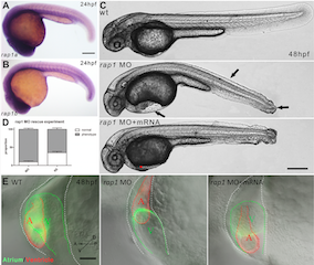
Image:Abnormal heart and caudal fin development in zebrafish due to Rap 1 knock down[1]
Individual Assessment 3
Fetal Development - Time Line
Historic Findings
[1] [2] https://www.youtube.com/watch?v=WXLPxjJszio
Individual Assessment 4
Context
The use of umbilical chord derived stem cells for therapeutic purposes is certainly widespread and has served as an effective tool for the treatment of cancers, heart disease and many other conditions.
This particular study aims to further investigate the potential use of Mesenchymal stem cells (MSC)derived from chord blood, this time looking at potential use as a promoter of wound healing in diabetic patients. In these patients incomplete healing of wounds is primarily associated with poor revascularization and decreased production of growth factors in the damaged area. Since MSCs are multipotent they hold a great promise for tissue regenration and .
The main advantage of investigation into such therapies lies in the fact that MSCs can be easily isolated and refined from chord blood as oppose to any other sources.
Study and Findings
This study was conducted on genetically diabetic mice who showed delayed would healing. A wound was induced followed by subcutaneous injection of Conditioned-MSCs (CM-MSC), UC-MSC (Umbilical cord derived MSCs) or control PBS (Phosphate buffer solution).
Wound healing was reported as a percentage of the initial would that had reepitheliasized. Time taken for complete reepitheliasized was accelerated from 14 days in the PBS group, down to 4 days; following the initial injection of CM-MSC. Histological examination of would margins at 14 days revealed that CM-MSC treated wounds had relatively enhanced repiehtelizsation, a thiner layer of dense granulation tissue and increased vascularisation. Further more Immunohistological staining also showed higher capillary density in CM-MSC treated wounds compared to both the UC-MSC and PBS treated groups. Finally the PCR analysis of RNA was extracted from the CM-MSC treated mice revealed significantly higher levels of factors promoting aniogenesis such as; VEGF, PDGF, and KGF. It is concluded that both the transplantation of UC-MSC and CM accelerate wound closure, increases angiogenesis and directly stimulate transrciption of vascular growth factors (VEGF, PDGF, and KGF.
Reference
<pubmed>3781996 </pubmed>
Pub Med
http://www.ncbi.nlm.nih.gov/pubmed/25084016
<pubmed>25084016</pubmed>
