User:Z3417458
Lab 8
1. Brief time course and overview of embryonic development of the human ovary.
- There are a series of stages in ovarian development these include; germ cell differentiation, continuous follicular growth and continuous follicular atresia
- At the early stages of development the gonads are the same in both sexes, the gonadal ridge forms into the ovary
- The essential genes for the process of ovarian development to occcur include Wnt-4 and DAX-1
- Granulosa cells are the cells that provide support in females and the follicle cells are located around the occytes
- follicle cells undergo proliferation as a result of follicular stimulating hormone among adult cells
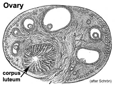
2. An image from the historic genital embryology section of the online notes in your description.
Lab 7
1. Recent research paper on the development of the Pancreas
The pancreas has two main functions, exocrine involving digestive enzymes and endocrine regulating glucose levels within the blood. There are 3 main stages of development in the pancreas. The primary stage involves initiation of morphogenesis with endodermal evagination[1] . Epithelial branching morphogenesis is the secondary stage; in which the basement membrane, differentiating islet progenitors delaminate. Finally at the apices of ductal structures, the formation of acinar cells is commenced. Throughout those stages specific waves of segregation occur with amplication to the endocrine compartment.
The increase in pancreatic endocrine cell population arise at embryonic and pancreatic post-natal growth and regeneraition. There is not much knowledge in the mechanism in endocrine cell expansion throughout the embryonic development. The study concluded through the investigation, that regulating pancreatic endocrine maturation and development is affected by the growth factor-beta (TGF-β) signaling. It is claimed that the process is mediated through the intracellular mediators of TGF- β signaling smad2, smad3 and smad7 expressed during early embryonic pancreatic epithelium. During genetic inactivation two events occur among the intracellular mediators, the smad2 and smad3 increase embryonic endocrine compartment whereas smad7 decrease in embryonic endocrine compartment[1].
From the results the function of smad7 was noted, showing that’s its important in differentiation and maturation of developmental endocrine. The suppression of smad2 &3 was identified as being regulated by smad7 due to their role in expansion of the pancreatic endocrine compartment. This knowledge highlights the mechanisms controlling β-cell development and also shows a connection among replication of pancreatic islets and developmental neogenesis of β-cell development [1]. Thus the study illustrated a regulatory point for β- cell expansion among a smad2, 3 & 7 complex. Such finds could have an effective impact on diabetes treatment, through the generation of β-cells and a target in regulating the cells proliferation.
Reference
2. Embryonic layers and tissues that contribute to the developing teeth
- Stages include week 6- lamina, week 7- placode, week-8 bud, week 11- cap, week 14- bell
- Contributions from neural crest mesenchyme, ectoderm and mesoderm
- Odontoblasts (secretion of predentin, calcifies for the formation of dentin)
- Ameloblasts (production of enamel)
- Periodontal ligament (CT , supports tooth, contains 'Sharpey’s fibers')
Lab 5 Assessment
Congenital Diaphragmatic hernia
A congenital diaphragmatic hernia (CDH) takes place when the diaphragm muscle is unable to close completely during embryo development. As a result the pleuroperitoneal foramen ‘Bochdalek’ (70-75%, posteriorly) fails to seal. That’s the most common hernia, although it may also occur anteriorly (23-28%) with the foramen of ‘Morgagni’. Therefore any contents of the abdominal viscera; such as the stomach, intestines, spleen or liver can pass through the thoracic cavity while constricting the lung. Due to this the lungs don’t have a large capacity to grow so their development is limited. This can lead to pulmonary hypoplasia or hypertension with further complications also arising. The incidence of CDH is 1-5:1000 births [1] , it’s a little more common in males.
In the development of the embryo the diaphragm forms during week 8 – 10, it is developed from several components; septum transversum, 3rd and 5th somite, ventral pleural sac, oesophageal mesentery and peritoneal membranes. The diaphragm is innervated by the phrenic nerve which branches of from C3-C5 nerves. The septum transversum anteriorly merges with the oesophageal mesentesry posteriorly connected by the pleuroperitoneal membrane in between them. CDH predominately occurs due to a failure in the fusion process and in past research clinical, genetic and experimental data has shown that during organogenesis there may be disturbances in the retinoid signaling pathway.
To manage the abnormality frequent ultrasonography is performed to observe any further changes throughout development. Prenatal treatments are limited as they can be invasive however there are several that have been developed. There are interuterine processes to allow progress in the lung development. A common strategy is the fetoscopic tracheal occlusion (FETO), involves stimulating the lungs to grow following the closure of the trachea. Postnatal treatment involves ensuring incubation, initiating mechanical ventilation with small volume, high frequency and reduced peak pressure. Attempts are made to repair the diaphragm surgically once cardio-respiratory functions are constant. An early diagnosis is always preferred to ensure prenatal and postnatal management can occur effectively.
References
- ↑ <pubmed>22214468</pubmed>
<pubmed>12566773</pubmed>
http://ult.sagepub.com.wwwproxy0.library.unsw.edu.au/content/12/1/8.full.pdf
Lab 4 Assessment
1. Paper on cord stem cells and it's findings
<pubmed>24074138</pubmed>
The paper investigates the nature of neonatal cord blood mesenchymal stem cells (CB-MSC) in the presence of serum, in patients who suffer from advanced heart failure. There are limitations in the capacity of regeneration for patients with heart failure, when using stem cells this is due to age and disease. The previous use of autologous somatic stem cells in cardiac cell therapy has had limitations, an alternative is juvenile cells which could potentially be more effective. There is some debate whether these juvenile cells would have the capacity to survive transplantation into a failing heart. Since juvenile MSC’s are inhibited in proliferation and their ability to clone as a result of the condition.
It was important to prove that in vitro neonatal cord blood mysenchymal stem cells could be functionally impaired by heart failure serum factors. The study was conducted among healthy volunteers and patients with heart failure, where human serum was required. Through the utilisation of TNF-a and IL-6 measures, the systemic quality of heart failure in sera was verified. The cultivated healthy CB-MSC from neonates were supplemented with protein-normalized human heart failure or control serum. The results illustrated that there was an increase cytokine concentration in heart failure sera, basic MSC properties were sustained when CB-MSC was under heart failure serum conditions. A significant decrease in clonogenic cells occurred with fewer colonies.
A possible treatment option for those who have heart failure is cardiac cell therapy that is still being considered. However it has been difficult to develop new treatments as most studies have been conducted on animals. Therapy in humans based on a cellular level hasn’t depicted major progress in the functioning of the heart. In conclusion the study demonstrated that CB- MSC’s biology is impacted upon through heart disease. This was depicted through the results in proliferation characteristics, stimulation of apoptosis and activation of stress signaling pathways. Furthermore in the experimental process it was found that MSC’s in serum from patients exhibiting heart failure, inhibits the cells proliferation. Future findings could establish specific cell therapies to each patient’s unique response.
2. Developmental Vascular shunts include;
- Ductus arteriosus, within the aortic arch, blood vessel connects aorta with the pulmonary artery
- Foramen ovale becomes fossa ovalis, located in the heart, in the septum of the atrium, allows blood to flow between the chambers
- Ductus venosus becomes the ligamentum venosum, located in the liver, connects the inferior vena cava to the umbilical vein
Lab 3 Assessment
Genital Abnormalities
--Z3417458 (talk) 21:01, 26 August 2014 (EST)
<<pubmed>24290348</pubmed>>
<<pubmed>25064170</pubmed>>
<<pubmed>23168057</pubmed>>
A review on spermatogenesis and cyptorchidism a common in males, results in an absence of testes either one or both.
<<pubmed>24829558</pubmed>>
--Mark Hill These are all relevant references, and a sentence description for selection would also help. Formatting >> should be a single >. (4/5)
Lab 2 Assessment
Human zygote
The stages in early development of a human zygote. All the zygote's matured under in vitro conditions. The original image was cropped to show only Figure A.
Reference
<pubmed>19924284</pubmed>
http://www.plosone.org/article/info%3Adoi%2F10.1371%2Fjournal.pone.0007844#pone-0007844-g004
--Mark Hill The image is suitable for this assessment, you have included information and formatting here that should only appear with the file summary. I have fixed some formatting issues (see page history for specific changes) and there should have been a better description of the figure identifying the acronyms used in the labelling. (4/5)
Lab 1 Assessment
Article 1
<pubmed>24206211</pubmed>
Summary
The research was carried out to identify if ethnicity effects in vitro fertilisation (IVF) treatment and its outcomes. It investigated a larger population (1517) over the period of 5 years, with no similar studies being conducted recently to this extent. A comparison was made between ethnic minority and white European groups and their live birth rate outcomes following IVF or intracytoplasmic sperm injection (ICSI) treatment.
Method
- The participants were women who were in completion of their first cycle of IVF or ISCI treatment occurring between 2006 -2011
- Each participant was required to identify their ethnic origin, there were several groups including South East Asians, Middle Eastern Asians, African Caribbeans and white Europeans.
- They were administered gonadotrophin (GnRH) releasing hormone agonists during the midluteal phase of their menstrual cycle.
- Transvaginal ultrasound and serum estradiol measurements were used to observe follicular development. Chorionic gonadotrophin was given if three follicles observed, had a measurement of 18mm or more.
- The occytes were retrieved and then fertilised by IVF or ICSI, following this step at least two embryos were then transferred into the uterus.
- Progesterone pessaries were used for luteal phase support and 16 days later a serum hCG level was used to measure the outcome.
- A transvaginal ultrasound was again used to finally confirm pregnancy and further tests were administered throughout the process, including an ultrasound scan.
Findings
The research found that there are significantly higher live birth, clinical pregnancy and implantation rates after IVF treatment in women from the white European group compared to the Ethnic group. From the sub ethnic groups, the South East Asian participants showed the lowest success rates. Further research needs to be conducted to support the results as there were less participants in the sub ethnic minority groups (14.95%) compared to the white European (85.1%).
Article 2
<pubmed>24040458</pubmed>
Summary
The paper highlighted the impact of endometriosis on IVF/ICSI treatment outcomes. There was an involvement of 1027 patients, 431 that suffered from infertility due to endometriosis. There have been particularly high risks of infertility due to aspects of endometriosis. This study’s main focus was to see if there were any significant differences in IVF/ICSI treatment outcomes and ovarian stimulation parameters, among women with endometriosis and women with tubal factors.
Method
- It included 1027 patients; 152 with stage I-II endometriosis, 279 with stage III-IV endometriosis, 596 patients with tubal factors were the control group.
The study was conducted between 2011 and 2012.
- Patients with endometriosis had complete removal of endometriosis lesion by laparoscopy before their IVF/ICSI treatment. The laparoscopy was also used to diagnose the control group with tubal infertility.
COH Protocol
- Endometriosis patients went through controlled ovarian hyperstimulation (COH) or GnRH-a prolonged protocol.
- Serum E2 level <pg/mL and serum LH level <2mIU/mL were administered in confirmation of pituitary suppression. The GnRH-a long protocol was given to the control group.
- Administration of 150 IU/d intramuscularly was commenced and ovarian stimulation with Puregon or recombinant FSH.
- Once two follicles reached a measurement of 18 mm, recombinant hCG was provided to trigger follicle maturation.
- Oocytes were retrieved transvaginally following hCG injection.
- The assessment of embryos had a few variables; cleavage rate, equality of blastomeres, degree of fragmentation, and mononuclearity in blastomeres.
- The embryos were classified into four categories according to the number of cells and fragmentation percentage.
- From the time of oocyte retrieval luteal phase support was given through 60mg injections of progesterone
Findings
The endometriosis patients responded ‘worse’ to the ovarian stimulation that tubal factor patients. Although it was shown that patients with endometriosis had more success rates compared to the patients with infertility as a result of tubal factors. The results depicted that IVF/ICSI is effective for patients who may have infertility due to endometriosis. This was aided by the administration of COH, pituitary suppression, and efficient surgery before the treatments. Without those factors, it is possible that different outcomes would have occurred. The results proved that both patients with endometriosis and tubal infertility had relatively equivalent pregnancy outcomes. Therefore IVF/ICSI (ART) could be an effective treatment for infertility in women due to endometriosis or tubal factors.
--Mark Hill These are appropriate references and good summaries. I have fixed the reference citation formatting. (5/5)
Lecture Reviews
Lecture 1
I thought the lecture was well divided for a first lecture of the semester. The information about the course and our requirements were well presented and easy to understand. I enjoyed the content about the history of embryology, I've never heard of the "Carnegie Stages of Development". I though that was really interesting to see the development of a zygote and for there to be such technology where you can see such detail is really amazing. I liked that we were shown video's, its always good to have lecture content presented in different ways, keeps me focused.
Lecture 2
I've previously learned about the process of meiosis and mitosis in high school biology, it was good to refresh my memory. I was familiar with most of the content since I completed histology in semester 1, so I happy that I could follow the information. The content on polar bodies was new to me, I didn't know what those were before the lecture. The stages of fertilisation for males and females were new to me. In this lecture I found the link between maternal age and the risks of Trisomy 21 surprising as I had no idea that there was a link. It is quite alarming how the risks increase so much with age.
Type in a Group
A combination of both of these;
Teamworker
A Teamworker is the oil between the cogs that keeps the machine that is the team running smoothly. They are good listeners and diplomats, talented at smoothing over conflicts and helping parties understand one other without becoming confrontational. Since the role can be a low-profile one, the beneficial effect of a Teamworker can go unnoticed and unappreciated until they are absent, when the team begins to argue, and small but important things cease to happen. Because of an unwillingness to take sides, a Teamworker may not be able to take decisive action when it is needed.
Completer Finisher
The Completer Finisher is a perfectionist and will often go the extra mile to make sure everything is "just right," and the things he or she delivers can be trusted to have been double-checked and then checked again. The Completer Finisher has a strong inward sense of the need for accuracy, and sets his or her own high standards rather than working on the encouragement of others. They may frustrate their teammates by worrying excessively about minor details and by refusing to delegate tasks that they do not trust anyone else to perform.
Lab Attendance
Lab 1 --Z3417458 (talk) 12:45, 6 August 2014 (EST)
http://www.ncbi.nlm.nih.gov/pubmed
<pubmed>25084016</pubmed> Lab 2 --Z3417458 (talk) 11:11, 13 August 2014 (EST)
Lab 3 --Z3417458 (talk) 11:15, 20 August 2014 (EST)
Lab 4 --Z3417458 (talk) 11:08, 27 August 2014 (EST)
Lab 5 --Z3417458 (talk) 11:03, 3 September 2014 (EST)
Lab 6 --Z3417458 (talk) 11:08, 10 September 2014 (EST)
Lab 7 --Z3417458 (talk) 11:08, 17 September 2014 (EST)
Lab 8 --Z3417458 (talk) 11:17, 24 September 2014 (EST)
Lab 9 --Z3417458 (talk) 11:26, 8 October 2014 (EST)
Lab 10
Lab 11
Lab 12

