Talk:Basic - Embryonic Heart Divisions: Difference between revisions
From Embryology
(First content added) |
No edit summary |
||
| Line 3: | Line 3: | ||
==Content== | ==Content== | ||
The internal heart undergoes significant changes in order to form the atria and ventricles of the adult heart. These can be summarised as follows: | |||
[[Image:HeartILP_draft_atrialsept1.jpg|thumb|center|Early atrial septation]] | {| style="width:50%" align=center | ||
|- style="background:lightsteelblue" | |||
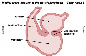
|Cells from the dorsal and ventral (back and front) walls of the heart grow and form two protrusions called the endocardial cushions. These grow towards each other and fuse to form the left and right atrioventricular canals. | |||
|[[Image:HeartILP_draft_AVcanalsept.jpg|thumb|Division of the atrioventricular canal]] | |||
|- | |||
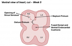
|Within the primordial atrium, a septum (the septum primum) grows towards the endocardial cushions. The space between the cushions and septum is known as the foramen primum. As the foramen primum decreases in size, a second opening forms in the septum – the foramen secundum. | |||
|[[Image:HeartILP_draft_atrialsept1.jpg|thumb|center|Early atrial septation]] | |||
|-style="background:lightsteelblue" | |||
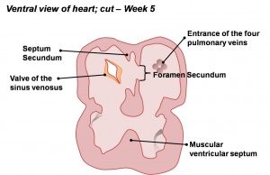
|A second septum (septum secundum) develops on the right of the septum primum. | |||
|[[Image:HeartILP_draft_atrialsept2.jpg|thumb|center|Atrial and ventricular septation]] | |||
|- | |||
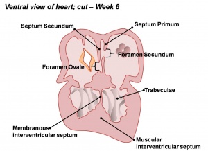
|A primordial muscular ridge exists in the floor of the ventricle. As the left and right ventricles grow, their medial (midline) walls fuse to form the interventricular septum. | |||
|[[Image:HeartILP_draft_aandvsept.jpg|thumb|center|Atrial and ventricular septation]] | |||
|-style="background:lightsteelblue" | |||
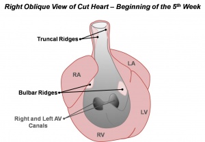
|Within the bulbus cordis and truncus arteriosus, which form the outflow tract, small ridges develop. They are continuous throughout the outflow tract and form a spiral shape. | |||
|[[Image:HeartILP_draft_outflowtract1.jpg|thumb|center|Development of the conotruncal ridges]] | |||
|- | |||
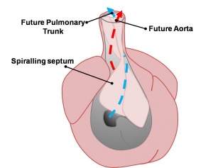
|As these ridges fuse, they create a spiral shaped septum throughout the outflow tract. The original outflow tract is therefore separated into both the aorta and pulmonary trunk. | |||
|[[Image:HeartILP_draft_outflowtract2.jpg|thumb|center|Division of the outflow tract forms the aorta and pulmonary trunk]] | |||
|} | |||
Thus the heart begins to resemble the adult heart in that it has two atria, two ventricles and the aorta forming a connection with the left ventricle, while the pulmonary trunk forms a connection with the right ventricle. | |||
Revision as of 09:28, 6 October 2009
--Phoebe Norville 09:14, 29 September 2009 (EST) Content added:
Content
The internal heart undergoes significant changes in order to form the atria and ventricles of the adult heart. These can be summarised as follows:
| Cells from the dorsal and ventral (back and front) walls of the heart grow and form two protrusions called the endocardial cushions. These grow towards each other and fuse to form the left and right atrioventricular canals. | |
| Within the primordial atrium, a septum (the septum primum) grows towards the endocardial cushions. The space between the cushions and septum is known as the foramen primum. As the foramen primum decreases in size, a second opening forms in the septum – the foramen secundum. | |
| A second septum (septum secundum) develops on the right of the septum primum. | |
| A primordial muscular ridge exists in the floor of the ventricle. As the left and right ventricles grow, their medial (midline) walls fuse to form the interventricular septum. | |
| Within the bulbus cordis and truncus arteriosus, which form the outflow tract, small ridges develop. They are continuous throughout the outflow tract and form a spiral shape. | |
| As these ridges fuse, they create a spiral shaped septum throughout the outflow tract. The original outflow tract is therefore separated into both the aorta and pulmonary trunk. |
Thus the heart begins to resemble the adult heart in that it has two atria, two ventricles and the aorta forming a connection with the left ventricle, while the pulmonary trunk forms a connection with the right ventricle.
