Talk:ANAT2341 Lab 1 - Gametogenesis
Cell Division
- Cell Division Milestones, Recent Nobel Prizes
Features Two Mechanical Processes
- Mitosis - microtubule based segregation of chromosomes and formation of 2 nuclei
- Cytokinesis - microfilament based splitting of the cell cytoplasmic contents as a whole into 2 daughter cells
Features Two Types
- Mitosis - occurs in all cells, producing genetically identical progeny.
- Meiosis - occurs only in germ cells (sperm=spermatozoa and egg=oocyte), producing genetically different progeny.
- progeny = daughter cells, offspring
Cell Changes
Nucleus
- Chromosome condensation
- Nuclear envelope breakdown
Cytoplasm
- Cytoskeleton reorganization
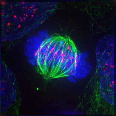
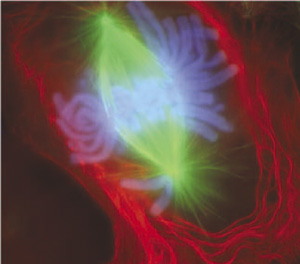
- Spindle formation (microtubule, MT) Contractile ring (microfilament, MF)
- Organelle redistribution
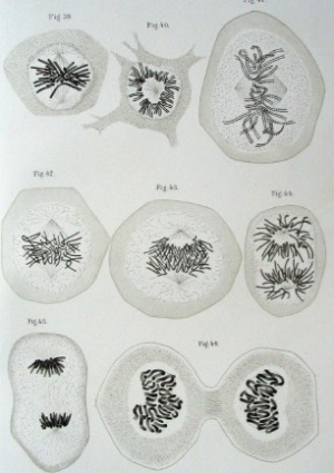
Mitosis
Mitosis Movie[1] See also MCB Movie - The stages of mitosis and cytokinesis in an animal cell
Note that DNA duplication has occurred earlier in the S Phase of the cell cycle. |
|
1. Prophase
- Chromosome DNA has been earlier duplicated (S Phase)
- Chromosomes begin condensing
- Chromosome pairs (chromatids) held together at centromere
- Microtubules disassemble
- Mitotic spindle begins to form
- Prophase ends when nuclear envelope breaks down
2. Prometaphase
- Microtubules now enter nuclear region
- Nuclear envelope forms vesicles around mitotic spindle
- Kinetochores form on centromere attach to some MTs of spindle
- Prometaphase ends when chromosomes move to metaphase plate
MCB Movie - Centromeric attachment of microtubules
3. Metaphase
- Kinetochore MTs align chromosomes in one midpoint plane
- Metaphase ends when sister kinetochores separate
4. Anaphase
- Separation of sister Kinetochores
- shortening of Kinetochore microtubules pulls chromosome to spindle pole
- Anaphase ends as nuclear envelope (membrane) begins to reform
5. Telophase
- Chromosomes arrive at spindle poles
- Kinetochore MTs lost
- Condensed chromosomes begin expanding
- Continues through cytokinesis
Cytokinesis
- Division of cytoplasmic contents
- Contractile ring forms at midpoint under membrane
- Microfilament ring Contracts forming cleavage furrow
- Eventually fully divides cytoplasm
Cell Organelles
- Mitochondria - Divide independently of cell mitosis, distributed into daughter cells
- Peroxisomes - localise at spindle poles
- Endoplasmic Reticulum - associated with the nuclear envelope vesicles.
- Golgi Apparatus- Golgi stack undergoes a continuous fragmentation process, fragments are distributed into daughter cells, then reassembled into new Golgi stacks
Meiosis
Meiosis Germ cell division (haploid)
- Reductive division generates the haploid gametes (egg, sperm)
- Each genetically distinct from the parent
- Genetic recombination (prophase 1)
- Exchanges portions of chromosomes maternal/paternal homologous pairs
- Independent assortment of paternal chromosomes (meiosis 1)
Homologous chromosomes pairing unique to meiosis
- Each chromosome duplicated and exists as attached sister chromatids before pairing occurs
- Genetic Recombination shown by chromosomes part red and part black
- chromosome pairing in meiosis involves crossing-over between homologous chromosomes
Meiosis I and II
- Meiosis I - separates the pairs of homologous chromosomes, reduces the cell from diploid to haploid.
- Meiosis II - separates each chromosome into two chromatids (chromosome behavior in meiosis II is like that of mitosis).
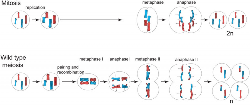
Figure 14.32. Comparison of meiosis and mitosis
Prophase I - The homologous chromosomes pair and exchange DNA to form recombinant chromosomes. Prophase I is divided into five phases:
- Leptotene - chromosomes start to condense.
- Zygotene - homologous chromosomes become closely associated (synapsis) to form pairs of chromosomes consisting of four chromatids (tetrads).
- Pachytene - crossing over between pairs of homologous chromosomes to form chiasmata (form between two nonsister chromatids at points where they have crossed over)
- Diplotene - homologous chromosomes begin to separate but remain attached by chiasmata.
- Diakinesis - homologous chromosomes continue to separate, and chiasmata move to the ends of the chromosomes.
Prometaphase I - Spindle apparatus formed, and chromosomes attached to spindle fibres by kinetochores.
Metaphase I - Homologous pairs of chromosomes (bivalents) arranged as a double row along the metaphase plate. The arrangement of the paired chromosomes with respect to the poles of the spindle apparatus is random along the metaphase plate. (This is a source of genetic variation through random assortment, as the paternal and maternal chromosomes in a homologous pair are similar but not identical. The number of possible arrangements is 2n, where n is the number of chromosomes in a haploid set. Human beings have 23 different chromosomes, so the number of possible combinations is 223, which is over 8 million.)
Anaphase I - The homologous chromosomes in each bivalent are separated and move to the opposite poles of the cell.
Telophase I - The chromosomes become diffuse and the nuclear membrane reforms.
Cytokinesis I - Cellular cytoplasmic division to form two new cells, followed by Meiosis II.
Prophase II - Chromosomes begin to condense, nuclear membrane breaks down and spindle forms.
Metaphase II - Spindle fibres attach to chromosomes, chromosomes align in cell centre.
Anaphase II - Chromosomes separate and move to the opposite poles of the cell.
Telophase II - Chromosomes reach spindle pole ends and the nuclear membrane reforms.
Cytokinesis - Cellular cytoplasmic division to form new cells.
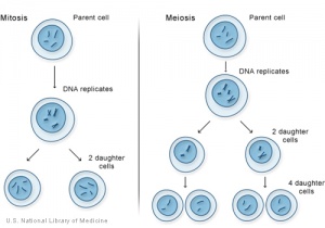
Comparison of Meiosis/Mitosis
McGraw-Hill Animation comparing Mitosis and Meiosis
- After DNA replication 2 nuclear (and cell) divisions required to produce haploid gametes
- Each diploid cell in meiosis produces 4 haploid cells (sperm) 1 haploid cell (egg)
- Each diploid cell mitosis produces 2 diploid cells
- ↑ <pubmed>12105179</pubmed>
