Unused files
From Embryology
The following files exist but are not embedded in any page. Please note that other web sites may link to a file with a direct URL, and so may still be listed here despite being in active use.
Showing below up to 250 results in range #501 to #750.
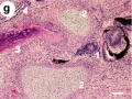
 Testis, young H&E reproductive system, male, convoluted seminiferous tubules x10.jpg 1,280 × 1,024; 396 KB
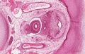
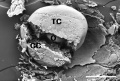
 Mouse follicle in vitro 02.jpg 600 × 701; 146 KB
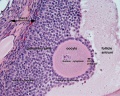
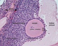
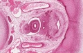
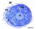
 Mouse antral follicle.jpg 600 × 705; 168 KB
 Mouse adult lymph node 07.mov ; 309 KB
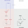
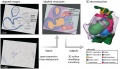
 Model coupling hematopoiesis with osteopoiesis.jpg 486 × 600; 44 KB
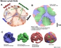
 Lymph node histology01.jpg 800 × 680; 282 KB
 Pig lung alveolarization.jpg 600 × 389; 38 KB
 Cytoplasmic lattices in oocytes and two-cell embryos.jpg 753 × 1,000; 226 KB
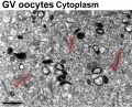 Cytoplasmic lattices in GV oocyte cytoplasm.jpg 1,048 × 846; 291 KB
 Mouse antral follicle 01.jpg 932 × 1,095; 374 KB
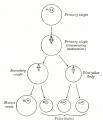
 Model for granulocytic nuclear lobulation.jpg 600 × 930; 85 KB
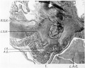
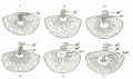
 Sabin1909 fig17.jpg 645 × 545; 80 KB
 Sabin1909 fig13.jpg 640 × 547; 114 KB
 Sabin1909 fig16.jpg 512 × 438; 120 KB
 Human zygote two pronuclei 02.png 433 × 422; 121 KB
 Canine embryo E21 image002.jpg 600 × 450; 48 KB
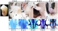
 Mouse olfactory development E12-E15.jpg 600 × 1,764; 323 KB
 Bronchial epithelial bridge.jpg 397 × 482; 28 KB
 Stage22 bf2a.jpg 600 × 800; 36 KB
 Stage22 bf2b.jpg 450 × 600; 22 KB
 Stage22 bf2c.jpg 300 × 400; 11 KB
- Intestinal rotation.mov ; 36 KB
 BGDA PracManual 2011 Practical 3.pdf ; 349 KB
 Mouse ovary 01.jpg 602 × 482; 69 KB
 Rat ovary follicle development 01.jpg 1,200 × 1,035; 201 KB
 Human fetal ovary SMAD6 expression.jpg 711 × 535; 167 KB
 Mouse oocyte and zona pellucida EM01b.jpg 600 × 600; 133 KB
 Mouse oocyte and zona pellucida EM01c.jpg 400 × 400; 60 KB
 Mouse inner ear development 01.jpg 600 × 373; 64 KB
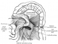
 Gray0054.jpg 800 × 513; 71 KB
 Bat embryo stage 10-13.jpg 547 × 767; 23 KB
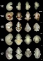 Bat embryo stage 12-17.jpg 548 × 767; 30 KB
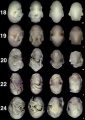 Bat embryo stage 18-24.jpg 518 × 734; 32 KB
 Lens-neural crest signaling 02.jpg 521 × 522; 22 KB
 Pyloric atresia 01.jpg 800 × 580; 35 KB
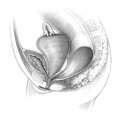 Hydrocolpos.jpg 375 × 361; 23 KB
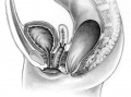 Perineal fistula.jpg 800 × 596; 82 KB
 Gray1042.jpg 969 × 745; 140 KB
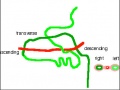 Midgut rotation icon.jpg 322 × 241; 17 KB
 Pancreas histology 10he.jpg 300 × 400; 73 KB
 Pancreas histology 40he.jpg 300 × 400; 42 KB
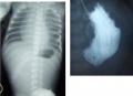
 Spina bifida.jpg 800 × 633; 77 KB
 Liver plasmodium infection cartoon.jpg 1,000 × 450; 78 KB
 Ovary histology 061a.jpg 800 × 640; 200 KB
 Ovary histology 061c.jpg 400 × 320; 56 KB
 Stage 22 image 210a.jpg 1,000 × 644; 310 KB
 Stage 22 image 210b.jpg 800 × 515; 207 KB
 Stage 22 image 210c.jpg 400 × 257; 52 KB
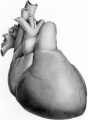
 Platypus right auricle 01.jpg 800 × 640; 124 KB
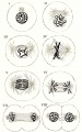
 Hertwig1892-plate01.jpg 919 × 1,475; 257 KB
 Human fetal uterus myometrium.jpg 500 × 554; 86 KB
 Ovarian follicular steroid synthesis.jpg 1,200 × 645; 89 KB
 Stage16-17-limbs01a.jpg 800 × 458; 55 KB
 Stage16-17-limbs01b.jpg 600 × 344; 35 KB
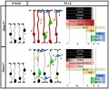
 Neural crest precursor differentiation.jpg 600 × 576; 42 KB
 Role of REX1 in stem cells.jpg 600 × 292; 30 KB
 Human embryonic stem cell defined conditions 01.jpg 1,200 × 1,132; 372 KB
 Human embryonic stem cell defined conditions 02.jpg 1,091 × 411; 187 KB
 Human embryonic stem cell defined conditions 03.jpg 500 × 376; 73 KB
 Macaque Xi at interphase 01.jpg 1,200 × 615; 157 KB
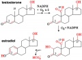 Estradiol synthesis.jpg 600 × 441; 40 KB
 Journal.pone.0018973.g001.jpg 570 × 499; 23 KB
 Merkel cell EM 02.jpg 984 × 685; 166 KB
- UK Growth Chart-A4 Girls NICM.pdf ; 138 KB
- UK Growth Chart-A4 Boys NICM.pdf ; 138 KB
- Course outline final 2011.pdf ; 2.07 MB
 Cervicovaginal mucus rheology.jpg 800 × 438; 74 KB
- Error creating thumbnail: File missingEctopic Pregnancies- United-States 1997–2006.jpg 800 × 332; 19 KB
 Uterus Serpine2 expression.jpg 600 × 620; 158 KB
 Human trisomy chromosome 7 and 19.jpg 1,000 × 804; 154 KB
 Stage21A.jpg 413 × 555; 23 KB
 Abnormal cytokinesis and apoptosis in UBE3A knockdown cells.jpg 430 × 647; 138 KB
 Stage19 bf14.jpg 1,200 × 900; 85 KB
 Stage19 bf15.jpg 1,003 × 1,000; 100 KB
 Pone.0006379.g003.jpg 484 × 591; 204 KB
 Human- fetal week 10 bf02.jpg 1,200 × 900; 91 KB
 Untitled.jpg 386 × 388; 13 KB
 Heart-histology-102.jpg 1,280 × 1,024; 242 KB
 Cleft Palate.jpg 698 × 600; 71 KB
 Table 3.png 1,008 × 630; 21 KB
 Clouds.jpg 2,560 × 1,920; 307 KB
 Clouds 01.jpg 1,200 × 900; 96 KB
 Pone 0012349 g001.jpg 662 × 521; 164 KB
 The frequency of SD cells for RB1 and SNRPN.jpg 600 × 769; 97 KB
- Hippocampal formation.pdf ; 149 KB
 Image 1.JPG 457 × 537; 67 KB
 Image 2.JPG 496 × 605; 44 KB
 1752-1947-1-94-1.jpg 600 × 198; 38 KB
 Histological Comparison of Cleft Palate in Mice.jpg 256 × 256; 7 KB
 Pone.0014375.g001.gif 100 × 87; 9 KB
 Viaat mutants exhibit cleft palate and umbilical hernia.jpg 256 × 256; 7 KB
 Gall bladder histology 005.gif 600 × 450; 683 KB
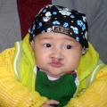
 A 12 year old PWS patient and a 4 year old AS patient.jpg 358 × 966; 141 KB
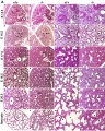 Mouse lung development 01.jpg 1,000 × 1,254; 791 KB
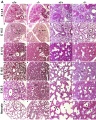 Mouse lung development 01a.jpg 800 × 1,003; 495 KB
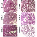 Mouse lung development 02.jpg 922 × 922; 239 KB
 Chicken heart 3D reconstruction from sections.jpg 1,000 × 571; 104 KB
 Abnormal stats.xls ; 15 KB
 Mouse telencephalon radial glia model.jpg 1,000 × 816; 80 KB
 Mouse ventral body wall development 01.jpg 1,200 × 635; 130 KB
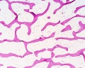 Bone-bon02he.jpg 1,280 × 1,024; 348 KB
 Rat karyotype.jpg 600 × 450; 24 KB
 Adhesion regulation blood or vessel differentiation.jpg 600 × 788; 42 KB
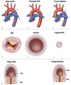 Aortic arch and ductus arteriosus.jpg 600 × 720; 68 KB
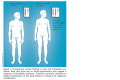 Klinefelter's Syndrome symptoms.png 1,008 × 675; 157 KB
 DiGeorge 2.jpg 518 × 368; 41 KB
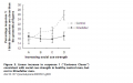 Emotional Response to 'cartoon' test Klinefelter Syndrome.png 1,008 × 630; 174 KB
- Lab7 2011 part 1.pdf ; 2.03 MB
- L7 part2 2011.pdf ; 1.55 MB
 Cleft lip 2.jpg 500 × 500; 160 KB
 TGF-beta ligands.jpg 798 × 1,000; 104 KB
 Blood test results.jpg 879 × 345; 54 KB
 DiGeorge Facial Features.jpg 1,280 × 1,024; 25 KB
 Tetralogy of Fallot Image.JPG 640 × 569; 24 KB
 UBE3A ubiquitylation pathway.jpg 830 × 365; 34 KB
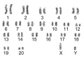
 Karyotype of Klinefelter's Syndrome.png 768 × 600; 222 KB
 Origin of the craniofacial skeleton.jpg 2,304 × 2,039; 319 KB
 Variations of Cleft Lip or Palate.jpg 951 × 1,050; 445 KB
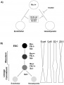
 Imprinting Inheritance in Familial AS.jpeg 186 × 160; 5 KB
 Tetralogy Of Fallot.PNG 1,238 × 525; 735 KB
 Fragile x inheritance..jpg 1,074 × 654; 66 KB
 Polysialic acid cell interactions.jpg 600 × 748; 67 KB
 Renal - S-shaped body stage.jpg 1,155 × 432; 99 KB
 Renal - early glomerulus.jpg 1,155 × 432; 52 KB
 Origin of the craniofacial skeleton-1.jpg 585 × 549; 123 KB
 Elastin (ELN) Gene on Chromosome 7.jpeg 419 × 179; 13 KB
 TBX1.jpeg 181 × 185; 6 KB
 NKX2-5.jpeg 469 × 173; 14 KB
 JAG1.jpeg 196 × 139; 6 KB
 The frataxin gene on chromosome 9.jpg 689 × 909; 40 KB
 Friedreich's Ataxia Pedigree.jpg 1,424 × 588; 80 KB
 HD Interview questions.png 204 × 135; 12 KB
 Point vs frameshift mutation of DMD gene.png 1,689 × 1,000; 874 KB
 Key cellular pathogenic mechanisms in HD.jpg 663 × 517; 47 KB
 TurnerSyndromeXChromosomeFormations.jpg 718 × 1,436; 107 KB
 HD Gene.jpg 945 × 424; 76 KB
 HD future research.jpg 687 × 869; 78 KB
 Stage5 bf1.jpg 800 × 618; 213 KB
 Stage5 bf11.jpg 618 × 800; 213 KB
 Stage5 bf11a.jpg 464 × 600; 134 KB
 Stage5 bf2.jpg 800 × 516; 122 KB
 Stage5 bf22.jpg 516 × 800; 123 KB
 Franklin Mall 02.jpg 767 × 1,000; 94 KB
 Human embryo skin 8-9 week EGA desmosomes.jpg 800 × 198; 40 KB
 Fragile X Inheritance..jpg 1,074 × 654; 105 KB
 Tinsley et al. utrophin results.jpg 452 × 449; 114 KB
 Experiment groups for utrophin upregulation.JPG 932 × 128; 20 KB
- 2011 Lecture - Neural Development.pdf ; 3.08 MB
 Dystrophin in the muscle fibre membrane.jpg 1,840 × 1,355; 415 KB
 Role of FXN Gene in Friedriech Ataxia.jpg 514 × 356; 51 KB
 Species brain sizes.jpg 600 × 664; 87 KB
 Model of cortical neurogenesis.jpg 1,000 × 565; 99 KB
 Regions of the brain.jpg 1,280 × 1,239; 348 KB
- ANAT2341 2011 Heart Dunwoodie.pdf ; 1.3 MB
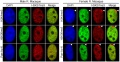
 Morula cell lines express ES markers.jpg 1,280 × 831; 205 KB
 Early lineage markers in morulae and blastocysts.jpg 1,280 × 868; 114 KB
 Embryology qrurl.jpg 328 × 328; 3 KB
 Site map qrurl.jpg 392 × 392; 5 KB
 Medicine qrurl.jpg 392 × 392; 5 KB
 Science qrurl.jpg 392 × 392; 5 KB
 Cord blood induced stem cells 01.jpg 800 × 787; 157 KB
 Cord blood induced stem cells 02.jpg 700 × 966; 189 KB
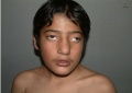
 Lowered Ear Lobes.jpg 1,078 × 828; 72 KB
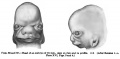
 Kollmann499.jpg 550 × 367; 40 KB
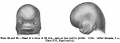
 Kollmann500.jpg 548 × 344; 38 KB
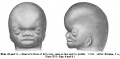
 Kollmann497-498.jpg 762 × 277; 45 KB
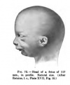
 Kollmann565.jpg 703 × 571; 79 KB
 Kollmann566-569.jpg 815 × 1,074; 112 KB
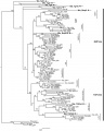
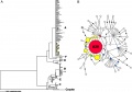 Dog mitochondrial DNA variations.jpg 1,000 × 703; 76 KB
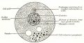
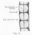 Kollmann051.jpg 327 × 354; 16 KB
 Microtiter plate - 96 wells.jpg 530 × 351; 67 KB
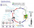 Opisthorchiasis.jpg 739 × 598; 84 KB
 Human preimplantation embryos 01.jpg 1,280 × 629; 261 KB
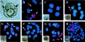
 Mouse interdigit apoptosis 02.jpg 764 × 764; 61 KB
- Bones.pptx ; 64 KB
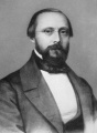 Rudolf Virchow.jpg 480 × 656; 170 KB
- 2011 Wiki Stats.xls ; 21 KB
 Gray0001.jpg 2,048 × 1,052; 185 KB
 Gray0002.jpg 963 × 1,536; 111 KB
 Gray0004.jpg 2,048 × 1,205; 199 KB
 Gray0005.jpg 1,314 × 1,536; 66 KB
 Bedford01.jpg 734 × 1,000; 82 KB
 Bedford02.jpg 1,069 × 911; 266 KB
 Stage23 bf7.jpg 600 × 906; 23 KB
 Uterus secretory phase 01.jpg 1,280 × 1,024; 318 KB
 Uterus secretory phase 02.jpg 1,280 × 1,024; 359 KB
 Model BMP satellite cell differentiation.jpg 800 × 473; 75 KB
 Stage23 bf8.jpg 578 × 872; 43 KB
 Venule microvessel EM.jpg 600 × 626; 91 KB
 Mouse in vitro follicle 01.jpg 661 × 534; 72 KB
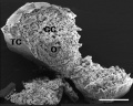
 Mouse in vitro follicle 02.jpg 800 × 639; 120 KB
 Mouse in vitro follicle 03.jpg 800 × 639; 99 KB
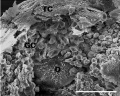
 Mouse in vitro follicle 04.jpg 799 × 537; 81 KB
 Mouse in vitro follicle 05.jpg 800 × 639; 82 KB
 Mouse in vitro follicle 06.jpg 800 × 639; 120 KB
 Keibel Mall 066-067.jpg 1,000 × 494; 43 KB
 Keibel Mall 068-069.jpg 1,000 × 358; 35 KB
 Keibel Mall 070-071.jpg 1,000 × 490; 45 KB
 Keibel Mall 072a.jpg 227 × 273; 9 KB
 Lymph node model dymamics.jpg 1,000 × 800; 231 KB
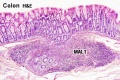 Colon MALT.jpg 500 × 333; 67 KB
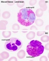 Neutrophil and eosinophil 01.jpg 480 × 600; 45 KB
 Spleen histology 12.jpg 600 × 450; 93 KB
 Spleen histology 13.jpg 600 × 450; 80 KB
 Erythrocyte and lymphocyte SEM01.jpg 800 × 522; 74 KB
 IBooks-icon.jpg 179 × 179; 8 KB
 Fetal liver erythroblasts 01.jpg 905 × 534; 69 KB
 Mouse neonatal ovary oocyte EM01.jpg 677 × 1,000; 266 KB
 Mouse neonatal ovary oocyte EM02.jpg 790 × 792; 179 KB
 Mouse neonatal ovary oocyte EM03.jpg 790 × 792; 187 KB
 Mouse neonatal ovary oocyte EM04.jpg 790 × 792; 235 KB
 Mouse-oocyte-d.jpg 790 × 792; 217 KB
 Mouse neonatal ovary oocyte EM05.jpg 790 × 792; 217 KB
 Mouse neonatal ovary oocyte EM06.jpg 790 × 792; 163 KB
 Mouse neonatal ovary oocyte EM07.jpg 790 × 792; 183 KB
 Monocyte EM01.jpg 923 × 1,000; 221 KB
 Spermatozoa tail cross-section cartoon.jpg 429 × 429; 43 KB
- The Carnegie Staged Embryos.pdf ; 3.87 MB
 Carnegie-book-qr-ISBN9780733431487.jpg 500 × 500; 10 KB
- Mouse face microCT 01.m4v ; 723 KB
- Mouse face microCT 02.mov ; 197 KB
 Streeter1920 05.jpg 737 × 807; 34 KB
 Streeter1920 06.jpg 1,000 × 339; 20 KB
 Streeter1920 07.jpg 495 × 800; 57 KB
 Streeter1920 08.jpg 514 × 800; 56 KB
 Streeter1920 09.jpg 679 × 690; 104 KB
 Streeter1920 10.jpg 1,000 × 404; 26 KB
 Streeter1920 11.jpg 1,000 × 495; 33 KB
 Streeter1921 fig27.jpg 949 × 1,000; 138 KB
 Johnson1917 fig09.jpg 700 × 636; 78 KB
- NIH - Stem Cell primer 2009.pdf ; 1.89 MB
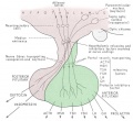 Hypothalamus pituitary cartoon.jpg 653 × 600; 81 KB
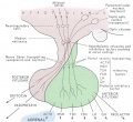 Hypothalamus pituitary adrenal cartoon.jpg 653 × 600; 82 KB
 Pituitary histology 008.jpg 1,280 × 1,024; 340 KB
 Image1183.jpg 800 × 351; 29 KB
 Image1184.jpg 800 × 378; 36 KB
- Human embryo stage22.mov ; 1 MB
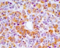
 Germ cell tumor 01.jpg 800 × 525; 161 KB
 Germ cell tumor 02.jpg 800 × 599; 168 KB
 Placenta previa and increta 01.jpg 960 × 326; 153 KB
- NIH Stem Cells 2001.pdf ; 4.84 MB
 Palate cartoon 01.jpg 700 × 539; 77 KB


_Gene_on_Chromosome_7.jpeg)


