Search results
From Embryology
Page title matches
File:John W. Saunders Jr.jpg ==John W. Saunders Jr (1919 - 2015)==(400 × 587 (13 KB)) - 12:51, 7 September 2017

Page text matches
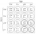
File:Morgan 1925 fig26.jpg yellow, G \ wrinkled, w, and round, W.(1,000 × 958 (144 KB)) - 08:10, 20 October 2014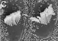
File:Cochlea stereocilia bundle.jpg Reference: Yang S-M, Chen W, Guo W-W, Jia S, Sun J-H, et al. (2012) '''Regeneration of Stereocilia of Hair Cells(482 × 342 (31 KB)) - 09:34, 3 October 2012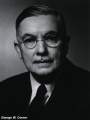
File:George Corner.jpg ==George W. Corner== Prof. George W. Corner (1889 - 1981) was the third director of the Carnegie Institution of(704 × 939 (110 KB)) - 18:19, 7 May 2016
File:LandacreAmstutz1929 table1.jpg | W 2355 | W 2221(1,000 × 1,347 (360 KB)) - 21:57, 15 December 2016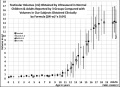
File:Male puberty testicular volume graph.jpg The formula (W-ss)3 x 0.64 is equivalent to the US equation W x H x L x 0.52. * width (W) without the scrotal skin (W-ss)(1,140 × 826 (126 KB)) - 14:28, 18 May 2019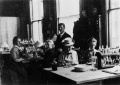
File:Hilfer1990 Fig07.jpg ==Figure 7. Leonard W. Williams teaching c1900== ...laboratory class at the Marine Biological Laboratory, circa 1900. Leonard W. Williams (standing, center) was the instructor. From its inception, course(1,200 × 845 (103 KB)) - 09:44, 28 August 2014
File:Fawcett1975 fig31.jpg From D. W. Fawcett, D. S. Friend, M. Price and R. W. Linck, Proc. 8th Intemat. Congr. Electron Microscopy, Canberra, 1974(1,280 × 403 (128 KB)) - 10:03, 20 August 2017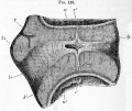
File:Foster135.jpg ...; li. ligamentum intervertebrale ; , k r . epiphysis of vertebra ; w. and w f . anterior and posterior vertebrae ; c. intervertebral dilatation of noto(965 × 815 (230 KB)) - 09:14, 20 June 2018
File:Wen1928-Fig01b.jpg ...yo H1093 (fig. 1, B) was obtained by Dr. Garrette Van Sweringen and Dr. W. W. Duemling, of Fort Wayne, Indiana. The clinical record gives the following(471 × 759 (31 KB)) - 15:51, 21 April 2016
File:Gray1110a.jpg ...ary or testis is formed. ug. Sinus urogenitalis. W. Left Wolffian body. w, w. Right and left Wolffian ducts.(578 × 600 (40 KB)) - 10:34, 5 June 2013
File:Foster034.jpg ...of the yolksac, o.p. line of junction between opaque and pellucid areas ; w. palisade-like yolk spheres which constitute the germinal wall.(1,003 × 717 (153 KB)) - 08:47, 11 January 2011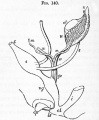
File:Foster140.jpg ...ody ; x. part at apex from which coni vasculosi are afterwards developed ; w. Wolffian duct ; m. Miillerian duct ; gc. genital cord consisting of Wolffi(679 × 824 (82 KB)) - 18:36, 12 January 2011
File:Minot1897 fig088.jpg (after W. His)(753 × 919 (90 KB)) - 15:44, 7 April 2014
File:Minot1897 fig085.jpg (after W. His)(1,061 × 1,052 (0 bytes)) - 15:35, 7 April 2014
File:Minot1897 fig081.jpg (after W. His)(837 × 1,036 (205 KB)) - 15:29, 7 April 2014
File:Mall1917 fig05.jpg Illustrating Group 5. Ovum containing a cylindrical embryo; No. 839 from Dr. W. S. Miller, Madison, Wis. X li(939 × 715 (74 KB)) - 19:54, 5 November 2013
File:Foster001.jpg * w. y. white yolk. This consists of a central flask-shaped mass and a number o * w. albumen consisting of alternate denser and more fluid layers(839 × 647 (111 KB)) - 11:05, 22 December 2012
File:Keibel Mall 418.jpg * W - Wolffian body(450 × 511 (43 KB)) - 19:35, 18 October 2012
File:Hilfer1990 Fig04.jpg ==Figure 4. W. Roux Surface views of frog embryos== Surface views of frog embryos from [[Embryology History - Wilhelm Roux|W. Roux]] (1888). His Figures 5 and 6 show normal embryos at early and late s(1,855 × 2,000 (238 KB)) - 17:03, 29 August 2014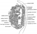
File:Keith1921 fig017.jpg (Modified by F. W. Jones from figures given by Peters and Selenka.)(1,095 × 1,000 (236 KB)) - 10:02, 22 December 2014


