Search results
From Embryology
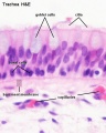
File:Trachea histology 01.jpg ==Trachea Histology== The trachea is lined by an cells described as a '''pseudostratified epithelium'''. (Sta(480 × 600 (47 KB)) - 10:41, 23 February 2013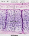
File:Hyaline cartilage 03.jpg ==Respiratory Trachea - Layers== {{Trachea histology links}}(500 × 626 (92 KB)) - 14:37, 10 March 2013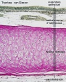
File:Hyaline cartilage 04.jpg ==Respiratory Trachea - Layers== {{Trachea histology links}}(500 × 626 (101 KB)) - 14:37, 10 March 2013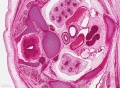
File:Stage 22 image 200.jpg * trachea :'''Links:''' [[Respiratory Development]] | [[Thymus Development]](1,200 × 877 (563 KB)) - 12:01, 14 June 2016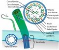
File:Cilium cartoon.jpg # '''Motile cilia''' (respiratory epithelia, trachea, uterine tube, ependymal cells) move and constantly beat in a single direct {{Respiratory Histology}}(800 × 682 (114 KB)) - 15:46, 27 March 2019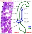
File:Stage14 respiratory tract.jpg Excerpts of the histology sections and their approximate level are shown in the cartoon of the embryo * initial bifurcation of foregut (oesophagus) and respiratory (trachea).(406 × 431 (76 KB)) - 21:13, 24 February 2019


