Search results
From Embryology
File:BeatonAnson1940 fig03-4.jpg ==Figs. 3 and 4. Reconstruction of human stapes== {{Stapes}} semidiagrammatic. x 45. 262(1,576 × 2,050 (374 KB)) - 11:20, 12 July 2019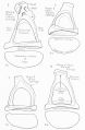
File:BeatonAnson1940 fig05-8.jpg ==Figs. 5 to 8. Excised specimens of human stapes== {{Stapes}} (see footnote 4). x 20.(1,623 × 2,482 (383 KB)) - 11:27, 12 July 2019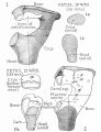
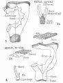
File:BeatonAnson1940 fig01-2.jpg ==Figs. 1 and 2. Reconstructions of human stapes== Human {{Stapes}} semi-diagrammatic (see footnote 4) x 45. 260(1,652 × 2,147 (652 KB)) - 11:14, 12 July 2019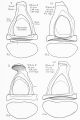
File:BeatonAnson1940 fig09-12.jpg :'''Links:''' {{Stapes}} [[Category:Hearing]][[Category:Middle Ear]][[Category:Stapes]][[Category:Fetal]](1,635 × 2,458 (422 KB)) - 11:42, 12 July 2019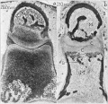
File:Anson1948 fig02.jpg ==Fig. 2. Photomicrographs of 202 mm and 310 mm fetus stapes and incus== Photomicrographs of the neck and head of the stapes and of the lentieular process of the incus, showing erosion of the cartilag(1,280 × 1,232 (241 KB)) - 08:55, 15 October 2017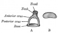
File:Gray0918.jpg ==Middle Ear - Stapes== A. Left stapes. B. Base of stapes, medial surface.(500 × 283 (17 KB)) - 17:24, 18 May 2015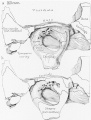
File:Anson1948 fig09.jpg ...al base and of the basal extremities of the crura. The dotted lines on the stapes indicates the approximate limits of the ossifying area; to either side of t(1,280 × 1,686 (235 KB)) - 20:43, 16 October 2017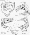
File:Anson1948 fig12.jpg ==Fig. 12. Drawings of a reconstruction of the stapes in the 180 mm Fetus== ...n the 180 mm. (21 week) fetus (Wisconsin series 45 B); X 7: (a) the entire stapes, in superolateral view; (1)) segment 1 (see inset), viewed from below (that(1,280 × 1,490 (289 KB)) - 20:52, 16 October 2017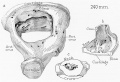
File:Anson1948 fig14.jpg ==Fig. 14. Drawings of a reconstruction of the stapes in a 240 mm== ...d of the base seen as though from the obturator space; (c) the neck of the stapes (capital portion removed) and the adjacent portions of the two crura, in la(1,280 × 872 (190 KB)) - 21:28, 16 October 2017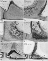
File:Anson1948 fig01.jpg ==Fig. 1. Photomicrographs of the base and crus of the stapes, showing progressive stages in the removal of cartilage and the formation o ...l) bone which forms one of the two constituent lamellas in the base of the stapes.(1,280 × 1,635 (310 KB)) - 17:02, 13 October 2017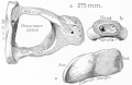
File:Anson1948 fig15.jpg ==Fig. 15. Drawings of a reconstruction of the stapes in a 275 mm== The stapes has attained adult form; of the crura, now deeply channeled, the posterior(1,280 × 829 (158 KB)) - 21:34, 16 October 2017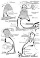
File:Anson1948 fig06.jpg ...right ear; all represent the transverse level of the posterior crus of the stapes. ...the incus. Destruction of periosteal bone on the obturator surface of the stapes keeps pace with the formation of endochondral bone within the capital and b(1,028 × 1,488 (224 KB)) - 20:16, 16 October 2017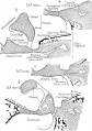
File:Anson1948 fig03.jpg ==Fig. 3. Drawings of the stapes and the adjacent fissular region of the otic capsule== Drawings (semi-diagrammatic) from Edinger tracings of the stapes and the adjacent fissular region of the otic capsule, showing developmenta(1,280 × 1,815 (358 KB)) - 18:46, 18 November 2017
File:Anson1948 fig10.jpg ...zone. In I) of figure 10 the hummocks exposed in the basal portion of the stapes are composed chiefly of calcified cartilage; their summits are capped by e(1,191 × 1,236 (204 KB)) - 21:00, 16 October 2017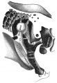
File:Gray0919.jpg ...cavity by ligaments: three for the malleus, and one each for the incus and stapes. ...estra vestibuli by a fibrous ring, the annular ligament of the base of the stapes (lig. annulare baseos stapedis).(500 × 731 (93 KB)) - 17:25, 18 May 2015
File:Anson1948 fig05.jpg ...19, through the middle of the fissular tract and the posterior crus of the stapes; c, through the vestibular orifice of the fissula, and d to- f, from sect ...er way; bone is formed on the obturator aspect of the base and crus of the stapes and on the tympanic and vestibular walls of the otic capsule. Concurrently,(1,280 × 1,797 (362 KB)) - 09:27, 15 October 2017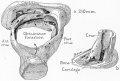
File:Anson1948 fig13.jpg ==Fig. 13. Drawings of a reconstruction of the stapes in a 210 mm== Drawings of a reconstruction of the stapes in a 210 mm. (23 week) fetus (Wisconsin series 51) ; X 7: (a) reconstructio(1,239 × 841 (203 KB)) - 21:17, 16 October 2017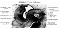
File:HansonAnson1962 fig01.jpg The malleus, like the incus and stapes, develops from a mesenchymal blastema located within the first two branchia ...mates the portion of the stapedial ring destined to become the head of the stapes.(1,280 × 643 (218 KB)) - 10:17, 7 January 2019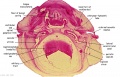
File:Stage 22 image 062.jpg ...um of tympanic membrane (L). Manubrium of malleus (L). Meckel's cartilage. Stapes (R). Auditory tube. Basal turn of cochlea duct (L). Endolymphatic sac (R).(1,000 × 645 (135 KB)) - 14:52, 5 October 2011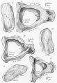
File:AnsonKarabinMartin1939 fig07-12.jpg ==Figs. 7 to 12. Reconstructions of the stapes; superior and medial views== In these and in the succeeding figures ant. crus. indicates anterior crus of stapes; cart. or cartil., cartilage; far. c., facial canal ; fen. cart, fenestral(1,280 × 1,880 (444 KB)) - 08:39, 22 October 2017


