Search results
From Embryology
Page title matches
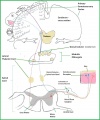
File:Somatosensory Map.JPG ...p the lateral spinothalamic tract to the thalamus before being sent to the somatosensory cotex where it is processed.(1,248 × 1,493 (163 KB)) - 21:06, 2 October 2012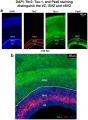
File:Somatosensory cortex of E20 rat.jpeg ==Coronal sections of somatosensory cortex of E20 rat==(2,003 × 2,722 (1.29 MB)) - 12:04, 9 November 2014
File:Somatosensory activation by corneal pain and blinking.gif ...rra An Approach to Localizing Corneal Pain Representation in Human Primary Somatosensory Cortex .PlosOne: 2012, Vol. 7, Issue 9, e44643, PMC3433421(129 × 80 (18 KB)) - 17:25, 14 September 2012


Page text matches
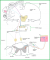
File:Somatosensory.png ...p the lateral spinothalamic tract to the thalamus before being sent to the somatosensory cotex where it is processed.(1,248 × 1,493 (722 KB)) - 20:51, 2 October 2012File:Somatosensory Map.JPG ...p the lateral spinothalamic tract to the thalamus before being sent to the somatosensory cotex where it is processed.(1,248 × 1,493 (163 KB)) - 21:06, 2 October 2012
File:Touch1.JPG '''Somatosensory: Touch'''(1,430 × 1,068 (374 KB)) - 02:11, 5 October 2012
File:Touch 2.JPG Somatosensory: Touch(2,592 × 1,936 (1.89 MB)) - 02:15, 5 October 2012File:Somatosensory activation by corneal pain and blinking.gif ...rra An Approach to Localizing Corneal Pain Representation in Human Primary Somatosensory Cortex .PlosOne: 2012, Vol. 7, Issue 9, e44643, PMC3433421(129 × 80 (18 KB)) - 17:25, 14 September 2012
File:Gray0937.jpg [[Category:Senses]] [[Category:Somatosensory]](700 × 288 (42 KB)) - 08:30, 19 August 2012
File:Gray0935.jpg [[Category:Senses]] [[Category:Somatosensory]](400 × 690 (79 KB)) - 08:26, 19 August 2012
File:Gray0938.jpg [[Category:Senses]] [[Category:Somatosensory]](800 × 274 (59 KB)) - 08:33, 19 August 2012
File:Homonculus Sensory and Motor Cortex .png This image shows the organisation of the primary motor and somatosensory cortex in the human cerebral cortex. The size of the body parts relates to(1,019 × 516 (153 KB)) - 14:30, 26 October 2017
File:Gray0936.jpg [[Category:Senses]] [[Category:Somatosensory]](602 × 450 (59 KB)) - 08:27, 19 August 2012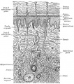
File:Gray0940.jpg [[Category:Senses]] [[Category:Somatosensory]] [[Category:Human]] [[Category:Integumentary]](618 × 700 (160 KB)) - 18:04, 25 September 2012File:Somatosensory cortex of E20 rat.jpeg ==Coronal sections of somatosensory cortex of E20 rat==(2,003 × 2,722 (1.29 MB)) - 12:04, 9 November 2014
File:Merkel cell EM 01.jpg ...ll (Merkel-Ranvier cell) integumentary (skin) receptor cell connected with somatosensory afferents.(984 × 738 (209 KB)) - 14:02, 24 July 2019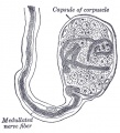
File:Gray0934.jpg [[Category:Senses]] [[Category:Somatosensory]](456 × 500 (63 KB)) - 08:26, 19 August 2012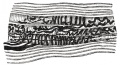
File:Gray0939.jpg [[Category:Senses]] [[Category:Somatosensory]] [[Category:Cat]](681 × 371 (90 KB)) - 08:35, 19 August 2012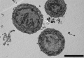
File:Merkel cell EM 02.jpg ...ll (Merkel-Ranvier cell) integumentary (skin) receptor cell connected with somatosensory afferents.(984 × 685 (166 KB)) - 13:25, 23 February 2013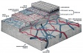
File:Gray0942.jpg [[Category:Senses]] [[Category:Somatosensory]] [[Category:Human]] [[Category:Integumentary]](800 × 515 (127 KB)) - 13:43, 25 September 2012
File:Telencephalon- Netrin-1 signaling thalamocortical projections.jpg ...uditory area; GE, ganglionic eminence; M1, primary motor area; S1, primary somatosensory area; V1, primary visual area.(454 × 800 (76 KB)) - 18:40, 7 November 2010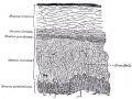
File:Gray0941.jpg [[Category:Senses]] [[Category:Somatosensory]] [[Category:Human]] [[Category:Integumentary]](700 × 524 (106 KB)) - 23:38, 19 August 2012


