Search results
From Embryology
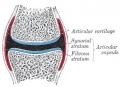
File:Gray0299.jpg ==Synovial Joint Cartoon== :'''Links:''' [[Musculoskeletal System - Joint Development|Joint Development]] | [[Cartilage Histology]](373 × 269 (34 KB)) - 07:44, 6 September 2011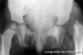
File:Congenital dislocation hip.jpg This X-ray shows incomplete development of the femur head within the pelvis joint. ...s|Limb Abnormalities]] | [[Musculoskeletal_System_-_Limb_Development|Limb Development]](400 × 265 (8 KB)) - 14:05, 22 May 2013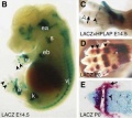
File:Joint development 02.jpg == Joint Development== (A) A 140-kb BAC from the Gdf5 locus was modified by inserting Cre-IRES-hPLAP into the translation start site of Gdf5 and used to make transgenic mi(454 × 403 (22 KB)) - 23:05, 21 March 2018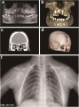
File:Cleidocranial dysplasia 01.jpg ...primary teeth, eruption failure of the permanent teeth, and impaired root development. ...lavicles and structural abnormalities occurring in the right shoulder peak joint.(518 × 700 (65 KB)) - 15:03, 13 February 2017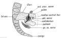
File:Keith1902 fig243.jpg ...in the ventral line. The cotyloid bone — os acetabuli — is formed in the Y-shaped cartilage between the three elements. It ossifies in the 13th year. P ...Musculoskeletal System - Pelvis Development|Pelvis Development]] | [[Fetal Development]](1,000 × 631 (98 KB)) - 12:26, 11 March 2018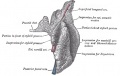
File:Gray1022.jpg ...the angle of the mandible. The remainder of the gland is irregularly wedge-shaped, and extends deeply inward toward the pharyngeal wall. ...to the posterior part of the mandibular fossa behind the temporomandibular joint. The deep surface is in contact with the internal and external carotid arte(629 × 400 (45 KB)) - 08:50, 11 May 2014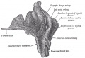
File:Gray1023.jpg ...the angle of the mandible. The remainder of the gland is irregularly wedge-shaped, and extends deeply inward toward the pharyngeal wall. ...to the posterior part of the mandibular fossa behind the temporomandibular joint. The deep surface is in contact with the internal and external carotid arte(585 × 400 (47 KB)) - 10:09, 11 May 2014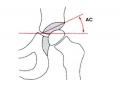
File:Acetabular angle.jpg ...ular rim. It is used clinically as the "acetabular index" to determine hip development and associated displasias. ...tabular antetorsion may be present causing anatomic instability of the hip joint.(600 × 433 (22 KB)) - 16:39, 26 June 2019


