Search results
From Embryology
Page title matches
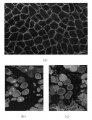
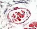
File:Normal control muscle (a) vs. Duchennes muscular dystrophy muscle (b).jpg ...ttered anti-AQP4 immunonegative fibers (asterisks in (b)) are noted in DMD muscle. Scale bar in (a)–(c) = 50μm. Normal control muscle (a) vs. Duchennes muscular dystrophy muscle (b).jpg(423 × 550 (131 KB)) - 16:11, 24 October 2011
File:Muscle development 1-Talk1 muscle development.pdf (2.75 MB) - 13:43, 31 March 2012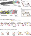
File:Muscle elongation.png ==Cartoon of WT embryo showing the three phases of muscle elongation== ...cells intercalating by extending protrusions that are subsequently filled. Muscle precursor cells exhibit protrusive activity in all directions. A1: Magnific(518 × 600 (194 KB)) - 10:29, 31 May 2017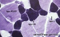
File:Muscle fiber types.jpg ==Muscle Fibre Types== Muscle fibre types identified by ATPase staining. Myosin binds and hydrolyzes ATP(400 × 250 (49 KB)) - 12:47, 7 October 2015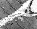
File:Skeletal Muscle EM05.jpg ==Skeletal Muscle EM==(2,000 × 1,569 (875 KB)) - 16:39, 16 April 2014
File:Skeletal Muscle EM04.jpg ==Skeletal Muscle EM==(2,400 × 1,720 (1.07 MB)) - 16:40, 16 April 2014
File:Cardiac muscle EM01.jpg ==Cardiac Muscle Electron Micrograph== ...is is a historic (1969) EM showing key features in cat cardiac ventricular muscle ultrastructure. Only the intercalated disc and some cross-striations can ty(1,072 × 735 (231 KB)) - 12:07, 20 March 2019
File:Skeletal Muscle EM03.jpg ==Skeletal Muscle EM== Image of muscle sarcomeres shows distinct banding pattern:(2,200 × 1,675 (976 KB)) - 17:00, 16 April 2014
File:Muscle- C2C12 differentiation.jpg '''(C)''' mRNA from the muscle-specific markers Csrp3, Hes6, Mef2a and Mef2d. Transcription levels are exp [[Category:Muscle]] [[Category:Graph]](600 × 889 (101 KB)) - 08:05, 28 September 2010
File:Cardiac muscle EM02.jpg ==Cardiac Muscle Electron Micrograph== ...inal historic (1969) EM has been relabeled to show key features in cardiac muscle ultrastructure. At the light microscope histology level, only the intercala(1,072 × 735 (224 KB)) - 12:08, 20 March 2019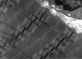
File:Skeletal Muscle EM02.jpg ==Skeletal Muscle EM==(2,191 × 1,590 (941 KB)) - 16:40, 16 April 2014
File:Lab7 muscle-1.jpg [[Category:Muscle]] [[Category:Histology]](1,000 × 844 (92 KB)) - 14:00, 31 March 2012
File:Muscle tutorial front.jpg (850 × 632 (130 KB)) - 16:11, 15 September 2010
File:Cardiac Muscle EM05.jpg ==Cardiac Muscle Electron Micrograph== This is a historic (1969) EM showing key features in cardiac muscle ultrastructure. {{Osmium}}(992 × 733 (158 KB)) - 12:42, 23 February 2013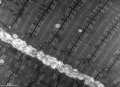
File:Skeletal Muscle EM01.jpg ==Skeletal Muscle EM==(2,200 × 1,586 (789 KB)) - 16:41, 16 April 2014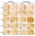
File:Lab7 muscle-2.jpg [[Category:Muscle]] [[Category:Histology]](1,000 × 1,007 (164 KB)) - 14:00, 31 March 2012File:Muscle Stem Cell.pdf (264 KB) - 14:31, 31 March 2012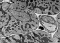
File:Cardiac muscle EM03.jpg ==Cardiac Muscle Electron Micrograph== ...erse section. This is a historic (1969) EM showing key features in cardiac muscle ultrastructure. Myofilaments (Mfl) are not grouped in discrete myofibrils,(849 × 615 (135 KB)) - 00:05, 5 October 2012
File:Lab7 muscle-1c.jpg [[Category:Muscle]] [[Category:Histology]](400 × 338 (23 KB)) - 14:00, 31 March 2012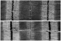
File:Cardiac muscle EM04.jpg ==Cardiac Muscle Electron Micrograph== This is a historic (1969) EM showing key features in cardiac muscle ultrastructure. {{Osmium}}(1,000 × 680 (191 KB)) - 11:48, 27 March 2019
_vs._Duchennes_muscular_dystrophy_muscle_(b).jpg)


Page text matches
File:Skeletal muscle histology 010.jpg ==Muscle Spindle Histology== * skeletal muscle spindle(1,280 × 1,024 (207 KB)) - 21:49, 15 August 2016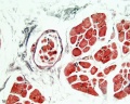
File:Skeletal muscle histology 009.jpg ==Muscle Spindle Histology== * skeletal muscle spindle(1,280 × 1,024 (274 KB)) - 21:50, 15 August 2016
File:Skeletal muscle histology 004.jpg ==Skeletal Muscle Histology== * Guinea pig skeletal muscle, muscle-tendon junction (myotendinous junction)(1,280 × 1,024 (242 KB)) - 15:33, 6 March 2012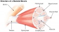
File:Skeletal muscle structure cartoon.jpg ==Skeletal Muscle Structure Cartoon== Shows the different levels of Connective Tissue (CT) associated with the muscle.(520 × 286 (46 KB)) - 14:29, 22 March 2018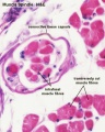
File:Skeletal muscle histology 015.jpg ==Muscle Spindle Histology== * skeletal muscle spindle(600 × 750 (84 KB)) - 17:41, 2 October 2011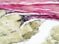
File:Skeletal muscle histology 444.jpg * Guinea pig skeletal muscle, muscle-tendon junction (myotendinous junction) * skeletal muscle, dense regular connective tissue(934 × 701 (125 KB)) - 15:34, 6 March 2012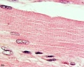
File:Skeletal muscle histology 005.jpg ==Human Skeletal Muscle Histology== * Human skeletal muscle(1,280 × 1,024 (288 KB)) - 14:13, 23 February 2013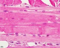
File:Skeletal muscle histology 002.jpg ==Human Skeletal Muscle Histology== * Human skeletal muscle(1,280 × 1,024 (352 KB)) - 14:11, 23 February 2013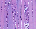
File:Skeletal muscle histology 006.jpg ==Skeletal Muscle Histology== * Human skeletal muscle(1,280 × 1,024 (237 KB)) - 17:07, 2 October 2011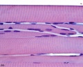
File:Skeletal muscle histology 007.jpg ==Skeletal Muscle Histology== * Human skeletal muscle(1,280 × 1,024 (253 KB)) - 17:07, 2 October 2011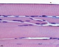
File:Skeletal muscle histology 077.jpg ==Skeletal Muscle Histology== * Human skeletal muscle(1,280 × 1,024 (286 KB)) - 17:06, 2 October 2011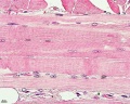
File:Skeletal muscle histology 022.jpg ==Human Skeletal Muscle Histology== * Human skeletal muscle(1,280 × 1,024 (471 KB)) - 14:10, 23 February 2013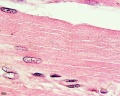
File:Skeletal muscle histology 055.jpg ==Human Skeletal Muscle Histology== * Human skeletal muscle(1,280 × 1,024 (333 KB)) - 14:13, 23 February 2013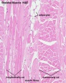
File:Skeletal muscle histology 014.jpg ==Human Skeletal Muscle Histology== * Human skeletal muscle(600 × 750 (127 KB)) - 18:23, 23 February 2013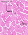
File:Skeletal muscle histology 011.jpg ==Human Skeletal Muscle Histology== * Human skeletal muscle(600 × 750 (132 KB)) - 17:30, 2 October 2011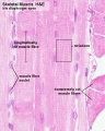
File:Skeletal muscle histology 012.jpg ==Human Skeletal Muscle Histology== * Human skeletal muscle(600 × 750 (121 KB)) - 17:33, 2 October 2011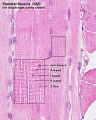
File:Skeletal muscle histology 013.jpg ==Human Skeletal Muscle Histology== * Human skeletal muscle(600 × 750 (185 KB)) - 17:34, 2 October 2011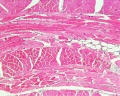
File:Skeletal muscle histology 001.jpg ==Human Skeletal Muscle Histology== * Human skeletal muscle(1,280 × 1,024 (562 KB)) - 14:11, 23 February 2013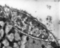
File:Muscle satellite cell EM01.jpg ==Muscle Satellite Cell Electron Micrograph== Transverse section of a skeletal muscle fiber from the rat sartorius (courtesy of Dr. G. Palade).(1,000 × 804 (142 KB)) - 00:54, 17 April 2014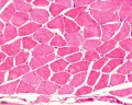
File:Skeletal muscle histology 003.jpg ==Human Skeletal Muscle Histology== * Human skeletal muscle(1,280 × 1,024 (411 KB)) - 14:11, 23 February 2013


