Search results
From Embryology
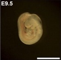
File:Mouse- embryo E9.5.jpg ==Mouse Embryo E9.5== Mouse- embryo E9.5.jpg(325 × 323 (10 KB)) - 12:46, 24 November 2010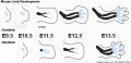
File:Mouse limb skeleton cartoon.jpg ==Mouse Limb Bud Development== Fore- and hind-limb buds for stages E9.5 to E13.5. Hindlimbs are morphologically delayed by about half a day.(1,000 × 487 (64 KB)) - 14:17, 13 February 2013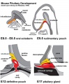
File:Mouse-pituitary development.jpg ==Mouse Pituitary Development== This cartoon is based upon and adapted from a review article on pituitary development.(660 × 800 (85 KB)) - 10:24, 8 September 2010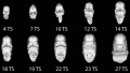
File:Mouse CT E9.5-E12 head.jpg ==Mouse Embryo Computed Tomography== ...1A of the original paper figure. See also animation based on these images [Mouse Stages Face microCT Movie]].(1,000 × 568 (56 KB)) - 13:16, 18 August 2014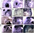
File:Mouse thymus development 01.jpg ==Expression of Transcription Factors in the pharyngeal region at E9.5 and E10.5== ...gion at E9.5 (20–23 somites) and E10.5 (30–33 somites) as detected by whole-mount in situ hybridization.(600 × 584 (69 KB)) - 13:15, 20 February 2012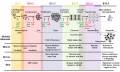
File:Mouse limb tissue development.jpg ==Mouse Limb Tissue Development== Change in cell types and tissue formation as a function of mouse developmental stage.(1,280 × 767 (161 KB)) - 13:26, 7 October 2014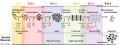
File:Mouse limb bone development timeline.jpg ==Mouse Limb Bone Development Timeline== Change in cell types and bone formation as a function of mouse developmental stage.(1,256 × 469 (107 KB)) - 10:29, 7 October 2014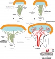
File:Mouse- placenta Hox13 expression.jpg ==Sites of HOXA13 expression during Mouse Placental labyrinth development== ...HOXA13-expressing endothelial progenitors contribute to the developing feto-placental vessels, which mature to form the umbilical artery (UA), chorionic(1,000 × 1,070 (150 KB)) - 12:48, 16 October 2010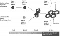
File:Mouse thyroid Hes1 model.jpg ==Proposed roles of Hes1 during murine thyroid development and function== * '''E9.5''' - Hes1 controls the number of thyrocyte progenitors in the median anlage(600 × 364 (33 KB)) - 09:17, 23 September 2011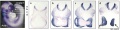
File:Mouse E9.5 neural crest - Crabp1.jpg ==Mouse E9.5 Neural Crest - Crabp1== ...ment]] | [[Developmental Signals - Retinoic acid|Retinoic acid]] | [[Mouse Development]](2,489 × 626 (217 KB)) - 11:08, 9 September 2015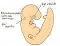
File:Day 9.5 Formation of posterior neuropore and forelimb bud.JPG :'''Links:''' [[Mouse Development]] | [[Mouse Stages]] | [[2009_Group_Project_4|Student Project]] ...d upon Fig. 116, in Dr Karl Theiler’s ‘The House Mouse; Atlas of Embryonic Development’, Springer - Verlag New York Inc, New York, 1989(752 × 568 (37 KB)) - 12:57, 25 June 2014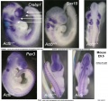
File:Mouse E9.5 neural crest - Crabp1, Sox10, Pax3.jpg ==Mouse E9.5 Neural Crest - Crabp1, Sox10, Pax3== ...mental Signals - Sox|Sox]] | [[Developmental Signals - Pax|Pax]] | [[Mouse Development]](1,323 × 1,233 (193 KB)) - 11:32, 9 September 2015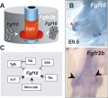
File:Mouse- respiratory development 04.jpg ==Mouse Respiratory Development Fibroblast Growth Factor Signaling== Signaling (Fgf10-Fgfr2b) in lung bud morphogenesis.(800 × 728 (73 KB)) - 12:58, 30 August 2016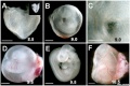
File:Mouse yolk sac 01.jpg ==Mouse Yolk Sac Development== Bright field images of wild-type WT embryos alone or embryos within their yolk sacs at the indicated sta(1,002 × 669 (115 KB)) - 16:56, 24 May 2013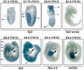
File:Mouse-Sp2 mRNA expression.png Sp2 mRNA expression in mouse embryos ...11, E8.0-TS12 and E9.5-TS15) with the indicated probes (Sp2, Sp2 sense, Nkx-2.5 and GATA4).(600 × 503 (877 KB)) - 13:16, 30 July 2010
File:Mouse E9.5 neural crest beta actin KO.jpg ==Mouse E9.5 Beta Actin Knock Out embryos ( Actb−/−) show Defects in Neural Crest On ...mental Signals - Sox|Sox]] | [[Developmental Signals - Pax|Pax]] | [[Mouse Development]](2,722 × 2,573 (732 KB)) - 11:09, 9 September 2015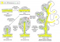
File:Mouse-pancreas duct formation.jpg ==Development and banching morphogenesis of the pancreas== ...linked by adherens junctions (E-cadherin), but lacking tight junctions (ZO-1).(1,000 × 709 (154 KB)) - 10:23, 16 November 2018
File:Melanoblast migration.png ==Directions of Melanoblast Migration in Embryonic Mouse Skin== (E9.5 to E17.5)(600 × 210 (40 KB)) - 14:00, 20 September 2016
File:Mouse face microCT 01.mov ==Mouse Face Development (micro-CT)== Ventral view of developing mouse face (E9.5-12.) series of μCT scans showing the range of shape and size variation.(197 KB) - 13:40, 6 April 2012
File:Mouse face microCT 01.m4v ==Mouse Face Development (micro-CT)== Ventral view of developing mouse face (E9.5-12.) series of μCT scans showing the range of shape and size variation.(723 KB) - 13:34, 6 April 2012


