Search results
From Embryology
Page title matches
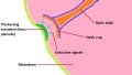
File:Formation of the lens 1.jpg ...ing the vital role of induction from the optic cup in the formation of the lens.(1,152 × 648 (102 KB)) - 17:42, 26 September 2012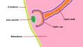
File:Formation of the lens 2.jpg ...erm, forming the lens vesicle. The combined structure of the optic cup and lens vesicle can now be referred to as the optic globe.(1,152 × 648 (94 KB)) - 17:17, 27 September 2012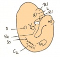
File:Day 10.5 Deep Lens Indentation.JPG Day 10.5: Deep lens indentation occurs. Tel = cerebral hemisphere, L = lens invagination, Hl = Hindlimb bud, Aa = forelimb bud, So = Somite 4, O = otic(514 × 483 (28 KB)) - 13:00, 25 June 2014
File:Lens-neural crest signaling 01.jpg ==Wnt mediates lens repression by neural crest cells and Transforming growth factor-β== # NCCs (blue) secrete TGF-βs, which signal to the non-lens ectoderm and dorsal optic vesicle.(300 × 400 (24 KB)) - 17:25, 31 August 2011
File:Lens-neural crest signaling 02.jpg ==Wnt mediates lens repression by neural crest cells and Transforming growth factor-β== Proposed molecular model to explain TGF-β- and Wnt-mediated lens restriction. Broken lines: interactions inferred from the literature.(521 × 522 (22 KB)) - 14:10, 29 April 2011
File:Day 11 Closure of lens vesicle.JPG Day 11: Closure of the lens vesicle occurs.(560 × 528 (27 KB)) - 13:19, 18 August 2014
File:Day 11.5 Lens Vesicle completely separated from surface.JPG Day 11.5: the lens vesicle is completely separated from the surface.(511 × 484 (29 KB)) - 13:01, 25 June 2014


Page text matches
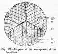
File:Hertwig268.jpg ==Fig. 268. Diagram of the arrangement of the lens-fibres== ...rior surface of the lens and their termination at the anterior star of the lens ; If", continuation of the same fibres Co the posterior star on the posteri(406 × 375 (39 KB)) - 21:48, 28 March 2012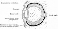
File:Bailey463.jpg ==Fig. 463. Diagram of developing lens and optic cup== ...ium. The mesodermal tissue between the latter and the anterior wall of the lens vesicle is the anlage of the substantia propria cornea.(679 × 345 (52 KB)) - 06:43, 31 August 2011
File:Keibel Mall 2 176.jpg ==Fig. 176. A lens at end of the third month formation of the lens sutures== ...the third month). The right upper quadrant has been removed. One sees the lens epithelium, its continuity with the lena fibres at the equator.(992 × 800 (134 KB)) - 11:34, 21 February 2014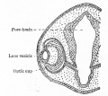
File:Bailey462.jpg ==Fig. 462. Showing somewhat later stage in development of optic cup and lens than is shown in Fig. 461== ...m, but about the eighth week a thin layer of mesoderm grows in between the lens vesicle and the surface ectoderm, completely separating them (Fig. 463).(463 × 411 (45 KB)) - 06:43, 31 August 2011
File:Gray0887.jpg ==Lens Epithelium== ...the margin of the lens, showing the transition of the epithelium into the lens fibers. (Babuchin.)(221 × 800 (67 KB)) - 22:32, 19 August 2012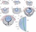
File:DevelopmentEye-PMC43029243.jpg '''Stages of lens formation in mouse embryos''' ...nchyme; 1° and 2° LFs, primary and secondary lens fibers; PLE, prospective lens ectoderm; RPE, retinal pigmented epithelium; SE, surface ectoderm.(654 × 594 (142 KB)) - 10:06, 19 November 2017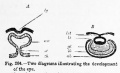
File:Hertwig264.jpg ...e between-brain (zh), is invaginated as a result of the development of the lens-pit (Ig). ...cup with double walls, an inner (ib) and an outer (ab) ; 1st, stalk of the lens ; gl, vitreous body.(653 × 395 (36 KB)) - 21:50, 28 March 2012
File:Stage 22 image 207.jpg ==Developing Lens - Human Embryo Carnegie stage 22== * lens (fibres)(1,200 × 753 (208 KB)) - 11:41, 14 June 2016File:Formation of the lens 2.jpg ...erm, forming the lens vesicle. The combined structure of the optic cup and lens vesicle can now be referred to as the optic globe.(1,152 × 648 (94 KB)) - 17:17, 27 September 2012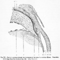
File:Hertwig267.jpg ...the eye ; I', transition of the lens epithelium into the lens-fibres ; Ic, lens-epithelium ; k, anterior chamber of the eye ; d, Descemet's membrane ; //,(700 × 701 (83 KB)) - 21:48, 28 March 2012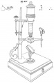
File:Meyer1932history7 fig02.jpg (8.) The Best lens, (b) Cuffs lens No. 4, (c) an English lens No. 0. The latter was used by Ledermuller and he says that it magnified eig(655 × 1,000 (85 KB)) - 21:28, 2 November 2015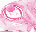
File:Stage 22 image 008-eye.jpg ...ages/Stage22/08-eye/Stage22-08-eye.html?zoom=4&lat=-2030&lon=2572&layers=B Lens and Cornea] ...ages/Stage22/08-eye/Stage22-08-eye.html?zoom=5&lat=-2242&lon=2982&layers=B Lens - anterior](1,200 × 1,059 (555 KB)) - 09:26, 17 December 2016
File:Bailey465.jpg ==Fig. 465. Successive stages in the development of the lens in the rabbit embryo== ...ngle layer of cuboidal cells, the anlage of the anterior epithelium of the lens (Figs. 463, 465, g, h, i).(806 × 931 (142 KB)) - 06:49, 31 August 2011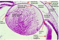
File:Stage 22 image 155.jpg ==Human Embryo Stage 22 Developing Lens== ...- Developing Retina]] | [[:File:Stage 22 image 155.jpg|Image - Developing Lens]](1,000 × 672 (239 KB)) - 07:46, 31 August 2011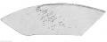
File:Keibel Mall 2 177.jpg ==Fig. 177. Transition of the lens epithelium into the lens fibres, in a fetus at the beginning of the fourth month==(965 × 389 (48 KB)) - 11:42, 21 February 2014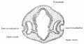
File:Bailey461.jpg ...epressed against the outet surface of the optic vesicle forming a distinct lens invagination (Fig. 461).(751 × 394 (54 KB)) - 06:42, 31 August 2011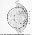
File:Hertwig266.jpg ...ch, choroidea ; If, lens-fibres ; le, lens-epithelium ; I' ', zone of the lens-fibre nuclei ; It, fundament of the cornea ; he, external corneal epitheliu(600 × 663 (83 KB)) - 21:48, 28 March 2012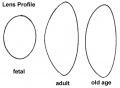
File:Gray0886.jpg ==Lens Profile== Profile views of the lens at different periods of life. 1. In the fetus. 2. In adult life. 3. In old(500 × 368 (17 KB)) - 22:33, 19 August 2012
File:McMurrich1914 fig277.jpg ...layer of optic cup; sf, spaces of Fontana; si, suspensory ligament of the lens; v, vitreous humor. - (Angelucci.)(1,000 × 715 (280 KB)) - 22:36, 31 January 2017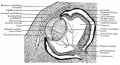
File:Bailey467.jpg ...d, the liquor Morgagni, may remain between the anterior epithelium and the lens fibers. ...the ''membrana pupillaris''. After the earlier and more rapid formation of lens fibers ceases, the hyaloid artery begins (about the seventh month) to under(946 × 515 (159 KB)) - 06:51, 31 August 2011


