Search results
From Embryology
Page title matches
File:Octopus bimaculatus embryonic development timeline.jpg ==Embryonic Development of Octopus bimaculatus== ...e table compares the embryonic development with the scale proposed for the development of L. pealii (Arnold 1965). *Observed in vivo.(1,280 × 1,394 (187 KB)) - 11:02, 21 May 2020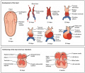
File:Progressive development of the Embryonic Heart.jpeg Development of the heart in the fetus and partitioning of the heart into four chambers ...gy, Connexions Web site. https://cnx.org/contents/FPtK1zmh@6.27:GxtDaPpg@3/Development-of-the-Heart/, Jun 19, 2013.(1,024 × 883 (154 KB)) - 11:30, 4 September 2018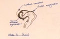
File:Week 6 embryonic development of CNS.jpg ==Week 6 embryonic development of CNS and emergence of pineal evagination.==(1,209 × 794 (165 KB)) - 12:43, 24 October 2014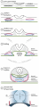
File:Development of Trilaminar Embryonic Disc .png (1) Trilaminalar embryonic disc after gastrulation; lateral plate mesoderm resides between the endoder (4) By folding the lateral edges of the embryonic disc move inwards, creating the anterior intestinal portal. The persisting(1,921 × 5,223 (1.24 MB)) - 10:44, 13 October 2017


Page text matches
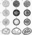
File:Hilfer1990 Fig16.jpg ==Figure 16. Townes and Holtfreter (1955) different embryonic layers in amphibians== ...o clumps and tended to take a position resembling that of normal embryonic development.(1,200 × 1,333 (247 KB)) - 10:43, 28 August 2014File:Octopus bimaculatus embryonic development timeline.jpg ==Embryonic Development of Octopus bimaculatus== ...e table compares the embryonic development with the scale proposed for the development of L. pealii (Arnold 1965). *Observed in vivo.(1,280 × 1,394 (187 KB)) - 11:02, 21 May 2020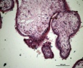
File:HillH52 chorionic villi 05.jpg Human early placental villi development at 4-5 weeks. (Hill H52, x20) {{HE}} scale bar - 100 μm) * Embryonic mesenchyme(1,200 × 992 (371 KB)) - 13:54, 22 March 2014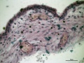
File:HillH52 chorionic villi 04.jpg Human early placental villi development at 4-5 weeks. (Hill H52, x40) {{HE}} scale bar - 50 μm) * Embryonic mesenchyme(1,200 × 900 (254 KB)) - 13:34, 22 March 2014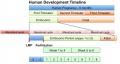
File:Human development timeline graph 02.jpg ==Human Development Timeline== |Simplified graphical view of human development timeline.(800 × 424 (61 KB)) - 09:37, 14 April 2016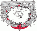
File:Gray0032.gif ...mbryonic ectoderm that will contribute to embryonic and placental membrane development ...storic term for what we today call endoderm that will contribute to embryo development(500 × 417 (57 KB)) - 11:13, 22 May 2011
File:Bat embryo stage 19.mov ==Bat Embryonic Development (stage 19)== ...Cancer Center, who provided images and stage information on the embryonic development of the bat.(30 KB) - 01:12, 29 April 2011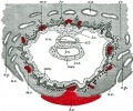
File:Gray0032.jpg ...ough showing the trilaminar embryo suggests late week 2 to early week 3 of development. Uterine cavity is shown at bottom of image. ...mbryonic ectoderm that will contribute to embryonic and placental membrane development(800 × 667 (159 KB)) - 08:51, 21 April 2013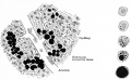
File:Bailey111.jpg ...the right are five individual cells showing stages of development from an embryonic cell to an adult fat cell.(916 × 567 (117 KB)) - 13:05, 18 January 2011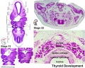
File:Stage13 and 22 thyroid development.jpg ==Thyroid Gland Embryonic Development Overview== Cross-sections of head and neck regions of embryonic stage 13 (week4-5) and stage 22 (week 8) are shown.(1,000 × 800 (283 KB)) - 11:49, 30 August 2011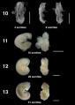
File:Bat embryo stage 10 to 13.jpg ==Bat Embryonic Development (stage 10-13)== ...Cancer Center, who provided images and stage information on the embryonic development of the bat.(800 × 1,122 (66 KB)) - 13:21, 6 July 2012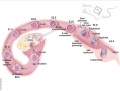
File:Mouse E0-E5.jpg ==Development of the Preimplantation Blastocyst in Mice== Cartoon shows an overview of mouse development from Embryonic Day 0 (E0) Through Day 5 (E5.0).(991 × 749 (90 KB)) - 10:08, 14 October 2016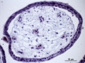
File:HillH52 chorionic villi 08.jpg Human early placental villi development at 4-5 weeks. (Hill H52, x40) {{HE}} scale bar - 50 μm) ...phoblast shell enclosing mesenchyme (extra-embryonic mesoderm), containing embryonic blood vessels.(1,200 × 900 (229 KB)) - 14:59, 3 July 2014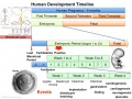
File:Human development timeline graph 01.jpg ==Human Development Timeline== This graph shows an overview of human development and details about embryonic development.(1,000 × 750 (141 KB)) - 08:49, 15 April 2014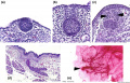
File:Mouse mammary development 01.jpg ==Embryonic Mouse Mammary Development== (a) [[:Category:Mouse E12.5|Embryonic day E12.5]] The epithelial cells have invaginated to form the initial bud,(1,200 × 773 (147 KB)) - 09:04, 20 March 2018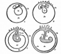
File:Kellicott 179.jpg From McMurrich (Development of the Human Body). ...ic ectoderm; the dotted line marks the line of the transition of the body (embryonic) ectoderm into that of the amnion.(893 × 800 (131 KB)) - 16:54, 23 December 2013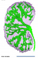
File:Human embryonic renal branching stage 22.jpg ==Embryonic Human Renal Urothelial Branching Development== ...he [[Kyoto Collection]] at Carnegie stage {{CS22}}. See also [[:File:Human embryonic renal branching 1.jpg|branching figure stages 14-22]].(500 × 756 (150 KB)) - 10:21, 18 January 2019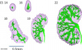
File:Human embryonic renal branching 1.jpg ==Embryonic Human Renal Urothelial Branching Development== ...{CS14}}, {{CS16}}, {{CS18}}, {{CS19}} and {{CS22}}. See also [[:File:Human embryonic renal branching stage 22.jpg|branching figure stage 22]].(1,280 × 779 (236 KB)) - 10:26, 18 January 2019- Error creating thumbnail: File with dimensions greater than 12.5 MP
File:Worm - embryonic cell lineage 01.jpg ==Worm - Embryonic Cell Lineage== Embryonic cell lineage developed by J .E. Sulston, E. Schierenberg, J. G. White, J. N(10,389 × 1,336 (598 KB)) - 11:25, 30 June 2012 
File:GladstoneHamilton1941 text-fig04.jpg ==Text-fig. 4. General .view of the embryonic disc at the anterior part of the primitive streak and groove== ...ion no. 57, the sections having been numbered from the anterior end of the embryonic disc. x 100.(1,280 × 633 (96 KB)) - 16:58, 26 February 2017


