Search results
From Embryology
Page title matches
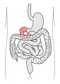
File:Duodenum cartoon.jpg ==Gastrointestinal Tract - Duodenum== ...egions starting from the stomach they are the [[:File:Duodenum cartoon.jpg|duodenum]], [[:File:Jejunum and ileum cartoon.jpg|jejunum and ileum]].(500 × 704 (42 KB)) - 09:35, 13 October 2016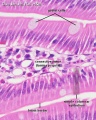
File:Duodenum histology 01.jpg ==Duodenum Histology == Duodenum (adult rat) {{HE}}(480 × 600 (83 KB)) - 12:27, 10 April 2013


Page text matches
File:Stage 22 image 186.jpg ==Duodenum and Pancreas== * Human embryonic pancreas and duodenum structure, [[Week 8]] [[Carnegie stage 22]].(1,000 × 658 (209 KB)) - 13:04, 17 April 2019
File:Stage 22 image 185.jpg ==Duodenum, Pancreas and Developing Adrenal Gland== ...xes of this image, see [[:File:Stage_22_image_186.jpg|Image - Pancreas and Duodenum]] and [[:File:Stage_22_image_187.jpg|Image - Developing Adrenal Gland]].(1,000 × 656 (109 KB)) - 14:03, 22 August 2011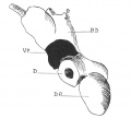
File:Odgers1930 fig01.jpg ==Fig. 1. Drawing of a model of the 5mm Human Embryo duodenum and pancreas== * D. duodenum(979 × 883 (106 KB)) - 12:01, 30 June 2015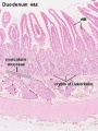
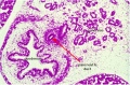
File:Intestine histology 006.jpg ==Duodenum== Histological section of the duodenum showing the villi and the intestinal glands (crypts of Lieberkühn).(400 × 533 (77 KB)) - 10:27, 13 October 2016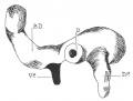
File:Odgers1930 fig02.jpg ==Fig. 2. Drawing of a model of 7.1 mm Human Embryo duodenum and pancreas== Viewed from below, in a 7-1 mm. embryo. D. duodenum; B.D. bile-duct; V.P. ventral pancreas; D.P. dorsal pancreas.(985 × 745 (119 KB)) - 12:02, 30 June 2015File:Duodenum cartoon.jpg ==Gastrointestinal Tract - Duodenum== ...egions starting from the stomach they are the [[:File:Duodenum cartoon.jpg|duodenum]], [[:File:Jejunum and ileum cartoon.jpg|jejunum and ileum]].(500 × 704 (42 KB)) - 09:35, 13 October 2016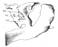
File:Odgers1930 fig03.jpg ==Fig. 3. Drawing of a model of 11.4 mm Human Embryo duodenum and ventral pancreas== Viewed from the right side, in an 11.4 mm. embryo. D. duodenum; B.D. bile-duct; V.P. ventral pancreas; W and W1 mark the two ventral pancr(926 × 755 (122 KB)) - 12:03, 30 June 2015File:Duodenum histology 01.jpg ==Duodenum Histology == Duodenum (adult rat) {{HE}}(480 × 600 (83 KB)) - 12:27, 10 April 2013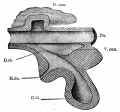
File:Bailey273.jpg ==Fig. 273. From a model of the duodenum and the primary evaginations of the liver and pancreas in a 5 mm sheep embr D.pan., Dorsal pancreas; Du., duodenum; D. ch., ductus choledochus; G. bl., gall bladder; H. du., hepatic duct.(560 × 522 (58 KB)) - 18:09, 24 January 2011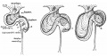
File:Frazer1915 fig05.jpg ==Fig. 5. Schemes to illustrate the formation of the curve of the duodenum== ...l of the small sac, turning to the right, and becoming continuous with the duodenum which passes down in front of the mesoduodenum.(1,000 × 529 (104 KB)) - 05:41, 9 January 2017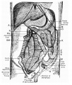
File:Reid1908 fig01.jpg ...the frontal plane, and about 2 cms. long, connected the second part of the duodenum to the posterior abdominal wall quite to the right of the superior mesenter(1,000 × 1,174 (252 KB)) - 18:57, 19 March 2018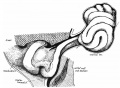
File:Frazer1915 fig10.jpg ...e and below the cut vitelline vein. The mesoduodenum is visible behind the duodenum and below the foramen of Winslow. Cf. fig. 8.(1,000 × 733 (155 KB)) - 05:42, 9 January 2017
File:Jejunum and ileum cartoon.jpg ...egions starting from the stomach they are the [[:File:Duodenum cartoon.jpg|duodenum]], [[:File:Jejunum and ileum cartoon.jpg|jejunum and ileum]]. | Duodenum (25 cm)(500 × 704 (45 KB)) - 12:04, 11 April 2019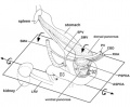
File:Human fetal pancreas anatomy cartoon.jpg Fetal topographical anatomy of the pancreatic head and duodenum with special reference to courses of the pancreaticoduodenal arteries. ...hesize a rotation along a left-right axis through the third portion of the duodenum in the later stage of development. In addition, the fourth portion is conne(455 × 376 (93 KB)) - 17:38, 6 April 2018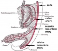
File:GIT blood supply.jpg # '''Foregut''' - celiac artery (Adult: pharynx, esophagus, stomach, upper duodenum, respiratory tract, liver, gallbladder pancreas) # '''Midgut''' - superior mesenteric artery (Adult: lower duodenum, jejunum, ileum, cecum, appendix, ascending colon, half transverse colon)(568 × 500 (47 KB)) - 12:48, 17 April 2019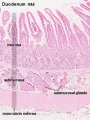
File:Intestine histology 005.jpg ==Duodenum==(400 × 533 (78 KB)) - 13:17, 3 April 2013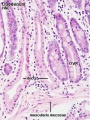
File:Intestine histology 007.jpg ==Duodenum==(400 × 533 (82 KB)) - 13:34, 3 April 2013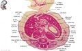
File:Embryo stage 22 F1L.jpg ** [[S#stomach|stomach]], [[D#duodenum|duodenum]], [[J#jejunum|jejunum]](619 × 389 (79 KB)) - 11:33, 31 May 2010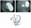
File:Duodenal atresia 02.jpg ('''a''') Upper gastrointestinal series showing a complete obstruction of the duodenum and contrast filling of anomalous bifurcated bile ducts (arrows). The small ...estinal series showing a complete obstruction at the second portion of the duodenum, and contrast was seen in the proximal jejunum which is located in the righ(765 × 682 (68 KB)) - 16:21, 16 April 2019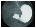
File:Duodenal atresia 01.jpg ...complete obstruction to the flow of contrast at the second portion of the duodenum. There is also contrast filling of the biliary tree above the duodenal bulb ...ructural developmental anomalies of duodenum] - ''Any congenital defect of duodenum that results from interference with the normal growth and differentiation o(750 × 592 (56 KB)) - 16:21, 16 April 2019


