Search results
From Embryology
File:Mall1917 fig02.jpg ...o. 12S9 from Dr. J. R. Cottell, Louisville, Ky. X 2. The picture shows the coelom filled mostly with granular magma.(629 × 525 (29 KB)) - 18:31, 5 November 2013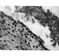
File:Wyburn1939-fig16.jpg ==Fig. 16. Photomicrograph of endothelial lining of umbilical cord coelom of embryo 42 mm== ...to cord “pillars” and continuous with rectus sheath. U.C. = umbilical cord coelom. L. = degenerating endothelial cells. Description in text.(848 × 800 (204 KB)) - 17:01, 14 September 2015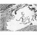
File:Wyburn1939-fig15.jpg ==Fig. 15. Photomicrograph of section of umbilical cord coelom of embryo 42 mm== ...to cord “pillars” and continuous with rectus sheath. U.C. = umbilical cord coelom. L. = degenerating endothelial cells. Description in text.(848 × 800 (189 KB)) - 17:02, 14 September 2015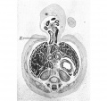
File:Wyburn1939-fig13.jpg Fig. 16. Photomicrograph of endothelial lining of umbilical cord coelom of embryo 42 mm. x circa 225. ...to cord “pillars” and continuous with rectus sheath. U.C. = umbilical cord coelom. L. = degenerating endothelial cells. Description in text.(848 × 800 (153 KB)) - 17:02, 14 September 2015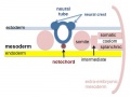
File:Mesoderm-cartoon4.jpg ...inar embryo showing the further development of the 3 layers and the space (coelom) that forms in the mesoderm (only the righthand side is shown, lefthand sid ...sion into somatic and {{splanchnic mesoderm}} separated by intra-embryonic coelom.(400 × 300 (20 KB)) - 10:09, 23 April 2020
File:Wyburn1939-plate04.jpg Fig. 15. Photomicrograph of section of umbilical cord coelom of embryo 42 mm. Site indicated by arrow in Fig. 14. x circa 75. Fig. 16. Photomicrograph of endothelial lining of umbilical cord coelom of embryo 42 mm. x circa 225.(1,700 × 2,247 (747 KB)) - 09:38, 15 September 2015
File:Wyburn1939-plate02.jpg ...I . =intercoelomic septum. D. = distal slit-like portion of umbilical cord coelom. S. = space in umbilical cord. U. = umbilical vein.(1,680 × 2,367 (786 KB)) - 17:13, 14 September 2015
File:Wyburn1939-text-fig08-10.jpg ...Horizontal lines = blood vessels. Black = mesoderm. U .C. = umbilical cord coelom. ...Horizontal lines = blood vessels. Black = mesoderm. U .C. = umbilical cord coelom.(1,588 × 1,400 (183 KB)) - 13:34, 15 September 2015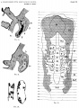
File:Dandy1910-plate02.jpg ...leural coelom; Coe, Peritoneal coelom; E.C., External Communication of the coelom: Pr, Pronephros; Ht, Projection of the heart in the pericardial cavity; X.(1,754 × 2,400 (951 KB)) - 12:10, 28 May 2017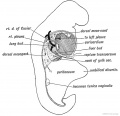
File:Keith1902 fig201.jpg ==Fig 201. The Form of the Coelom in a 3rd week Embryo as viewed from the right side==(826 × 800 (87 KB)) - 09:32, 20 January 2014
File:Bremer1914 plate05.jpg ...and mesothelial cord, leading from funnel (f.) to unlined space (a). Coe., coelom; h, smaller unlined space;c, endothelial cord. X circa 580. ...space, containing corpuscles. c and d, mesothelial cords (see text); Coe., coelom. X circa 800.(643 × 1,000 (144 KB)) - 21:50, 27 October 2015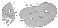
File:Minot1889 fig04.jpg V, vein; Ar, artery; All, allantois cavity; Coe, coelom; Y, yolk sack ; X 22 diams.(1,000 × 482 (153 KB)) - 11:52, 8 May 2018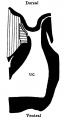
File:Wyburn1939-text-fig07.jpg .... Horizontal lines = blood vessel. Black = mesoderm. U.C. = umbilical cord coelom.(539 × 928 (46 KB)) - 13:32, 15 September 2015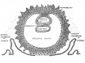
File:Keith1921 fig016.jpg ==Fig. 16. Showing the Origin of the Primitive Coelom, the Mesoblast and Cavity of the Amnion during the Development of the Human(1,075 × 800 (203 KB)) - 09:57, 22 December 2014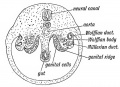
File:Keith1902 fig073.jpg ==Fig. 73 Diagrammatic section of the abdominal region of the coelom==(800 × 577 (97 KB)) - 10:07, 7 January 2014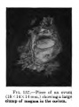
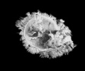
File:Keibel Mall 157.jpg Piece of an ovum (18x14x14 mm) showing a large clump of magma in the coelom.(300 × 406 (17 KB)) - 05:23, 6 September 2012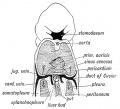
File:Keith1902 fig183.jpg ...183. Diagram to show the manner in which the Ducts of Cuvier encircle the Coelom at the junction of the Pericardial and Pleural Parts (Iter venous)==(879 × 800 (113 KB)) - 09:32, 20 January 2014
File:Wyburn1939-fig14.jpg ...to cord “pillars” and continuous with rectus sheath. U.C. = umbilical cord coelom. L. = degenerating endothelial cells. Description in text.(848 × 800 (164 KB)) - 17:02, 14 September 2015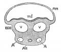
File:Minot1889 fig02.jpg ...ullary groove; v, v, veins; A, A, umbilical arteries; All, allantois; coe, coelom.(760 × 650 (91 KB)) - 11:51, 8 May 2018
File:Dandy1910-plate01.jpg '''Fig. 3.''' Section 76, X 50. Pr, Pronephros; Coe, Coelom; Pl, Pleural Cuelom; V, Umbilical vein; Br V, Branch of umbilical vein, onl ...r tip of somite IV on opposite side) ; Coe, Coelom; E.C., Communication of coelom with exterior; Ch, Chorda.(1,738 × 2,359 (541 KB)) - 12:09, 28 May 2017


