Search results
From Embryology
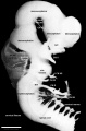
File:Human Stage16 neural02.jpg ==Human Carnegie Stage 16 Neural== ...x|Right lateral view of the central nervous system of embryo at [[Carnegie stage 16]]. Showing 5 secondary vesicles, 3 brain flexures, eye and otic vesicle,(1,352 × 2,048 (286 KB)) - 08:50, 13 October 2017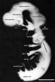
File:Human Stage14 neural02.jpg ==Human Carnegie Stage 14 Neural== ...h=500px|Lateral view of the central nervous system of embryo at [[Carnegie stage 14]]. Shown to the level of the myelencephalon.(1,375 × 2,048 (506 KB)) - 17:55, 14 April 2016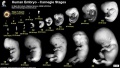
File:Human Carnegie stage 1-23.jpg | Carnegie stage 1 to 23, showing human external appearance and growth during the first 8 we Stage 1 is early zygote and not in scale. Has been enlarged to show pronuclei and(1,000 × 563 (98 KB)) - 11:32, 14 May 2017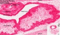
File:Stage 22 image 220.jpg ==Developing Pituitary - Human Embryo Carnegie stage 22== * Human embryo [[Week 8]], [[Carnegie stage 22]](1,200 × 699 (334 KB)) - 11:44, 14 June 2016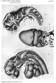
File:Gage1905-plate02.jpg ...tral nervous system and the strong cephalic flexure characteristic of this stage of development are evident. The mesoderm in the flexure has been removed. ...t brings out somewhat more clearly the grouping of folds into lobules (cf. Table II, in the text). The elevations in Fig. 3 correspond to the depressions in(1,007 × 1,500 (221 KB)) - 14:32, 18 August 2016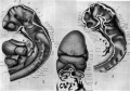
File:Gage1905-plate2.jpg ...tral nervous system and the strong cephalic flexure characteristic of this stage of development are evident. The mesoderm in the flexure has been removed. ...t brings out somewhat more clearly the grouping of folds into lobules (cf. Table II, in the text). The elevations in Fig. 3 correspond to the depressions in(2,149 × 1,500 (392 KB)) - 14:33, 18 August 2016


