Search results
From Embryology
Page title matches
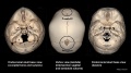
File:Skull CT normal sutures 02.jpg ==Skull Normal Sutures== Computed Tomography (CT) scan with 3D surface-rendered reconstructions.(1,000 × 559 (92 KB)) - 23:13, 21 March 2018

Page text matches
- ...ahly]] | [[:File:Skull CT abnormal 07.jpg|Trigonocephaly]] | [[:File:Skull CT abnormal 08.jpg|Oxycephaly]] | [[Computed Tomography]]<noinclude>[[Category867 bytes (106 words) - 10:17, 22 March 2016
- [[File:Fetal_head_lateral.jpg|thumb|alt=12 week fetal skull|300px|Fetal Head (12 weeks) showing cartilage (blue) and bone (red)]] ...{intramembranous ossification}}. While the bones that form the base of the skull are formed by {{endochondral ossification}}.31 KB (4,342 words) - 04:14, 5 July 2022
- | [[File:Stage16-18 face 02.jpg|right]] ! Historic - cartilaginous and membranous skull29 KB (4,105 words) - 09:18, 25 June 2018
- ...ive Tissue Components|CT Components]] | [[ANAT2241 Connective Tissue Types|CT Types]] | [[ANAT2241 Blood|Blood]] | [[ANAT2241 Bone, Bone Formation and Jo ...[[Birth]] | [[Neonatal_Development|Neonatal]] | [[Neonatal Diagnosis]] | [[Normal Development - Milk|Milk]] | [[Nutrition]] | [[Postnatal - Growth Charts|Gr234 KB (28,397 words) - 15:05, 10 April 2018
- ...urement of Abdominal Circumference (AC) is used to determine fetal age and normal development (small/large/abnormal) parameters. Measured at the outer edge o :A medical procedure in which a physiologic solution (such as normal saline) is infused into the uterine cavity to replace the amniotic fluid.95 KB (12,855 words) - 22:22, 1 January 2020