Search results
From Embryology
Page title matches
- ...ular bed]]<noinclude>[[Category:Template]][[Category:Term Link]][[Category:Placenta]][[Category:Cardiovascular]][[Category:Vein]][[Category:Artery]]</noinclude200 bytes (22 words) - 09:19, 20 November 2018
- ...CT_01.jpg|Image - computed tomography]] | [[Placenta - Vascular Beds]] | [[Placenta Development]]<noinclude>[[Category:Template]]</noinclude>448 bytes (58 words) - 14:21, 18 May 2013
Page text matches
- ...ular bed]]<noinclude>[[Category:Template]][[Category:Term Link]][[Category:Placenta]][[Category:Cardiovascular]][[Category:Vein]][[Category:Artery]]</noinclude200 bytes (22 words) - 09:19, 20 November 2018
- ...CT_01.jpg|Image - computed tomography]] | [[Placenta - Vascular Beds]] | [[Placenta Development]]<noinclude>[[Category:Template]]</noinclude>448 bytes (58 words) - 14:21, 18 May 2013
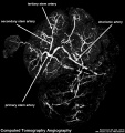
File:Human placenta vascular CT 01.jpg ==Human Placenta Vascular Bed - Computed tomography angiography (CTA)== Placenta is viewed from the fetal side of the placenta using CT imaging.(938 × 1,000 (126 KB)) - 23:14, 21 March 2018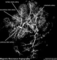
File:Human placenta vascular MRI 01.jpg ==Human Placenta Vascular Bed - Magnetic resonance angiography (MRA)== Placenta is viewed from the fetal side of the placenta using MRI imaging.(938 × 1,000 (151 KB)) - 13:41, 18 May 2013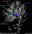
File:Human placenta vascular MRI 02.jpg ==Human Placenta Vascular Bed - Magnetic resonance angiography (MRA)== Placenta is viewed from the fetal side of the placenta using MRI imaging. Chorionic artery and its branches have been colour coded(938 × 1,000 (156 KB)) - 23:15, 21 March 2018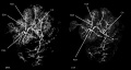
File:Human placenta vascular 01.jpg ==Human Placenta Vascular Bed== Placenta is viewed from the fetal side of the placenta using MRI and CT imaging.(1,200 × 644 (125 KB)) - 23:15, 21 March 2018- Placenta. 2006 Sep-Oct;27(9-10):939-58. Epub 2006 Feb 20. ...cental function is far from clear. Several features of the human placental bed are mirrored by processes in other species with haemochorial placentation,2 KB (266 words) - 09:34, 7 June 2017
- ...a vascular MRI 02.jpg|thumb|alt=Human placenta vascular MRI|Human placenta vascular MRI]] ...can be measured by doppler ultrasound. When considering placental vascular bed development, the associated changes in maternal decimal blood flow and deve6 KB (856 words) - 11:45, 23 April 2020
- {{Placenta Links}} =Radioangiographic studies of circulation in the maternal placenta of the rhesus monkey: preliminary report=12 KB (1,800 words) - 19:02, 4 June 2017
- =Placenta Embryology and Circulation= ...epage] | [http://www.a-s-a.com.au|Australian Sonographers Association] | [[Placenta Development]] | [[Main Page|Embryology]]20 KB (2,722 words) - 17:17, 11 April 2018
- ...e retired. In addition to her research articles she published 2 books "The Placenta of Laboratory Animals and Man" (1975) and "Placental Vasculature and Circul {{Placenta Links}}17 KB (2,515 words) - 11:22, 31 May 2019
- This page introduces an overview of aspects of the basic fetal subunit of the placenta, the placental villi development. In early placentation, each villi proceed {{Placenta Links}}12 KB (1,577 words) - 11:50, 10 April 2019
- ...e heterogeneous microscopic images representing histological slides of the placenta. The results presented in this study were obtained using a set of 50 images Histopathology; Image analysis; Image segmentation; Mathematical morphology; Placenta; Texture13 KB (1,821 words) - 09:34, 21 August 2018
- ...(19 somite) vascular distribution|thumb|Image of mouse embryo (19 somite) vascular distribution (about [[Carnegie stage 11|Human stage 12]])]] This lecture has 2 parts; firstly introducing an overview of early vascular development, secondly the key events in heart development.21 KB (2,924 words) - 09:45, 7 September 2017
- ...mesoderm. These blood islands extend and fuse together making a primordial vascular network. Within these islands the peripheral cells form endothelial cells w See also the related pages: {{artery}}, {{vein}}, {{placenta vascular bed}}, {{coronary circulation}}.23 KB (3,085 words) - 09:39, 4 December 2019
- ...can surgeon;''' '''D.'s windows = fat-free portions of mesentery framed by vascular arcades adjacent to the attached margin of the intestine. ...etrium invaded by the chorionic villi; unites with the chorion to form the placenta.5 KB (781 words) - 14:20, 16 February 2013
- This lecture is an introduction to the development and functions of the placenta. ...''plakuos'' = flat cake) named on the basis of this organs appearance. The placenta a mateno-fetal organ which begins developing at implantation of the blastoc23 KB (3,115 words) - 09:49, 21 August 2018
- ! [[Placenta Development|Placenta Terms]] (expand to view) * '''after-birth''' - term used to describe the delivery of placenta and placental membranes following birth of the child.25 KB (3,408 words) - 12:47, 5 June 2019
- =Early Vascular Development= [[File:Mouse embryo vascular.png|thumb|Mouse embryo (19 somite) vascular distribution]]28 KB (4,084 words) - 17:49, 30 May 2012
- =Early Vascular Development= [[File:Mouse embryo vascular.png|thumb|Mouse embryo (19 somite) vascular distribution]]28 KB (4,102 words) - 09:02, 18 September 2014


