Search results
From Embryology
Page title matches
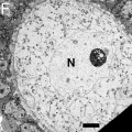
File:Mouse neonatal ovary oocyte EM07.jpg ==Electron Micrographs of Oocytes in Neonatal Mouse Ovaries== * '''F''' - Oocyte with in primary follicle with mitochondria and ER evenly distributed throug(790 × 792 (183 KB)) - 12:04, 4 March 2012

Page text matches
File:Mouse neonatal ovary oocyte EM06.jpg ==Electron Micrographs of Oocytes in Neonatal Mouse Ovaries== * '''F''' - Oocyte with in primary follicle with mitochondria and ER evenly distributed throug(790 × 792 (163 KB)) - 07:29, 3 March 2012File:Mouse neonatal ovary oocyte EM07.jpg ==Electron Micrographs of Oocytes in Neonatal Mouse Ovaries== * '''F''' - Oocyte with in primary follicle with mitochondria and ER evenly distributed throug(790 × 792 (183 KB)) - 12:04, 4 March 2012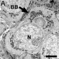
File:Mouse neonatal ovary oocyte EM02.jpg ==Electron Micrographs of Oocytes in Neonatal Mouse Ovaries== * '''A''' - Micrograph of an oocyte within a germline cyst from PND1 showing a well defined Balbiani body (arro(790 × 792 (179 KB)) - 12:22, 19 June 2014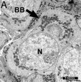
File:Mouse oocyte balbini body EM01.jpg ==Mouse Oocyte Balbini Body== Electron micrograph of oocytes in neonatal ovary micrograph of an oocyte within a germline cyst from PND1 showing a well defined Balbiani body (arro(695 × 700 (155 KB)) - 12:54, 19 April 2018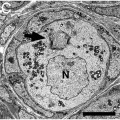
File:Mouse neonatal ovary oocyte EM04.jpg ==Electron Micrographs of Oocytes in Neonatal Mouse Ovaries== * '''C''' - Micrograph of an oocyte within a primordial follicle at PND7 again showing a Balbiani body (arrow).(790 × 792 (235 KB)) - 07:28, 3 March 2012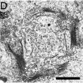
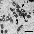
File:Mouse neonatal ovary oocyte EM05.jpg ==Electron Micrographs of Oocytes in Neonatal Mouse Ovaries== * '''D''' - Higher-power view of Golgi from oocyte in C showing stacks of vesicles arranged in a circular structure.(790 × 792 (217 KB)) - 07:29, 3 March 2012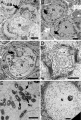
File:Mouse neonatal ovary oocyte EM01.jpg ==Electron Micrographs of Oocytes in Neonatal Mouse Ovaries== * [[:File:Mouse neonatal ovary oocyte EM02.jpg|'''A''']] - Micrograph of an oocyte within a germline cyst from PND1 showing a well defined Balbiani body (arro(677 × 1,000 (266 KB)) - 07:35, 3 March 2012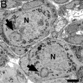
File:Mouse neonatal ovary oocyte EM03.jpg ==Electron Micrographs of Oocytes in Neonatal Mouse Ovaries== ...tes from PND3 (top oocyte in a newly formed primordial follicle and bottom oocyte still within a germline cyst) with Balbiani bodies (arrows).(790 × 792 (187 KB)) - 12:09, 4 March 2012


