Search results
From Embryology
Page title matches
File:Keibel Mall 2 297-299.jpg (After Mall.) d, branch of the portal vein ; a, branch of the hepatic vein. File:Keibel Mall 2 297.jpg|Fig. 297.(1,280 × 786 (135 KB)) - 08:50, 18 November 2018

Page text matches
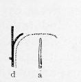
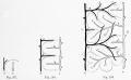
File:Keibel Mall 2 297.jpg (After Mall.) d, branch of the portal vein ; a, branch of the hepatic vein. File:Keibel Mall 2 297.jpg|Fig. 297.(354 × 359 (13 KB)) - 08:48, 18 November 2018File:Keibel Mall 2 297-299.jpg (After Mall.) d, branch of the portal vein ; a, branch of the hepatic vein. File:Keibel Mall 2 297.jpg|Fig. 297.(1,280 × 786 (135 KB)) - 08:50, 18 November 2018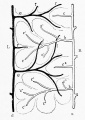
File:Keibel Mall 2 299.jpg (After Mall.) d, branch of the portal vein ; a, branch of the hepatic vein. File:Keibel Mall 2 297.jpg|Fig. 297.(733 × 1,034 (120 KB)) - 08:48, 18 November 2018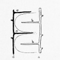
File:Keibel Mall 2 298.jpg (After Mall.) d, branch of the portal vein ; a, branch of the hepatic vein. File:Keibel Mall 2 297.jpg|Fig. 297.(599 × 605 (40 KB)) - 08:48, 18 November 2018- ...ion 1]] | [[:File:Keibel Mall Table-Skeleton 5.jpg|Upper Limb Ossification 2]] | [[Musculoskeletal_System_-_Bone_Development_Timeline|Bone Development T1 KB (138 words) - 23:56, 24 March 2014
- File:Keibel_Mall_002.jpg|Fig. 2. Almost Mature Human Ovum File:Keibel_Mall_026.jpg|Fig. 2. 6Section of the Embryo17 KB (2,474 words) - 21:35, 14 November 2013
- {{Human Embryology Manual 2 TOC}} Keibel_Mall_2_002.jpg|Fig. 2. Wall of the neural tube in a human embryo about two weeks old27 KB (4,015 words) - 09:37, 22 December 2018
- ...4-u1.0-B978-1-4377-2002-0..00016-3&isbn=978-1-4377-2002-0&uniqId=330028653-2#4-u1.0-B978-1-4377-2002-0..00016-3 Chapter 16 – Development of Limbs] (ch ...mulates under the neural plate with thinner mesoderm laterally. This forms 2 thickened streaks running the length of the embryonic disc along the rostro27 KB (3,913 words) - 14:35, 21 November 2019
- {{Human Embryology Manual 2 17}} In the embryo of 2.5 mm which Thompson described (Fig. 236, p. 311), the {{liver}} is in a ver64 KB (10,400 words) - 16:22, 4 February 2019
- ...the head resembling those of the primitive segments been observed (see p. 2 70) . ...KO, which is a 10.2 mm embryo of the His collection); XII being an embryo 2.1 mm; XVIII of 7mm; XIX of 5 mm; II of 7mm; IX of 17mm; XLIII of 15mm; VI o31 KB (4,938 words) - 18:51, 9 July 2012
- ...artly rescues Sox9 expression. These data reveal instructive roles of CaV1.2 in limb development, and more generally expand our understanding of how mod ...mulates under the neural plate with thinner mesoderm laterally. This forms 2 thickened streaks running the length of the embryonic disc along the rostro49 KB (7,059 words) - 10:08, 18 December 2021
- ...h. 1911, p. 173 ; Keibel and Mall's Manual of Human Embryology, 1912, vol. 2, p. 291. '''Fig. 274.''' The Alimentary System of a Human Embryo 2'5 mm. long, and near the commencement of the 4th week of development. (Prof78 KB (12,834 words) - 18:16, 29 December 2014
- ...have not been available for study. The other two are reproduced as figures 2 and 3 of this article. </ref> ...form a vessel that resembles the adrenolumbar vein of quadrupeds) ; V’. 1, 2,3, 4, left renal veins; V. 4, fourth right renal vein: X, common trunk of r47 KB (7,707 words) - 04:36, 29 July 2019
- ==Chapter 2. Care and Utilization of the Collection== ...pecimens and an objective description recorded on the form shown in figure 2, giving first the dimensions of the entire mass, then the measurements and56 KB (7,365 words) - 04:08, 19 February 2020
- ...ears. E. Acetabular region of hip-bone at fourteen years of age. 1. ilium; 2, ischium: 3, pubis; 4. os acetabuli; 5. bony nodules between ilium and isch ...old. D. At about the fifth year. E. Near the age of puberty. 1, diaphysis; 2, distal epiphysis; 3, head; 4, great trochanter; 5, small trochanter.59 KB (8,942 words) - 14:20, 6 October 2017
- 2. ‘ Splenic ’ or post-ileal . . . . . . . . 15 05 5. Post-caecal and retrocolic l 2076 69-2 6. Ectopic 1 1 0035 |52 KB (8,641 words) - 17:44, 2 March 2020
- ...nchymal cells later migrate. The view generally accepted, that of Fleming, Mall, Spalteholz, and Meves, is that the primitive connective tissue fibers are ...endo plasm and an outer distinct hyaline layer of ectoplasm (Fig. 291 ^4) (Mall, Amer. Jour. Anat., vol. 1, 1902). In the ectoplasm fibrils appear, derived50 KB (8,001 words) - 11:41, 13 September 2012
- ...in the embryonic Age Groups into three periods: (1) cloacal development; (2) the development of a urogenital sinus and rectum; and (3) the establishmen ...e. (3) Top three photographs are of sections through the caudal end of the 2-3 somite embryo, {{CE7650}}. In the center photograph epihiast and hy,-L-eh35 KB (5,551 words) - 09:48, 11 March 2018
- ...to record my indebtedness to [[Embryology History - Franz Keibel|Professor Keibel]] of Freiburg, and Professor Felix, of Zurich, for the courtesies extended ...which can readily be seen as a light- colored strand having a diameter of 2 to 3 mm., running perpendicularly to the direction of the fibers of the ven43 KB (7,137 words) - 12:58, 4 March 2017
- ...elopment of the egg previous to the formation of the first polar spindle, (2) the formation of the first polar body, (3) the condition of the egg at o\n ...ed the stage of development included in either Groups II, III or IV (figs. 2, 3 and 4). The nuclei of the eggs of this third group show a marked change.49 KB (8,451 words) - 11:36, 15 December 2019


