Search results
From Embryology
Page title matches
File:Early Development of Heart Tube.jpg ==Early Development of Heart Tube== Dorsal and lateral views of the earliest stages of cardiac development in the human embryo.(1,475 × 1,099 (132 KB)) - 10:20, 12 November 2015

Page text matches
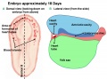
- [[Image:Basic Heart Development Timeline.jpg|center|800px]] ...heart initially forms from two tubes located bilaterally (on either side) of the '''trilaminar embryo''' in the '''cranial''' (head) region. The image4 KB (598 words) - 10:19, 12 November 2015
- <Flowplayer height="564" width="720" autoplay="false">Heart folding 002.flv</Flowplayer> Text: introduction; Picture: location of heart tubes; Text + animation: basic folding;4 KB (626 words) - 19:20, 26 February 2013
- [[Image:Basic Heart Development Timeline.jpg|center|800px]] ...y Heart Tube.jpg|thumb|right|upright=1.5|22 day embryo showing segments of heart tube]]8 KB (1,255 words) - 08:34, 20 April 2020
- [[File:Hedgehog.jpg|thumb|300px|An image of a hedgehog, the animal of which the Hh proteins are named after.]] ...arying levels within different tissues in the body, and also act as a form of redundancy to an extent between one another.65 KB (10,013 words) - 09:33, 7 August 2018