Search results
From Embryology
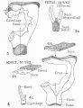
File:BeatonAnson1940 fig03-4.jpg ==Figs. 3 and 4. Reconstruction of human stapes== {{Stapes}} semidiagrammatic. x 45. 262(1,576 × 2,050 (374 KB)) - 11:20, 12 July 2019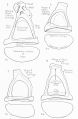
File:BeatonAnson1940 fig05-8.jpg ==Figs. 5 to 8. Excised specimens of human stapes== {{Stapes}} (see footnote 4). x 20.(1,623 × 2,482 (383 KB)) - 11:27, 12 July 2019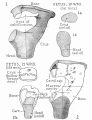
File:BeatonAnson1940 fig01-2.jpg ==Figs. 1 and 2. Reconstructions of human stapes== Human {{Stapes}} semi-diagrammatic (see footnote 4) x 45. 260(1,652 × 2,147 (652 KB)) - 11:14, 12 July 2019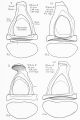
File:BeatonAnson1940 fig09-12.jpg :'''Links:''' {{Stapes}} [[Category:Hearing]][[Category:Middle Ear]][[Category:Stapes]][[Category:Fetal]](1,635 × 2,458 (422 KB)) - 11:42, 12 July 2019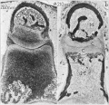
File:Anson1948 fig02.jpg ==Fig. 2. Photomicrographs of 202 mm and 310 mm fetus stapes and incus== Photomicrographs of the neck and head of the stapes and of the lentieular process of the incus, showing erosion of the cartilag(1,280 × 1,232 (241 KB)) - 08:55, 15 October 2017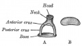
File:Gray0918.jpg ==Middle Ear - Stapes== A. Left stapes. B. Base of stapes, medial surface.(500 × 283 (17 KB)) - 17:24, 18 May 2015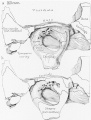
File:Anson1948 fig09.jpg ...al base and of the basal extremities of the crura. The dotted lines on the stapes indicates the approximate limits of the ossifying area; to either side of t(1,280 × 1,686 (235 KB)) - 20:43, 16 October 2017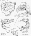
File:Anson1948 fig12.jpg ==Fig. 12. Drawings of a reconstruction of the stapes in the 180 mm Fetus== ...n the 180 mm. (21 week) fetus (Wisconsin series 45 B); X 7: (a) the entire stapes, in superolateral view; (1)) segment 1 (see inset), viewed from below (that(1,280 × 1,490 (289 KB)) - 20:52, 16 October 2017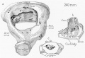
File:Anson1948 fig14.jpg ==Fig. 14. Drawings of a reconstruction of the stapes in a 240 mm== ...d of the base seen as though from the obturator space; (c) the neck of the stapes (capital portion removed) and the adjacent portions of the two crura, in la(1,280 × 872 (190 KB)) - 21:28, 16 October 2017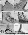
File:Anson1948 fig01.jpg ==Fig. 1. Photomicrographs of the base and crus of the stapes, showing progressive stages in the removal of cartilage and the formation o ...l) bone which forms one of the two constituent lamellas in the base of the stapes.(1,280 × 1,635 (310 KB)) - 17:02, 13 October 2017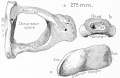
File:Anson1948 fig15.jpg ==Fig. 15. Drawings of a reconstruction of the stapes in a 275 mm== The stapes has attained adult form; of the crura, now deeply channeled, the posterior(1,280 × 829 (158 KB)) - 21:34, 16 October 2017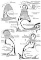
File:Anson1948 fig06.jpg ...right ear; all represent the transverse level of the posterior crus of the stapes. ...the incus. Destruction of periosteal bone on the obturator surface of the stapes keeps pace with the formation of endochondral bone within the capital and b(1,028 × 1,488 (224 KB)) - 20:16, 16 October 2017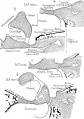
File:Anson1948 fig03.jpg ==Fig. 3. Drawings of the stapes and the adjacent fissular region of the otic capsule== Drawings (semi-diagrammatic) from Edinger tracings of the stapes and the adjacent fissular region of the otic capsule, showing developmenta(1,280 × 1,815 (358 KB)) - 18:46, 18 November 2017
File:Anson1948 fig10.jpg ...zone. In I) of figure 10 the hummocks exposed in the basal portion of the stapes are composed chiefly of calcified cartilage; their summits are capped by e(1,191 × 1,236 (204 KB)) - 21:00, 16 October 2017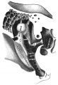
File:Gray0919.jpg ...cavity by ligaments: three for the malleus, and one each for the incus and stapes. ...estra vestibuli by a fibrous ring, the annular ligament of the base of the stapes (lig. annulare baseos stapedis).(500 × 731 (93 KB)) - 17:25, 18 May 2015
File:Anson1948 fig05.jpg ...19, through the middle of the fissular tract and the posterior crus of the stapes; c, through the vestibular orifice of the fissula, and d to- f, from sect ...er way; bone is formed on the obturator aspect of the base and crus of the stapes and on the tympanic and vestibular walls of the otic capsule. Concurrently,(1,280 × 1,797 (362 KB)) - 09:27, 15 October 2017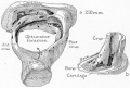
File:Anson1948 fig13.jpg ==Fig. 13. Drawings of a reconstruction of the stapes in a 210 mm== Drawings of a reconstruction of the stapes in a 210 mm. (23 week) fetus (Wisconsin series 51) ; X 7: (a) reconstructio(1,239 × 841 (203 KB)) - 21:17, 16 October 2017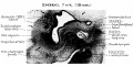
File:HansonAnson1962 fig01.jpg The malleus, like the incus and stapes, develops from a mesenchymal blastema located within the first two branchia ...mates the portion of the stapedial ring destined to become the head of the stapes.(1,280 × 643 (218 KB)) - 10:17, 7 January 2019
File:Stage 22 image 062.jpg ...um of tympanic membrane (L). Manubrium of malleus (L). Meckel's cartilage. Stapes (R). Auditory tube. Basal turn of cochlea duct (L). Endolymphatic sac (R).(1,000 × 645 (135 KB)) - 14:52, 5 October 2011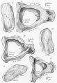
File:AnsonKarabinMartin1939 fig07-12.jpg ==Figs. 7 to 12. Reconstructions of the stapes; superior and medial views== In these and in the succeeding figures ant. crus. indicates anterior crus of stapes; cart. or cartil., cartilage; far. c., facial canal ; fen. cart, fenestral(1,280 × 1,880 (444 KB)) - 08:39, 22 October 2017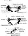
File:HansonAnsonBast1959 fig01.jpg ...is grooved by the facial nerve. Part of this mass is the primordium of the stapes. However, it is still separate from the aggregation of which will become th ...a point on the hyoid bar (at II) just distal to the blastemal lobe of the stapes (fig. 1c). The primordia of the malleus and incus are not yet distinct stru(1,280 × 1,634 (149 KB)) - 14:25, 27 December 2017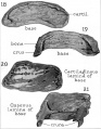
File:AnsonKarabinMartin1939 fig18-21.jpg ...structions of the osseous and the cartilaginous portion of the base of the stapes== In these and in the succeeding figures ant. crus. indicates anterior crus of stapes; cart. or cartil., cartilage; far. c., facial canal ; fen. cart, fenestral(1,280 × 1,643 (180 KB)) - 09:01, 22 October 2017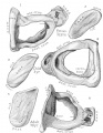
File:AnsonKarabinMartin1939 fig01-06.jpg ==Fig. 1 to 6. Reconstructions of the stapes - superior and medial views== In these and in the succeeding figures ant. crus. indicates anterior crus of stapes; cart. or cartil., cartilage; far. c., facial canal ; fen. cart, fenestral(1,280 × 1,650 (324 KB)) - 08:34, 22 October 2017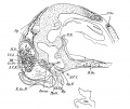
File:Watson1915 Fig08.jpg ...F.E., foramen endolymphaticum ; Mall., malleus; S.Oc, supraoccipital; Si., stapes; Sy.N., sympathetic nerve; FJJ, posterior, hyomandibular branch of the faci(1,183 × 1,000 (211 KB)) - 23:16, 30 October 2013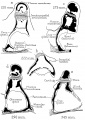
File:Anson1948 fig07.jpg ...d neck and head of the stapes (345 mm., e). Thus, with regard to form, the stapes is essentially an adult ossicle in the 290 mm. fetus (d) ; with respect to(1,280 × 1,801 (219 KB)) - 18:48, 13 May 2018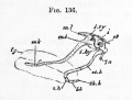
File:Foster136.jpg ...nanubrium or handle of the malleus ; tjy. tegmen tympani ; ?'. incus ; st. stapes ; i.hy. interhyal ligament ; st.h. stylohyal cartilage ; h.h. hypohyal ; b.(556 × 420 (33 KB)) - 09:19, 20 June 2018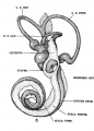
File:Streeter1917-fig06.jpg abuts against the stapes it will be noted that it also is beginning to spread over(539 × 749 (93 KB)) - 14:49, 17 September 2015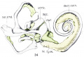
File:Bast1933 fig34.jpg ...sti., scala vestibuli; Sc. tym., scala tympani; imp. Sta.p., impression of stapes; Sac. end., endolyrnphatic duct; Sm:., sacculus; Ut1'z'c., utriculus; Duct.(1,000 × 677 (73 KB)) - 13:38, 13 December 2018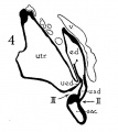
File:Anson-1934 fig04.jpg ...., medullary tube; sae., saccule; s.s.d., superior semicircular duct; st., stapes; utr., utricle; vest, vestibular part of vesicle; u.e.d., utriculo-endolymp(484 × 543 (34 KB)) - 12:37, 2 February 2017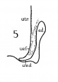
File:Anson-1934 fig05.jpg ...., medullary tube; sae., saccule; s.s.d., superior semicircular duct; st., stapes; utr., utricle; vest, vestibular part of vesicle; u.e.d., utriculo-endolymp(335 × 470 (15 KB)) - 13:13, 2 February 2017
File:Anson-1934 fig07.jpg ...., medullary tube; sae., saccule; s.s.d., superior semicircular duct; st., stapes; utr., utricle; vest, vestibular part of vesicle; u.e.d., utriculo-endolymp(324 × 336 (11 KB)) - 13:14, 2 February 2017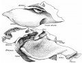
File:Frazer1914 fig06.jpg ...ossula rotunda. which lies over the pouch. Above this the facial nerve and stapes are seen cut, their other parts remaining on the other portion of the model(1,000 × 769 (176 KB)) - 06:26, 9 January 2017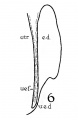
File:Anson-1934 fig06.jpg ...., medullary tube; sae., saccule; s.s.d., superior semicircular duct; st., stapes; utr., utricle; vest, vestibular part of vesicle; u.e.d., utriculo-endolymp(376 × 580 (20 KB)) - 13:14, 2 February 2017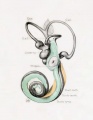
File:Streeter028.jpg ...shown in green. Although the greater part of the cistern abuts against the stapes, it will be noted that it is also begiiming to spread over the liorsal surf * Impressio staped., area in contact with base of stapes(774 × 1,000 (69 KB)) - 17:32, 18 May 2015
File:Ananomy of Ear.JPG Middle ear: Ossicles - Malleus, Incus and Stapes(566 × 405 (23 KB)) - 12:14, 19 September 2012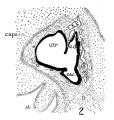
File:Anson-1934 fig02.jpg ...., medullary tube; sae., saccule; s.s.d., superior semicircular duct; st., stapes; utr., utricle; vest, vestibular part of vesicle; u.e.d., utriculo-endolymp(569 × 600 (98 KB)) - 12:36, 2 February 2017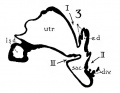
File:Anson-1934 fig03.jpg ...., medullary tube; sae., saccule; s.s.d., superior semicircular duct; st., stapes; utr., utricle; vest, vestibular part of vesicle; u.e.d., utriculo-endolymp(527 × 408 (25 KB)) - 12:37, 2 February 2017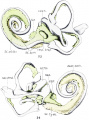
File:Bast1933 fig33-34.jpg ...sti., scala vestibuli; Sc. tym., scala tympani; imp. Sta.p., impression of stapes; Sac. end., endolyrnphatic duct; Sm:., sacculus; Ut1'z'c., utriculus; Duct.(1,000 × 1,353 (145 KB)) - 16:06, 22 October 2017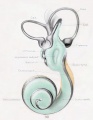
File:Streeter030.jpg The cartilaginous stapes was removed from this model and the oval impression that it makes on the ci * Impressio staped., area in contact with base of stapes(774 × 1,000 (78 KB)) - 17:33, 18 May 2015
File:Anatomy of the Ear.JPG Middle ear: Ossicles - Malleus, Incus and Stapes(566 × 405 (23 KB)) - 16:41, 28 September 2012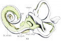
File:Bast1933 fig33.jpg ...sti., scala vestibuli; Sc. tym., scala tympani; imp. Sta.p., impression of stapes; Sac. end., endolyrnphatic duct; Sm:., sacculus; Ut1'z'c., utriculus; Duct.(1,000 × 673 (73 KB)) - 11:18, 24 October 2017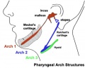
File:Pharyngeal arch cartilages.jpg ...art of the hyoid cartilage. The ends of this "U" shaped cartilage form the stapes of the middle ear ossicles.(400 × 324 (26 KB)) - 00:40, 16 July 2015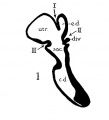
File:Anson-1934 fig01.jpg ...., medullary tube; sae., saccule; s.s.d., superior semicircular duct; st., stapes; utr., utricle; vest, vestibular part of vesicle; u.e.<l., utriculo-endolym(524 × 578 (24 KB)) - 09:36, 2 February 2017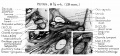
File:HansonAnson1962 fig02.jpg ...is. The long crus of the incus approximates the future capital part of the stapes but is separated from the manubrium by the chorda tympani. The loose mesenc(1,280 × 601 (233 KB)) - 10:17, 7 January 2019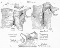
File:Anson1948 fig11.jpg ...the otic capsule in a 180 mm. (21 week) fetus (Wisconsin series 45-b) with stapes removed; X 7: (a) viewed from a posterosuperior position, that is, looking(1,220 × 981 (230 KB)) - 21:08, 16 October 2017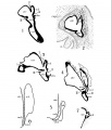
File:Anson-1934 fig01-07.jpg ...., medullary tube; sae., saccule; s.s.d., superior semicircular duct; st., stapes; utr., utricle; vest, vestibular part of vesicle; u.e.d., utriculo-endolymp(1,280 × 1,506 (210 KB)) - 12:44, 2 February 2017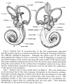
File:Streeter1917-fig06-07.jpg abuts against the stapes it will be noted that it also is beginning to spread over(1,337 × 1,671 (639 KB)) - 14:49, 17 September 2015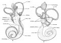
File:Streeter1917-fig08-09.jpg ...th (Carnegie Collection, No. 1018) enlarged 9 diameters. The cartilagenous stapes was removed from this model and the oval impression that it makes on the ci(1,200 × 853 (218 KB)) - 14:39, 17 September 2015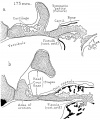
File:Anson1948 fig04.jpg The stapes is now excavated on the obturator surface of the base (a and b), at the bas(1,280 × 1,541 (247 KB)) - 09:21, 15 October 2017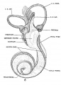
File:Streeter1917-fig08.jpg ...th (Carnegie Collection, No. 1018) enlarged 9 diameters. The cartilagenous stapes was removed from this model and the oval impression that it makes on the ci(610 × 853 (110 KB)) - 15:12, 17 September 2015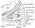
File:Keith1902 fig039.jpg ...atinous tissue is absorbed, so that the malleus and incus and developing {{stapes}}, with the chorda tympani, become surrounded by the hypoblasts lining of t(751 × 600 (114 KB)) - 19:35, 17 April 2018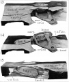
File:AnsonKarabinMartin1939 fig13-15.jpg In these and in the succeeding figures ant. crus. indicates anterior crus of stapes; cart. or cartil., cartilage; far. c., facial canal ; fen. cart, fenestral(1,280 × 1,519 (171 KB)) - 08:46, 22 October 2017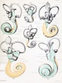
File:Streeter028-30.jpg * Impressio staped. - area in contact with base of stapes ...shown in green. Although the greater part of the cistern abuts against the stapes, it will be noted that it is also begiiming to spread over the liorsal surf(748 × 1,000 (134 KB)) - 17:30, 18 May 2015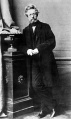
File:Karl Bogislaus Reichert.jpg ...#cartilage|cartilage]] band. The dorsal ends form the middle ear ossicle (stapes) and temporal bone styloid process, the ventral part ossifies to form hyoid(357 × 600 (40 KB)) - 17:31, 13 September 2016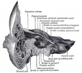
File:Gray0913.jpg ...ex border is upward. In the recent state it is occupied by the base of the stapes, the circumference of which is fixed by the annular ligament to the margin(671 × 600 (98 KB)) - 07:02, 19 August 2012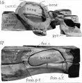
File:AnsonKarabinMartin1939 fig16-17.jpg In these and in the succeeding figures ant. crus. indicates anterior crus of stapes; cart. or cartil., cartilage; far. c., facial canal ; fen. cart, fenestral(1,280 × 1,301 (163 KB)) - 08:54, 22 October 2017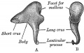
File:Gray0917.jpg ...lar process is tipped with cartilage, and articulates with the head of the stapes(600 × 379 (44 KB)) - 17:24, 18 May 2015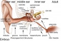
File:Adult hearing embryonic origins.jpg ** Stapes - Pharyngeal Arch 2 cartilage Neural crest (ectoderm)(1,000 × 675 (80 KB)) - 14:26, 8 May 2018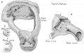
File:Anson1948 fig16.jpg ==Fig. 16. Drawings of a reconstruction of the stapes in a fetus at term==(1,280 × 817 (133 KB)) - 11:07, 14 October 2017
File:HansonAnsonBast1959 fig02.jpg ...be marle: the medial (mel larger Pllrt the lobe lI'I'lI gil'e rise 10 the stapes; the pm'llale"al to Ihe nerve wiLL become the latero- h!Jllle; Ihe iniercol(1,280 × 824 (130 KB)) - 14:14, 1 January 2018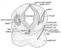
File:Keith1902 fig035.jpg ...lato-quadrate bar (cartilaginous skeleton of maxillary process), while the stapes is an independent formation developed round the stapedial artery. It may be(800 × 633 (125 KB)) - 12:46, 28 June 2016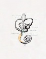
File:Streeter027.jpg * Impressio staped., area in contact with base of stapes(774 × 1,000 (51 KB)) - 17:31, 18 May 2015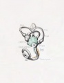
File:Streeter026.jpg * Impressio staped., area in contact with base of stapes(774 × 1,000 (45 KB)) - 17:31, 18 May 2015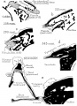
File:Anson1948 fig08.jpg ...the newborn (c), the capsule has acquired an adult appearance, as has the stapes.(1,280 × 1,764 (207 KB)) - 20:25, 16 October 2017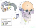
File:Cranial neural crest skeletal fate 01.jpg * '''ST''' - stapes(800 × 633 (59 KB)) - 22:36, 11 May 2019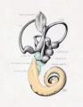
File:Streeter029.jpg * Impressio staped., area in contact with base of stapes(774 × 1,000 (74 KB)) - 17:32, 18 May 2015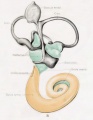
File:Streeter031.jpg * Impressio staped., area in contact with base of stapes(774 × 1,000 (79 KB)) - 17:34, 18 May 2015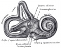
File:Gray0921.jpg ...is the fenestra vestibuli, closed, in the fresh state, by the base of the stapes and annular ligament. On its medial wall, at the forepart, is a small circu(640 × 500 (95 KB)) - 07:30, 19 August 2012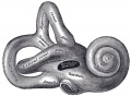
File:Gray0920.jpg ...is the fenestra vestibuli, closed, in the fresh state, by the base of the stapes and annular ligament. On its medial wall, at the forepart, is a small circu(600 × 438 (69 KB)) - 07:28, 19 August 2012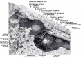
File:Gray0914.jpg ...ex border is upward. In the recent state it is occupied by the base of the stapes, the circumference of which is fixed by the annular ligament to the margin(708 × 500 (102 KB)) - 07:13, 19 August 2012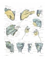
File:Macklin-plate05.jpg ...oint of view is almost parallel with the plane of the paper. The ring-like stapes is seen and to it is attached the tendon of the stapedius muscle, with the(2,331 × 3,061 (925 KB)) - 13:48, 24 April 2014


