Search results
From Embryology
Page title matches
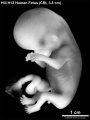
File:HillH13 Fetus bf01.jpg ==Human Fetus (Week 9)== HillH13 fetus week 9 CRL 3.8 cm left view.(1,200 × 1,600 (171 KB)) - 08:41, 14 December 2014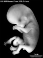
File:HillH13 Fetus bf02.jpg ==Human Fetus (Week 9)== HillH13 fetus week 9 CRL 3.8 cm left view.(1,200 × 1,600 (170 KB)) - 08:40, 14 December 2014
File:Ultrasound Fetus 01.mp4 (3.45 MB) - 16:40, 12 March 2013File:Ultrasound Fetus 02.mp4 (782 KB) - 16:12, 16 March 2013
File:Dog fetus day 28.jpg ==Dog Fetus day 28==(400 × 375 (24 KB)) - 16:38, 25 September 2012
File:Dog fetus day 30.jpg ==Dog Fetus day 28==(400 × 375 (32 KB)) - 16:39, 25 September 2012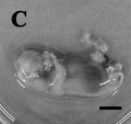
File:Dog fetus day 32.jpg ==Dog Fetus day 28==(400 × 375 (28 KB)) - 16:40, 25 September 2012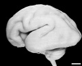
File:Human Fetus CRL240mm brain.jpg ==Human Fetus Brain== [[Category:Human Fetus]][[Category:Neural]](1,280 × 1,030 (129 KB)) - 16:05, 27 March 2018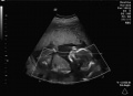
File:Ultrasound Fetus 01-icon.jpg (659 × 480 (39 KB)) - 16:40, 12 March 2013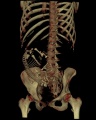
File:Fetus 35 week CT.jpg [[Category:Human Fetus]] [[Category:Computed Tomography]](700 × 874 (62 KB)) - 14:21, 24 August 2010
File:Human week 10 fetus 11.jpg ==Human Female Fetus - Sacrum (10 week)== {{Human Female Fetus Week 10 gallery}}(1,200 × 900 (304 KB)) - 15:24, 25 May 2016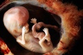
File:Fetus week 9-10-icon.jpg (220 × 147 (12 KB)) - 12:57, 24 April 2014
File:Human week 10 fetus 01.jpg ==Human Female Fetus (10 week)== {{Human Female Fetus Week 10 gallery}}(2,300 × 1,327 (448 KB)) - 11:57, 30 May 2016
File:Human week 10 fetus 12.jpg ==Human Female Fetus - Olfactory Nerve (10 week)== {{Human Female Fetus Week 10 gallery}}(1,200 × 900 (349 KB)) - 14:38, 25 May 2016
File:Human week 10 fetus 02.jpg ==Human Female Fetus (10 week)== See also [[:File:Human week 10 fetus 01.jpg|'''Large Image Version''']](800 × 462 (83 KB)) - 14:38, 11 October 2015
File:Human week 10 fetus 03.jpg ==Human Female Fetus - Pelvic Region (10 week)== {{Human Female Fetus Week 10 gallery}}(1,600 × 1,200 (370 KB)) - 17:21, 25 May 2016
File:Human week 10 fetus 04.jpg ==Human Female Fetus - Oral Cavity (10 week)== {{Human Female Fetus Week 10 gallery}}(1,600 × 1,200 (534 KB)) - 15:48, 25 May 2016
File:Human week 10 fetus 05.jpg ==Human Female Fetus - Heart (10 week)== {{Human Female Fetus Week 10 gallery}}(1,600 × 1,200 (612 KB)) - 16:39, 25 May 2016
File:Human week 10 fetus 06.jpg ==Human Female Fetus - Midgut Herniation (10 week)== {{Human Female Fetus Week 10 gallery}}(1,200 × 900 (251 KB)) - 17:01, 25 May 2016
File:Human week 10 fetus 23.jpg ==Human Female Fetus - Pelvic Region (10 week)== {{Human Female Fetus Week 10 gallery}}(1,600 × 1,200 (393 KB)) - 17:21, 25 May 2016
File:Human week 10 fetus 07.jpg ==Human Female Fetus - Spleen (10 week)== {{Human Female Fetus Week 10 gallery}}(1,200 × 900 (283 KB)) - 11:56, 30 May 2016
File:Human week 10 fetus 26.jpg ==Human Female Fetus - Midgut Herniation (10 week)== {{Human Female Fetus Week 10 gallery}}(1,200 × 900 (262 KB)) - 17:03, 25 May 2016
File:Human week 10 fetus 08.jpg ==Human Female Fetus Epiglottis (10 week)== {{Human Female Fetus Week 10 gallery}}(1,200 × 900 (323 KB)) - 21:03, 8 October 2015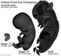
File:Size comparison embryo-fetus actual.jpg ==Size Comparison of the Embryo to Fetus== Original file name: EFsizeactual.jpg (Size comparison embryo-fetus actual.jpg)(194 × 178 (14 KB)) - 17:45, 6 May 2011
File:Human week 10 fetus 09.jpg ==Human Female Fetus - Atlas and Axis (10 week)== {{Human Female Fetus Week 10 gallery}}(1,200 × 900 (345 KB)) - 14:54, 25 May 2016
File:Human week 10 fetus 10.jpg ==Human Female Fetus - Pituitary and Lamina Terminalis (10 week)== {{Human Female Fetus Week 10 gallery}}(1,200 × 900 (291 KB)) - 09:21, 20 February 2019
File:Human fetus skeleton x-ray 03.jpg ==Human Fetus Skeleton X-ray== Fetus with limb deformities (indicated by arrows), which include bowing of the ra(628 × 934 (81 KB)) - 17:04, 22 April 2014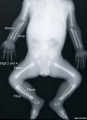
File:Human fetus skeleton x-ray 01.jpg ==Human Fetus Skeleton X-ray== {{Human fetus skeleton x-ray links}}(662 × 910 (54 KB)) - 17:05, 22 April 2014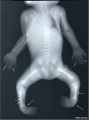
File:Human fetus skeleton x-ray 02.jpg ==Human Fetus Skeleton X-ray== Fetus with limb deformities including bowing of the digits, bowing and hypoplasia(686 × 928 (70 KB)) - 17:05, 22 April 2014
File:2015BGDA Lecture - Development of the Embryo-Fetus 1.pdf ==BGDA Lecture - Development of the Embryo-Fetus 1== [[BGDA Lecture - Development of the Embryo/Fetus 1|Lecture Page]](2.65 MB) - 11:15, 7 May 2015File:2017 BGDA Lecture - Development of the Embryo-Fetus 2.pdf PDF version of 2017 lecture page: [[BGDA Lecture - Development of the Embryo/Fetus 2]](3.23 MB) - 08:44, 15 May 2017File:2018 BGDA Lecture - Development of the Embryo-Fetus 2.pdf [[BGDA Lecture - Development of the Embryo/Fetus 2]](5.67 MB) - 21:58, 13 May 2018File:2019 BGDA Lecture - Development of the Embryo-Fetus 1.pdf ==BGDA Lecture - Development of the Embryo/Fetus 1== [[BGDA Lecture - Development of the Embryo/Fetus 1]](1.93 MB) - 22:19, 2 May 2019File:BGDA Lecture 2014 - Development of the Embryo-Fetus 1.pdf ==BGDA Lecture - Development of the Embryo/Fetus 1==(869 KB) - 14:24, 7 May 2014File:2019 BGDA Lecture - Development of the Embryo-Fetus 2.pdf (3.59 MB) - 09:35, 13 May 2019File:2017 BGDA Lecture - Development of the Embryo-Fetus 1.pdf (3.39 MB) - 16:38, 3 May 2017File:2018 BGDA Lecture - Development of the Embryo-Fetus 1.pdf (8.38 MB) - 08:08, 4 May 2018


Page text matches

File:Koala fetus.jpg ==Koala Fetus== Koala fetus near birth.(559 × 1,000 (77 KB)) - 16:32, 1 December 2010File:HillH13 Fetus bf01.jpg ==Human Fetus (Week 9)== HillH13 fetus week 9 CRL 3.8 cm left view.(1,200 × 1,600 (171 KB)) - 08:41, 14 December 2014File:HillH13 Fetus bf02.jpg ==Human Fetus (Week 9)== HillH13 fetus week 9 CRL 3.8 cm left view.(1,200 × 1,600 (170 KB)) - 08:40, 14 December 2014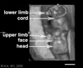
File:Ultrasound12wk 3D image.jpg ...ultrasound static image of the 12 week fetus shows a ventral view with the fetus upside down, with the head down and cord to the top. ...ategory:Genetic Abnormalities]] [[Category:Human Embryo]] [[Category:Human Fetus]](301 × 248 (8 KB)) - 15:10, 11 October 2009
File:HillH13 Fetus.gif ==Human Fetus (Week 9)== HillH13 fetus week 9 CRL 3.8 cm left view. (stereo pair animated gif)(450 × 600 (274 KB)) - 08:40, 14 December 2014File:Size comparison embryo-fetus actual.jpg ==Size Comparison of the Embryo to Fetus== Original file name: EFsizeactual.jpg (Size comparison embryo-fetus actual.jpg)(194 × 178 (14 KB)) - 17:45, 6 May 2011
File:Lineback1920 fig06-7.jpg ===Fig. 6. Cross-section of the ascending colon of a human fetus=== Cross-section of the ascending colon of a human fetus 105 mm. CR. length, showing the three tseniaj in a triangular position in t(1,200 × 808 (177 KB)) - 09:13, 17 January 2013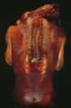
File:Spina Bifida 1.jpg ==Spina Bifida Fetus== Dorsal view of fetus with extensive spina bifida and anencephaly.(393 × 599 (49 KB)) - 10:28, 11 May 2016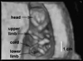
File:Ultrasound12wk 3D image2.jpg ==Ultrasound Fetus (12 week)== Image from near end of movie showing ventral view of fetus head to top, upper limbs, lower limbs and umbilical cord visible.(362 × 264 (8 KB)) - 09:06, 6 November 2012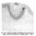
File:Keibel Mall 211.jpg ==Fig. 211 Human Fetus Integumentary Gland==(427 × 436 (43 KB)) - 13:01, 24 August 2012File:Human week 10 fetus 02.jpg ==Human Female Fetus (10 week)== See also [[:File:Human week 10 fetus 01.jpg|'''Large Image Version''']](800 × 462 (83 KB)) - 14:38, 11 October 2015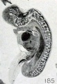
File:Mall Meyer1921 fig185.jpg ==Fig. 185. Fetus in sagittal section showing maceration== Fetus in sagittal section showing maceration, especially of the nervous system. N(341 × 500 (36 KB)) - 09:23, 13 December 2012
File:Fetal corpus cavernosum and corpus spongiosum 01.jpg * A - Corpus cavernosum, fetus of 13 WPC * C - Corpus cavernosum, fetus of 22 WPC(1,795 × 2,082 (919 KB)) - 11:42, 7 September 2014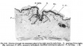
File:Keibel Mall 212.jpg ==Fig. 212 Human Fetus Integumentary Gland== Fetus 8 months.(650 × 371 (39 KB)) - 13:00, 24 August 2012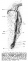
File:Keibel Mall 209.jpg ==Fig. 209 Human Fetus Integumentary Hair== Fetus Month 8.(458 × 990 (89 KB)) - 13:02, 24 August 2012
File:Mall Meyer1921 fig180.jpg ==Fig. 180. Fetus showing continuity of epidermis across the mouth== Fetus showing continuity of epidermis across the mouth, with obliteration of the(442 × 478 (32 KB)) - 09:20, 13 December 2012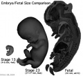
File:Size comparison embryo-fetus.jpg ==Size comparison of the Embryo to Fetus (week 10)== Original file name: EFsize.jpg (Size comparison embryo-fetus.jpg)(327 × 300 (29 KB)) - 17:46, 6 May 2011
File:Bardeen1914-fig04.jpg ==Fig. 4. Gastrointestinal Tract Fetus 35 mm lateral view == ...ch, small and large intestines, rectum, bursa omentalis and mesentery of a fetus 35 mm long (No. 8, Wisconsin Collection).(840 × 1,019 (93 KB)) - 08:30, 29 April 2017
File:Bardeen1914-fig03.jpg ==Fig. 3. Gastrointestinal Tract Fetus 35 mm ventral view == ...ch, small and large intestines, rectum, bursa omentalis and mesentery of a fetus 35 mm long (No. 8, Wisconsin Collection).(405 × 1,014 (55 KB)) - 08:31, 29 April 2017File:Human week 10 fetus 07.jpg ==Human Female Fetus - Spleen (10 week)== {{Human Female Fetus Week 10 gallery}}(1,200 × 900 (283 KB)) - 11:56, 30 May 2016
File:Simkins1928 plate09.jpg 38 Peripheral portion of an ovary from a seven-month fetus. X 750. 39 Peripheral portion of an ovary from a full-term fetus. cl.g., genitaloid cell. X 7 50.(1,548 × 2,096 (263 KB)) - 16:58, 31 January 2018
File:Anson1948 fig01.jpg (a) Base at posterior crus, left ear; 167 mm. fetus (Wisconsin series 105, slide 19, section 9). (b) Base and anterior crus, left ear; 175 mm. fetus (Wisconsin series 104, slide 17, section 5).(1,280 × 1,635 (310 KB)) - 17:02, 13 October 2017
File:Bardeen1914-fig06.jpg ==Fig. 6. Lateral view fetus 40 mm== ...he stomach, small and large intestines, bursa omentalis and mesentery of a fetus 40 mm. long (No. 8, Wisconsin Collection).(721 × 1,000 (88 KB)) - 09:52, 3 October 2017File:2019 BGDA Lecture - Development of the Embryo-Fetus 1.pdf ==BGDA Lecture - Development of the Embryo/Fetus 1== [[BGDA Lecture - Development of the Embryo/Fetus 1]](1.93 MB) - 22:19, 2 May 2019File:2015BGDA Lecture - Development of the Embryo-Fetus 1.pdf ==BGDA Lecture - Development of the Embryo-Fetus 1== [[BGDA Lecture - Development of the Embryo/Fetus 1|Lecture Page]](2.65 MB) - 11:15, 7 May 2015
File:Bardeen1914-fig05.jpg ==Fig. 5. Ventral view fetus 40 mm== ...he stomach, small and large intestines, bursa omentalis and mesentery of a fetus 40 mm. long (No. 8, Wisconsin Collection). In [[:File:Bardeen1914-fig06.jpg(465 × 1,000 (61 KB)) - 07:05, 15 November 2015
File:Wallaby embryo 05.jpg ==Tammar Wallaby Embryo and Fetus== ...ld within the folds of the yolk sac, which has become highly vascular. The fetus has well developed fore limbs ready for the climb to the pouch and the tong(996 × 1,100 (127 KB)) - 11:06, 29 July 2019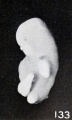
File:Mall Meyer1921 fig133.jpg ==Fig. 133. Marked macerated and deformed fetus from the same case== Fig. 133. Marked macerated and deformed fetus from the same case. X4.(365 × 610 (40 KB)) - 09:12, 6 December 2012
File:Mall Meyer1921 plate19.jpg Fig. 218. A fetus and cord showing marked maceration changes. No. 797. X1.35. Fig. 219. An older fetus, showing bleb-formation and curvature in extremities, due to maceration and(939 × 1,200 (237 KB)) - 18:44, 23 November 2012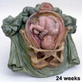
File:Galletti1770 week 24.jpg Fetus 24 weeks(450 × 450 (40 KB)) - 15:12, 13 October 2009
File:B050966-01.jpg ==Female Human Fetus 14cm==(2,000 × 992 (681 KB)) - 11:22, 25 June 2015
File:Frazer006 bw600.jpg ==Fetus during Third Trimester in Uterus compared to Non-pregnant Uterus== [[Category:Historic Embryology]] [[Category:Human Fetus]] [[Category:Third Trimester]] [[Category:Cartoon]](600 × 575 (47 KB)) - 17:30, 1 June 2013File:Human fetus skeleton x-ray 02.jpg ==Human Fetus Skeleton X-ray== Fetus with limb deformities including bowing of the digits, bowing and hypoplasia(686 × 928 (70 KB)) - 17:05, 22 April 2014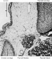
File:Boyd1950 fig09.jpg ==Fig. 9. Section fetus 145 mm to show aberrant portion of thyroid tissue lying between thyroid and {{Online Editor}} - [[Fetal Development|Fetus]] 145mm is about 16 weeks ({{GA}} 18 weeks).(801 × 900 (163 KB)) - 08:29, 21 March 2017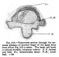
File:Keibel Mall 216.jpg ==Fig. 216 Human Fetus Finger== Transverse section through the terminal phalanx of another finger of the same fetus from which [[:File:Keibel Mall 215.jpg|Fig. 215]] is taken. The basal cell(366 × 356 (30 KB)) - 12:43, 13 January 2019File:Human week 10 fetus 01.jpg ==Human Female Fetus (10 week)== {{Human Female Fetus Week 10 gallery}}(2,300 × 1,327 (448 KB)) - 11:57, 30 May 2016File:Human fetus skeleton x-ray 03.jpg ==Human Fetus Skeleton X-ray== Fetus with limb deformities (indicated by arrows), which include bowing of the ra(628 × 934 (81 KB)) - 17:04, 22 April 2014
File:Mall Meyer1921 plate14.jpg Fig. 145. Enlarged fetus from same, showing maceration effects. X2. Fig. 150. Macerated, distorted normal cat fetus 10 mm. long. (After Kunz.)(941 × 1,200 (260 KB)) - 19:21, 23 November 2012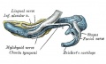
File:Gray0179.jpg ...outer aspect 95mm fetus]] | [[:File:Gray0181.jpg|Image - inner aspect 95mm fetus]](617 × 368 (47 KB)) - 18:34, 27 August 2012
File:Lineback1920 fig04-5.jpg ==Cross-section of the colon of a human fetus== Fig. 4. Cross-section of the colon of a human fetus 52 mm. CR. length.(1,200 × 756 (150 KB)) - 09:16, 17 January 2013
File:Gray0181.jpg ...outer aspect 95mm fetus]] | [[:File:Gray0181.jpg|Image - inner aspect 95mm fetus]] | [[Fetal Development]](617 × 368 (52 KB)) - 18:35, 27 August 2012File:Human Fetus CRL240mm brain.jpg ==Human Fetus Brain== [[Category:Human Fetus]][[Category:Neural]](1,280 × 1,030 (129 KB)) - 16:05, 27 March 2018
File:Gray0178.jpg ...outer aspect 95mm fetus]] | [[:File:Gray0181.jpg|Image - inner aspect 95mm fetus]](617 × 368 (44 KB)) - 18:33, 27 August 2012
File:Gray0180.jpg ...outer aspect 95mm fetus]] | [[:File:Gray0181.jpg|Image - inner aspect 95mm fetus]] | [[Fetal Development]](617 × 368 (48 KB)) - 18:34, 27 August 2012
File:Anson1948 fig02.jpg ==Fig. 2. Photomicrographs of 202 mm and 310 mm fetus stapes and incus== ...0 (a) 202 mm fetus (Wisconsin series 70 slide 37, section 6); (b) 310 mm fetus (Wisconsin series 51, slide 38, section 6).(1,280 × 1,232 (241 KB)) - 08:55, 15 October 2017
File:Anson1948 fig07.jpg ==Fig. 7. Otic capsule developmental stages 210, 222, 275, 290 and 345 mm Fetus== ...fetus (Vlfisconsin series 59, slide 36, section 5); (e) 345 mm. (38 week) fetus (Wisconsin series 61,. slide 42, section 1).(1,280 × 1,801 (219 KB)) - 18:48, 13 May 2018
File:Streeter01.jpg '''A''', grade 1, fetus No. 1183; CR length 60 mm., weight 19.5 grams '''B''', grade 2, fetus No. 1282b, CR length 65.5 mm., weight I5 grams(800 × 634 (120 KB)) - 22:00, 12 May 2016
File:Mall Meyer1921 fig234.jpg ==Fig. 234. A very softened, macerated fetus==(331 × 793 (46 KB)) - 11:54, 3 December 2012
File:Lowsley1912 fig05.jpg ==Fig. 5. Human Fetus 16 cm Prostate==(1,641 × 1,083 (425 KB)) - 21:56, 16 June 2016
File:Human- fetal week 10 urogenital C.jpg ==Human Fetus (week 10) Female== Female fetus, 10 week, 40 mm CRL, early fetal, sagittal section, pelvic region(600 × 450 (105 KB)) - 17:42, 28 May 2011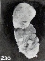
File:Mall Meyer1921 fig230.jpg ==Fig. 230. A decidedly mummified fetus==(281 × 379 (21 KB)) - 11:50, 3 December 2012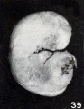
File:Mall Meyer1921 fig39.jpg ==Fig. 39. Macerated firmly rolled-up young fetus== The specimen consists of a rather poorly preserved young fetus, measuring 16.5 mm. CR. The ventral abdominal wall had been seriously injur(414 × 535 (41 KB)) - 07:11, 4 December 2012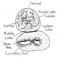
File:Lowsley1912 fig02.jpg ==Fig. 2. Human Fetus 7.5 cm==(759 × 740 (102 KB)) - 21:52, 16 June 2016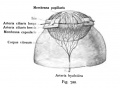
File:Kollmann722.jpg pupillaris sind nach einem menschlichen Fetus von 8 Monaten, die Membrana capsularis nach einem menschlichen Fetus von 6 Wochen eingezeichnet; +:(533 × 391 (28 KB)) - 10:52, 21 October 2011
File:Mall1917 fig11-13.jpg '''Fig. 11''' Stunted fetus with a large hernia in umbilical cord, also spina bifida. '''Fig. 13''' Normal fetus with hernia of midbrain.(1,000 × 599 (79 KB)) - 06:04, 12 November 2013
File:Mall Meyer1921 fig89.jpg ==Fig. 89. Normal fetus with hernia of mid-brain==(535 × 854 (90 KB)) - 08:24, 3 December 2012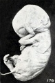
File:Mall Meyer1921 fig176.jpg ==Fig. 176. Fetus showing gluing of hand to face==(537 × 807 (70 KB)) - 09:18, 13 December 2012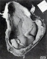
File:Mall Meyer1921 fig223.jpg ==Fig. 223. External appearance of fetus in situ==(562 × 705 (80 KB)) - 11:43, 3 December 2012
File:Lowsley1912 fig06.jpg ==Fig. 6. Human Fetus 19 cm Prostate==(1,327 × 916 (237 KB)) - 21:57, 16 June 2016
File:McMurrich1930 fig85.jpg ==Fig. 85. Representations of human fetus at term and of ungulate placenta== .... Where the uterus is also shown there is usually no indication of how the fetus is connected with it; only in QIII, 8 are the cotyledons represented, on th(1,280 × 1,698 (373 KB)) - 11:46, 20 April 2020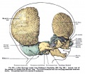
File:Keibel Mall 321.jpg ==Fig. 321 Lateral view of the cranium of a human fetus 80 mm long== Lateral view of the cranium of a human fetus 80 mm long.(746 × 651 (122 KB)) - 19:33, 4 September 2014
File:Mall Meyer1921 fig224.jpg ==Fig. 224. An older macerated fetus with extremities extended instead of folded==(440 × 551 (43 KB)) - 11:44, 3 December 2012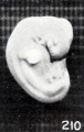
File:Mall Meyer1921 fig210.jpg ==Fig. 210. Normal well-preserved cat fetus==(240 × 379 (17 KB)) - 10:14, 3 December 2012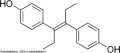
File:Diethylstilbestrol.jpg * Female fetus - increased risk abnormal reproductive tract and cancer. * Male fetus - abnormal genitalia.(600 × 263 (16 KB)) - 11:29, 6 March 2019
File:Humphrey1940 fig04.jpg ==Fig. 4. Olfactory and accessory olfactory formations in the 37 mm fetus== ...illustrate the olfactory and accessory olfactory formations in the 37 mm. fetus. Activated protargol preparation.(1,000 × 882 (101 KB)) - 17:50, 24 October 2017
File:Atwell1926 plate06.jpg 19 Wax—plalte reconstruction of the hypophysis from :1. 45-mm. human fetus (Huber collection, no. XVIII), viewed from above and in front. X 35. 20 Reconstruction of the epithelial hypophysis from a 55»mm. human fetus (U.B.E.C., no. 25), viewed from above and in front. X 35.(809 × 1,500 (136 KB)) - 15:09, 9 November 2016
File:Watson1918 plate01.jpg ...ion, 110. 1358e). A graphic reconstruction of these structures in the same fetus is shown in text figure 4. In figure 10 the left deferent duct is cut obl ..., estimated age 19 weeks (Carnegie Collection, no. 1049). This is the same fetus shown in text-figure 6. In figure 15 it will be noted that the left ampul(1,200 × 1,601 (398 KB)) - 09:21, 8 December 2016
File:BrauneB2.jpg ==The Position of the Uterus and Fetus at Term (1872)== ...tial section through the fetus shows the relative size and position of the fetus during pregnancy.(1,200 × 485 (143 KB)) - 22:57, 3 June 2015
File:Galletti1770 week 16.jpg ==Fetus 16 weeks==(450 × 450 (32 KB)) - 08:00, 27 May 2010
File:Simkins1928 plate03.jpg 14 Cross~section of the testis of a fetus, 190 mm. X 40. 15 Detail of the tubules of a 190-mm. fetus. X 750.(1,551 × 2,086 (293 KB)) - 16:45, 31 January 2018
File:BrauneA1.jpg ==The Position of the Uterus and Fetus at Term (1872)== ...tial section through the fetus shows the relative size and position of the fetus during pregnancy.(1,200 × 485 (141 KB)) - 17:13, 30 October 2012File:Human week 10 fetus 05.jpg ==Human Female Fetus - Heart (10 week)== {{Human Female Fetus Week 10 gallery}}(1,600 × 1,200 (612 KB)) - 16:39, 25 May 2016
File:Macklin-plate2a.jpg ==Plate 2. The skull of a human fetus of 43 millimeters greatest length==(1,142 × 1,500 (361 KB)) - 16:45, 23 April 2014
File:Macklin-plate1a.jpg ==Plate 1. The skull of a human fetus of 43 millimeters greatest length==(1,142 × 1,500 (374 KB)) - 16:45, 23 April 2014
File:Macklin-plate5a.jpg ==Plate 5. The skull of a human fetus of 43 millimeters greatest length==(1,142 × 1,500 (324 KB)) - 16:46, 23 April 2014
File:Macklin-plate3a.jpg ==Plate 3. The skull of a human fetus of 43 millimeters greatest length==(1,142 × 1,500 (321 KB)) - 16:46, 23 April 2014
File:Macklin-plate4a.jpg ==Plate 4. The skull of a human fetus of 43 millimeters greatest length==(1,142 × 1,500 (335 KB)) - 16:46, 23 April 2014
File:Lowsley1912 fig01.jpg ==Fig 1. Human Fetus 5 cm==(990 × 620 (29 KB)) - 21:53, 16 June 2016File:Human week 10 fetus 12.jpg ==Human Female Fetus - Olfactory Nerve (10 week)== {{Human Female Fetus Week 10 gallery}}(1,200 × 900 (349 KB)) - 14:38, 25 May 2016
File:Ultrasound Demonstrating Facial Features.jpg ...ere the face of the fetus is clearly shown. This is an example of a normal fetus.(1,280 × 1,024 (61 KB)) - 19:50, 5 October 2011
File:BrauneC2.jpg ==The Position of the Uterus and Fetus at Term (1872)== ...n through the maternal anatomy shows the relative size and position of the fetus during pregnancy.(1,200 × 461 (142 KB)) - 17:13, 30 October 2012
File:Watson1918 plate02.jpg ...th, estimated age 21 weeks (Carnegie Collection, no. 1171), being the same fetus shown in text figure 7. In figure 16 is shown the left anipulla and seminal '''18 to 21''' Transverse sections through the seminal tract in a human fetus 221 mm. crown-rump length, estimated age 25 weeks (Carnegie Collection, no.(1,200 × 1,628 (367 KB)) - 09:33, 8 December 2016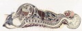
File:BrauneC3.jpg ==The Position of the Uterus and Fetus at Term (1872)== ...n through the maternal anatomy shows the relative size and position of the fetus during pregnancy.(1,200 × 461 (143 KB)) - 22:58, 3 June 2015
File:Mall Meyer1921 fig211.jpg ==Fig. 211. Normal poorly preserved cat fetus of approximately the same length==(212 × 470 (18 KB)) - 10:15, 3 December 2012
File:Anson1948 fig06.jpg ==Fig. 6. Otic capsule developmental stages 190 mm and 215 mm fetus== ...) fetus (Wisconsin series 129, slide 20, section 3); (d) 215 mm. (24 week) fetus (Wisconsin series 62, slide 28, section 4). Parts (1, b and d represent sec(1,028 × 1,488 (224 KB)) - 20:16, 16 October 2017
File:Fetal size change.jpg [[Category:Human Fetus]](337 × 443 (18 KB)) - 11:04, 16 August 2014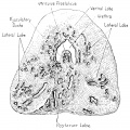
File:Lowsley1912 fig07.jpg ==Fig. 7. Human Fetus 27 cm Prostate==(1,437 × 1,435 (486 KB)) - 21:58, 16 June 2016
File:Streeter1919-fig02.jpg ...Topographical relations of the caudal end of the spinal cord in the human fetus from the eighth to the twenty-fifth week== ...long; 67-mm. fetus, 4.75 mm. long; 111-mm. fetus, 12.25 mm. long; 221-mm. fetus, 32 mm. long.(2,023 × 1,318 (499 KB)) - 22:24, 31 January 2019File:2018 BGDA Lecture - Development of the Embryo-Fetus 2.pdf [[BGDA Lecture - Development of the Embryo/Fetus 2]](5.67 MB) - 21:58, 13 May 2018
File:Mall Meyer1921 fig91.jpg From No. 230, a fetus compressus 57 mm. CR. X0.75.(352 × 191 (14 KB)) - 15:07, 15 January 2013
File:Atwell1926 plate05.jpg 16 Wax~platc reconstruction of the epithelial hypophysis from a. 25-mm. human fetus (U.B.E.C., no. 33), viewed from above and in front. X 65. 18 Reconstruction of the epithelial hypophysis from a 26-mm. human fetus (U.B.E.C., 110. 1), viewed from above and in front. X 50.(855 × 1,500 (135 KB)) - 16:30, 9 November 2016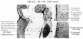
File:HansonAnson1962 fig04.jpg ==Fig. 4. Fetus 15 week 115 mm== '''a''', In the 15-week fetus (115 mm.) the malleus begins to ossify from a single center located medial(1,280 × 598 (201 KB)) - 10:31, 7 January 2019
File:Keibel Mall 070-071.jpg ==Fig. 68-69 Head of a Fetus 47.5 mm==(1,000 × 490 (45 KB)) - 21:14, 10 September 2012
File:Keibel Mall 068-069.jpg ==Fig. 68-69 Head of a Fetus 25mm==(1,000 × 358 (35 KB)) - 21:14, 10 September 2012
File:Grays Anatomy Embryology cover.jpg Cover image for iBook developed from [[:File:Gray0038.jpg|Figure 38]] - Fetus in Utero between Fifth and Sixth Months.(459 × 600 (25 KB)) - 18:45, 13 May 2013
File:Keibel Mall 285-288.jpg Fig. 285. Median section through the knee-joint of a fetus 13 cm long. a, patella; b. connective tissue over patella; c, lig Fig. 286-288. Hip-joint of a male fetus 25 cm. long, of a female child six years old, and of a male adult. a. regio(703 × 674 (72 KB)) - 02:38, 14 September 2012
File:Wallaby embryo 04.jpg ==Tammar Wallaby Embryo and Fetus== Fetus at day 23 of pregnancy, showing the increase in the vascular region and the(1,138 × 1,098 (110 KB)) - 13:17, 23 May 2012
File:Streeter002-3.jpg ==Fig. 2. Detail of the lateral semicircular duct in a human fetus 30 mm. long== ==Fig. 3. Detail of the lateral canal in a human fetus 16 mm. long==(607 × 800 (106 KB)) - 12:41, 15 February 2011File:Human week 10 fetus 08.jpg ==Human Female Fetus Epiglottis (10 week)== {{Human Female Fetus Week 10 gallery}}(1,200 × 900 (323 KB)) - 21:03, 8 October 2015
File:Keibel Mall 197.jpg ==Fig. 200 Human Fetus==(500 × 469 (46 KB)) - 07:15, 23 August 2012
File:Keibel Mall 198.jpg ==Fig. 198 Human Fetus==(500 × 443 (57 KB)) - 07:18, 23 August 2012
File:Keibel Mall 196.jpg ==Fig. 196 Human Fetus==(448 × 383 (35 KB)) - 07:18, 23 August 2012
File:Keibel Mall 202.jpg ==Fig. 202 Human Fetus==(481 × 500 (47 KB)) - 07:21, 23 August 2012
File:Keibel Mall 201.jpg ==Fig. 201 Human Fetus==(434 × 543 (65 KB)) - 07:24, 23 August 2012
File:Keibel Mall 200.jpg ==Fig. 200 Human Fetus==(781 × 645 (74 KB)) - 07:14, 23 August 2012
File:Kingsbury1932 plate04.jpg 24 Cat; 75-mm. fetus. Frontal section of the tonsillar region. Two lymphatic radicles filled wit 25 Cat; near term (130—mm. fetus). Frontal section of the tonsillar region. Lymphatic radicles filled with l(1,280 × 1,960 (426 KB)) - 21:45, 28 March 2017File:Human fetus skeleton x-ray 01.jpg ==Human Fetus Skeleton X-ray== {{Human fetus skeleton x-ray links}}(662 × 910 (54 KB)) - 17:05, 22 April 2014
File:Wislocki1920 plate 1.jpg Fig. 2. Gross appearance of a guinea-pig fetus with the amnion opened 36 hours after injection of trypan-blue into the amn ...ssium ferrocyanide and iron ammonium citrate into the amniotic cavity. The fetus was killed 30 minutes after injection and immersed in acid formalin.(1,145 × 1,200 (173 KB)) - 10:48, 16 June 2013File:Human week 10 fetus 10.jpg ==Human Female Fetus - Pituitary and Lamina Terminalis (10 week)== {{Human Female Fetus Week 10 gallery}}(1,200 × 900 (291 KB)) - 09:21, 20 February 2019
File:Mall Meyer1921 fig219.jpg ==Fig. 219. An older fetus, showing bleb-formation and curvature in extremities==(255 × 642 (28 KB)) - 11:41, 3 December 2012
File:Wislocki1920 plate 4.jpg ==Plate 4. Cat Fetus and Placenta== ===Fig. 13. Cat fetus measuring 6.8 cm surrounded by enraptured membranes===(935 × 1,000 (199 KB)) - 12:17, 27 January 2014
File:Mall Meyer1921 fig220.jpg ==Fig. 220. A fetus showing marked clubbing of the extremities and obliteration of the features(227 × 450 (17 KB)) - 11:42, 3 December 2012File:BGDA Lecture 2014 - Development of the Embryo-Fetus 1.pdf ==BGDA Lecture - Development of the Embryo/Fetus 1==(869 KB) - 14:24, 7 May 2014
File:Mall Meyer1921 plate04.jpg Fig. 36. Section of a similar fetus, to show structure. No. 675. X4.5. Fig. 39. Macerated, firmly rolled-up, young fetus. No. 921. X2.25.(945 × 1,200 (216 KB)) - 09:50, 6 December 2012
File:Anson1948 fig05.jpg ==Fig. 5. Otic capsule developmental stages 160 mm and 170 mm fetus== ...vestibular orifice of the fissula, and d to- f, from sections of the 170 mm. fetus which pass through similar levels of the fissula and ossicle (in successio(1,280 × 1,797 (362 KB)) - 09:27, 15 October 2017
File:Keibel Mall 207.jpg ==Fig. 207 Human Fetus==(800 × 538 (59 KB)) - 07:33, 23 August 2012
File:Keibel Mall 206.jpg ==Fig. 206 Human Fetus==(415 × 750 (79 KB)) - 07:33, 23 August 2012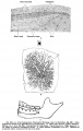
File:Keibel Mall 224.jpg ==Fig. 224 Human Fetus==(706 × 1,112 (105 KB)) - 08:08, 24 August 2012
File:Keibel Mall 224A.jpg ==Fig. 224A Human Fetus==(671 × 377 (38 KB)) - 08:10, 24 August 2012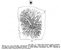
File:Keibel Mall 224B.jpg ==Fig. 224A Sheep Fetus==(685 × 563 (56 KB)) - 08:11, 24 August 2012
File:Keibel Mall 224C.jpg ==Fig. 224C Human Fetus==(706 × 306 (20 KB)) - 08:12, 24 August 2012
File:Lowsley1912 fig03.jpg ==Fig. 3. Human Fetus 12.5 cm==(1,897 × 1,601 (281 KB)) - 21:51, 16 June 2016
File:Cleft lip 01.jpg ==Human Fetus (18 week) Cleft Lip Ultrasound== ...ng of the lip which may also include cleft palate. The movie above shows a fetus (at 18 weeks gestation, 20 weeks obstetric) which has a facial cleft.(585 × 438 (34 KB)) - 15:46, 17 May 2014
File:Wallaby embryo 01.jpg ==Tammar Wallaby Embryo and Fetus== (c) Fetus at day 23 of pregnancy, showing the increase in the vascular region and the(1,026 × 1,000 (152 KB)) - 13:10, 23 May 2012
File:Gray0039.gif [[Category:Human Fetus]](500 × 384 (43 KB)) - 17:46, 18 August 2009
File:Gilmour1941 fig10.jpg ==Fig. 10. 444 mm fetus==(511 × 907 (105 KB)) - 11:58, 17 May 2018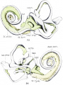
File:Bast1933 fig33-34.jpg ==Figs. 33 and 34. Human Fetus CRL 50 mm== Fig. 33. Fetus 86; size, 50 mm. crown-rump length; age, about 10 weeks. Anterior view of a(1,000 × 1,353 (145 KB)) - 16:06, 22 October 2017
File:Bardeen1914-plate02.jpg ...he stomach, small and large intestines, bursa omentalis and mesentery of a fetus 40 mm. long (No. 8, Wisconsin Collection). In figure 6 the kidney and adren ...uodenum, eaeeum, colon, rectum, bursa omentalis and rnesentery of the same fetus. 5 diameters. The specimen here illustrated may be compared with that descr(1,398 × 2,577 (275 KB)) - 07:06, 15 November 2015
File:Mall Meyer1921 fig87.jpg ==Fig. 87. Stunted fetus with large hernia in umbilical cord==(388 × 569 (40 KB)) - 06:04, 12 November 2013
File:Wislocki1920 plate 2.jpg Fig. 7. Kidney of guinea-pig fetus measuring 36 mm., showing trypan-blue in the convoluted tubules 72 hours af Fig. 8. Section of the umbilical cord of a guinea-pig fetus measuring 42 mm., showing vitally stained mucoid connective-tissue cells, 2(988 × 1,200 (245 KB)) - 10:47, 16 June 2013
File:Mall Meyer1921 fig280.jpg Showing disproportion between the chorionic vesicle and the fetus, sparse development of the villi, and some hydatiform degeneration. Xl.5.(754 × 512 (84 KB)) - 08:48, 12 December 2012
File:Bast1933 fig33.jpg ==Figs. 33 Human Fetus CRL 50 mm== Fig. 33. Fetus 86; size, 50 mm. crown-rump length; age, about 10 weeks. Anterior view of a(1,000 × 673 (73 KB)) - 11:18, 24 October 2017
File:Kollmann617.jpg ==Fig. 617. 30 cm long female Fetus (End of the 5th month)== ...delineation of individual lobes: lobes at a somewhat younger than Fig. 618 fetus To the most striking limitation of the island is by the fissura cerebri lat(688 × 395 (41 KB)) - 18:02, 18 October 2011
File:Nanagas1925-fig16.jpg * '''a''' - the normal fetus has a crown-heel length corresponding to the crown-heel length of the anenc * '''b''' - the normal fetus has a crown-heel length equal to that of the anencephalus as calculated fro(1,300 × 1,901 (451 KB)) - 13:11, 16 September 2015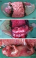
File:Canine embryo E35-38 image004.jpg ...rtilisation as bitches are ready for mating a few days prior to ovulation. Fetus located within the intact fetal membranes and placenta. The placenta is vie '''A.''' Fetus located within the intact fetal membranes and placenta. The placenta is vie(613 × 1,000 (169 KB)) - 23:24, 28 April 2011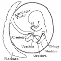
File:Windle1940 fig43.jpg .... 43. Relation of the bladder, amnion and vestigial allantois in the human fetus==(1,000 × 1,003 (119 KB)) - 11:33, 8 June 2019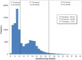
File:Gestational age distribution NIPT.jpg ...ure shows the frequency distribution by week of the gestational age of the fetus at the time testing. Percent of patients in each trimester is displayed in ...ategory:Genetic Abnormalities]] [[Category:Human Embryo]] [[Category:Human Fetus]][[Category:Molecular]][[Category:Graph]](671 × 493 (41 KB)) - 11:50, 6 November 2016File:Human week 10 fetus 04.jpg ==Human Female Fetus - Oral Cavity (10 week)== {{Human Female Fetus Week 10 gallery}}(1,600 × 1,200 (534 KB)) - 15:48, 25 May 2016
File:Keibel Mall 222.jpg ==Fig. 222 Pig Fetus==(684 × 543 (84 KB)) - 17:16, 25 February 2013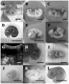
File:Control and parthenogenetic canine fetuses.jpg (A–C) The relative size and gross morphology of a control fetus on days 28, 30 and 32 of pregnancy, respectively. (H, I) A parthenogenetic fetus recovered on day 30 of pregnancy. Note that the vascular system is well dev(660 × 800 (111 KB)) - 16:32, 25 September 2012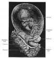
File:Gray0030.jpg ==Fig. 30. Fetus of about eight weeks, enclosed in the amnion==(600 × 644 (104 KB)) - 12:10, 22 April 2013
File:Atwell1926 plate07.jpg 22 Wax-plate reconstruction of the hypophysis from a 102-mm. human fetus (U.B.E.C., no. 17), viewed dorsally and from above. X 35. 24 R4-vonstrtwtion of tho lxypophyeis from 9. human fetus of 28.5 cm. (l.T.B.l'}.(?., no. 45), viewed from the left side. X 15.(1,000 × 1,033 (104 KB)) - 15:01, 9 November 2016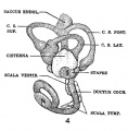
File:Streeter1917-fig04.jpg ==Fig. 4 lateral view of a model reconstructed from a human fetus 50 mm CR length== ...ces. This figure shows a lateral view of a model reconstructed from a human fetus 50 mm. CR length (Carnegie Collection, No. 84). The scala vestibuli is in t(536 × 544 (61 KB)) - 15:06, 17 September 2015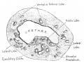
File:Lowsley1912 fig04.jpg ==Fig. 4. Human Fetus 12.5 cm==(1,483 × 1,108 (343 KB)) - 17:41, 8 November 2017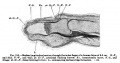
File:Keibel Mall 215.jpg ==Fig. 215 Human Fetus Finger==(727 × 379 (53 KB)) - 12:58, 24 August 2012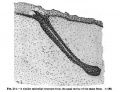
File:Keibel Mall 214.jpg ==Fig. 214 Human Fetus Integumentary Gland==(650 × 497 (56 KB)) - 12:55, 24 August 2012
File:Watson1918 fig08.jpg ...e seminal vesicles showing their relation to the deferent ducts in a human fetus 276 mm crown-rump length==(1,000 × 565 (71 KB)) - 20:10, 8 December 2016
File:Mall Meyer1921 plate07.jpg * [[:File:Mall_Meyer1921_fig87.jpg|Fig. 87. Stunted fetus with large hernia in umbilical cord. No. 1330. X0.9.]] * [[:File:Mall_Meyer1921_fig89.jpg|Fig. 89. Normal fetus with hernia of mid-brain. No. 1690. X6.75.]](929 × 1,200 (178 KB)) - 15:01, 15 January 2013
File:ZAnencephaly.jpg This is a picture of a fetus with anencephaly; a neural tube defect in which the cerebral hemispheres do(372 × 550 (16 KB)) - 12:09, 3 March 2014
File:Mall Meyer1921 plate18.jpg Fig. 187. Slight swelling of fetus from brief maceration. No. 2146. X2. Fig. 210. Normal, well-preserved cat fetus.(949 × 1,200 (220 KB)) - 18:59, 23 November 2012
File:Anson1948 fig08.jpg ==Fig. 8. Otic capsule developmental stages 205, 210, 290, 345, 345 mm Fetus and Newborn Infant== ...fetus (Wisconsi11 series 59, slide 36, section 5); (d) 345 mm. (38 week), fetus (Wisconsin series 61, slide 42, section 1); (e) newborn infant (Wisconsin s(1,280 × 1,764 (207 KB)) - 20:25, 16 October 2017
File:Keibel Mall 059-060.jpg The Marburg fetus No.21. A fetus measuring 25 mm. in its greatest length js shown from the left side and fro(1,000 × 586 (54 KB)) - 14:33, 24 March 2014
File:Galletti1770 week 10.jpg ==Fetus (week 10)==(450 × 450 (19 KB)) - 08:02, 27 May 2010
File:Keibel Mall 218.jpg ==Fig. 218 Human Fetus Nail Bed==(600 × 221 (25 KB)) - 12:59, 24 August 2012
File:Keibel Mall 217.jpg ==Fig. 217 Human Fetus Nail Bed==(600 × 198 (24 KB)) - 12:59, 24 August 2012
File:Watson1918 fig07.jpg ...e seminal vesicles showing their relation to the deferent ducts in a human fetus 178 mm crown-rump length==(1,000 × 570 (59 KB)) - 20:15, 8 December 2016
File:Watson1918 fig06.jpg ...e seminal vesicles showing their relation to the deferent ducts in a human fetus 171.4 mm crown-rump length==(1,000 × 518 (47 KB)) - 20:20, 8 December 2016
File:Kollmann052.jpg ==Fig. 52. Notochord and vertebrae of Human fetus 12 cm long, 3.5 months==(646 × 417 (36 KB)) - 14:33, 23 October 2011
File:HansonAnson1962 fig05.jpg ==Fig. 5. Fetus 17 week 133 mm== [[Category:Fetus]][[Category:Second Trimester]](1,280 × 604 (225 KB)) - 10:32, 7 January 2019
File:Mall Meyer1921 plate17.jpg Fig. 176. Fetus showing gluing of hand to face. No. 316. Fig. 180. Fetus showing continuity of epidermis across the mouth, with obliteration of the(926 × 1,200 (255 KB)) - 08:58, 13 December 2012
File:LeeHalpert1932 plate02.jpg ==Plate 2 Fetus 130 mm Gall Bladder== '''3''' Gall bladder of a 130 mm fetus; cross sections from the corpus (a) and from the neck (b). The surface patt(1,241 × 1,506 (314 KB)) - 14:12, 23 January 2019
File:HansonAnson1962 fig03.jpg ==Fig. 3. Fetus 11 week 62 mm== ...s to grow at a rate comparable to that of the malleus; as a result, in the fetus of 11 weeks the anterior process is dwarfed by this relatively large cartil(1,280 × 612 (215 KB)) - 10:21, 7 January 2019
File:Watson1918 fig04.jpg ...g the seminal vesicles and their relation to the deferent ducts in a human fetus 105 mm crown-rump length==(800 × 415 (40 KB)) - 19:37, 8 December 2016
File:Watson1918 fig05.jpg ...e seminal vesicles showing their relation to the deferent ducts in a human fetus 130 mm crown-rump length==(800 × 589 (39 KB)) - 19:40, 8 December 2016
File:Elephant ovary and uterus.jpg The uterus and ovaries of an elephant fetus at 17.5 month of gestation. The ovary on the left is still enclosed withi Ovary from a 20.2 month old elephant fetus on the left and a 6 month old calf on the right showing great shrinkage a(1,200 × 424 (61 KB)) - 11:02, 7 January 2015
File:Bast1931 plate01.jpg ==Plate 1 Model of internal ear of a 150 mm (C.R.) human fetus== ...a drawing of part of a model of the internal ear of a 150 mm (C.R.) human fetus, age about eighteen and one-half weeks. It is drawn to show the arterial bl(1,280 × 871 (113 KB)) - 07:55, 15 December 2018
File:Keibel Mall 213.jpg ==Fig. 213 Human Fetus Integumentary Gland==(670 × 547 (49 KB)) - 12:59, 24 August 2012
File:Human- fetal week 10 sagittal planes.jpg '''Human Fetus'''(600 × 250 (29 KB)) - 15:39, 27 April 2010
File:Fetal lung histology 02.jpg [[Category:Respiratory]] [[Category:Histology]] [[Category:Human Fetus]](450 × 600 (74 KB)) - 23:18, 28 February 2012
File:Gellhorn1904 fig03.jpg ==Fig. 3. Hymen of a fetus of seven months==(800 × 902 (123 KB)) - 14:48, 10 February 2017
File:McMurrich1930 fig84.jpg ...mbilical cord which is seen issuing from the ventral abdominal wall of the fetus.(1,280 × 1,819 (784 KB)) - 11:45, 20 April 2020
File:Kollmann614.jpg ==Fig. 614. Human fetus of 4 Month (10 cm CRL)== The cerebrum shows nowhere transient furrows. The fetus was immediately after the expulsion with formol 10: 100 injects, opened aft(679 × 502 (33 KB)) - 18:11, 18 October 2011
File:Keibel Mall 203-205.jpg ==Fig. 209 Human Fetus Integumentary Hair==(838 × 1,000 (129 KB)) - 13:03, 24 August 2012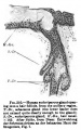
File:Keibel Mall 210.jpg ==Fig. 210 Human Fetus Integumentary Gland==(340 × 542 (41 KB)) - 13:01, 24 August 2012
File:Mouse- early conceptus.jpg Asymmetries between fetus and rest of conceptus and shape of regular tetrahedral 4-cell stage. ...to the base of the chorio-allantoic placenta (CAP). The right side of the fetus faces towards the placenta, as does the umbilical cord (UC). Its tail (FT)(661 × 311 (101 KB)) - 08:43, 5 April 2010
File:Bardeen1914-fig07.jpg ...tomach, duodenum, eaeeum, colon, rectum, bursa omentalis and rnesentery of fetus 40 mm long (No. 8, Wisconsin Collection). 5 diameters.(451 × 936 (63 KB)) - 09:53, 3 October 2017
File:Keibel Mall 2 369.jpg ==Fig. 369. — Blood-corpuscles from the vessels of a human fetus of eight months==(793 × 700 (55 KB)) - 11:07, 29 March 2014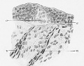
File:Keibel Mall 2 121.jpg Fig. 121. Frontal section through a papilla vallata of an 11 cm. human fetus. (After Graberg. Schwalbe's Morphol. Arbeiten, vol. 8, 1898, PI. 11, Fig 1.(600 × 475 (65 KB)) - 07:08, 3 March 2017
File:LeeHalpert1932 plate04.jpg '''5''' Gall bladder of a 370 mm fetus in longitudinal section. The active proliferation of the lining epithelium '''6''' Gall bladder of a 280 mm fetus in cross section. The coarse ramifications (plexus perimuseularis) of the(1,000 × 2,015 (271 KB)) - 14:15, 23 January 2019
File:Fetal MRI of cerebellum.png Sagittal MRI of fetal cerebellum. Gradient echo of a fetus at 33 weeks of gestation.(812 × 356 (260 KB)) - 15:27, 26 October 2017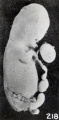
File:Mall Meyer1921 fig218.jpg ==Fig. 218. A fetus and cord showing marked maceration changes== ...gaping mouth, and locally edematous cord are also well shown in No. 797, a fetus 35 mm. long, represented in figure 218.(291 × 593 (31 KB)) - 05:22, 4 December 2012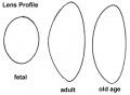
File:Gray0886.jpg Profile views of the lens at different periods of life. 1. In the fetus. 2. In adult life. 3. In old age. In the fetus, the lens is nearly spherical, and has a slightly reddish tint; it is soft(500 × 368 (17 KB)) - 22:33, 19 August 2012
File:Gellhorn1904 fig01.jpg ==Fig. 1. Hymen of a fetus of twenty-five weeks==(800 × 1,255 (187 KB)) - 06:37, 10 February 2017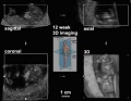
File:Ultrasound12wk 3D.jpg ==Ultrasound image 12 week Fetus showing Three Dimensional (3d) Axes== ...ategory:Genetic Abnormalities]] [[Category:Human Embryo]] [[Category:Human Fetus]](512 × 398 (18 KB)) - 09:53, 12 November 2018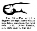
File:Keibel Mall 078.jpg Fig. 78 represents the middle finger of a fetus of 52 mm. from the side.(278 × 236 (18 KB)) - 01:55, 26 August 2012
File:Wilhelm Braune 1872 titlepage.jpg ==The Position of the Uterus and Fetus at Term (1872)== ...nach durchschnitten an Gefrornen Cadavern. (The position of the uterus and fetus end of pregnancy, according to frost-cut Cadavern)(677 × 1,000 (57 KB)) - 09:52, 31 October 2012
File:Keibel Mall 2 122.jpg Fig. 122. Frontal section through a papilla vallata of a 21.3 cm. human fetus. (After Graberg, Schwalbe's Morphol. Arbeiten, vol. 8, 1898, PI. 11, Fig. 4(532 × 477 (35 KB)) - 07:08, 3 March 2017File:Human week 10 fetus 09.jpg ==Human Female Fetus - Atlas and Axis (10 week)== {{Human Female Fetus Week 10 gallery}}(1,200 × 900 (345 KB)) - 14:54, 25 May 2016
File:Watson1918 fig03.jpg Human fetus 80.3 mm crown-rump length, estimated age 13 weeks ([[Carnegie Collection]],(800 × 409 (26 KB)) - 19:34, 8 December 2016
File:Humphrey1940 fig02.jpg ==Fig. 2. Developing olfactory and accessory olfactory formations 26 mm fetus== ...the developing olfactory and accessory olfactory formations in the 26 mm. fetus. Erythrosin and toluidin blue preparation.(1,000 × 480 (61 KB)) - 17:45, 24 October 2017
File:Kollmann608.jpg ==Fig. 608. Brain of a fetus in three monthly median section== Fig. 608. Gehirn eines 3 monatlichen Fetus im Medianschnitt.(697 × 563 (77 KB)) - 19:50, 17 October 2011
File:Gellhorn1904 fig02.jpg ==Fig. 2. Hymea of a fetus ol eight mouths==(700 × 964 (82 KB)) - 14:48, 10 February 2017
File:Arey1924 fig229.jpg ==Fig. 229. Sections of the integument from a three-months fetus==(1,000 × 625 (150 KB)) - 12:40, 23 October 2016
File:Keibel Mall 077.jpg Fig. 77 shows the sole of the right foot of a fetus of 44 mm. with its touch pads.(424 × 700 (33 KB)) - 01:54, 26 August 2012
File:Human- fetal week 10 heart ABCD.jpg '''Human Fetus'''(600 × 450 (133 KB)) - 16:10, 27 April 2010
File:Fetal week 14 head bone lateral 01.jpg [[Category:Human Fetus]] [[Category:Musculoskeletal]] [[Category:Bone]] [[Category:Head]] [[Catego(1,000 × 773 (107 KB)) - 14:53, 15 May 2013
File:Keibel Mall 2 584.jpg ==Fig. 584. Right kidney of a five months' human fetus seen from the ventral surface==(464 × 800 (68 KB)) - 09:56, 13 November 2018
File:Kollmann444.jpg Human fetus of 21 cm crown-rump length (CRL). After a fresh preparation. Menschlicher Fetus von 2i cm Kopfsteißlänge. Nach einem frischen Präparat.(600 × 472 (36 KB)) - 12:26, 20 October 2011
File:BrauneA.jpg ==The Position of the Uterus and Fetus at Term (1872)== ...tial section through the fetus shows the relative size and position of the fetus during pregnancy.(1,200 × 485 (141 KB)) - 16:09, 30 October 2012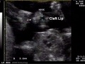
File:Cleft lip 03.jpg ...ng of the lip which may also include cleft palate. The movie above shows a fetus (at 18 weeks gestation, 20 weeks obstetric) which has a facial cleft.(585 × 439 (38 KB)) - 01:05, 18 May 2011
File:Gray0462.gif [[Category:Human Fetus]](350 × 403 (49 KB)) - 14:09, 6 July 2012
File:Fetal weight change.jpg [[Category:Human Fetus]](600 × 444 (34 KB)) - 05:08, 29 November 2011
File:Keith1902 fig029.jpg ...g. 29 The condition of the Eight and Left Doral Aortae in a 6th week human fetus==(872 × 600 (81 KB)) - 13:42, 27 January 2014
File:Kollmann631.jpg Menschlicher Fetus des 5. Monats. Von hinten gesehen.(604 × 338 (23 KB)) - 17:23, 17 October 2011File:Human week 10 fetus 11.jpg ==Human Female Fetus - Sacrum (10 week)== {{Human Female Fetus Week 10 gallery}}(1,200 × 900 (304 KB)) - 15:24, 25 May 2016
File:Anson1948 fig09.jpg ==Fig. 9. Otic capsule developmental stages 160 mm fetus== ...stapes and adjacent portions of the otic capsule in a 160 mm. (19.5 week) fetus (Wisconsin series 41); superior (cranial) views; X 7: (a) of the stapes ent(1,280 × 1,686 (235 KB)) - 20:43, 16 October 2017
File:Bardeen1914-plate01.jpg ...l and large intestines, rectum, bursa omentalis and mesentery, of :1 human fetus 27 mm. long (No. 6, Wisconsin Collection) 7 diameters. ...h, small and large intestines, rectum, bursa omentalis and mesentery of a. fetus 35 mm. long (No. 8, Wisconsin Collection). Owing to an artefact the coils i(1,347 × 2,089 (259 KB)) - 17:48, 14 November 2015
File:Kollmann684.jpg Frontalschnitt durch den Oberkiefer eines menschlichen Fetus von 18 cm Körper- dem knorpeligen Nasenseptum S. Bei dem menschlichen Fetus von 18 cm(741 × 590 (92 KB)) - 10:10, 21 October 2011
File:Fetal length and weight change.jpg [[Category:Human Fetus]] [[Category:Statistics]](600 × 444 (47 KB)) - 09:08, 6 June 2011
File:CHAOS.jpeg ...High Airway Obstruction Syndrome (CHAOS) discovered during postmortem of a fetus. The white arrow indicates tracheal atresia and hyperechoism of the lungs c ...the fetal period or not. the caption only shows that it was present in the fetus.(600 × 600 (77 KB)) - 14:43, 9 November 2014
File:Braune2 C1.jpg ==The Position of the Uterus and Fetus at Term (1872)== ...n through the maternal anatomy shows the relative size and position of the fetus during pregnancy.(3,123 × 1,200 (918 KB)) - 16:39, 30 October 2012
File:Congdon1922-1-16.jpg ...liest stage only the first arch is present, while in the last (a full-term fetus) the vessels have acquired nearly their adult form. The so-called fifth arc * Figure 16, full-term fetus.(980 × 1,000 (157 KB)) - 21:30, 6 November 2018
File:BrauneC1.jpg ==The Position of the Uterus and Fetus at Term (1872)== ...n through the maternal anatomy shows the relative size and position of the fetus during pregnancy.(1,200 × 461 (143 KB)) - 22:58, 3 June 2015
File:Anson1948 fig03.jpg ...1, section 9; (C) slide 19, section 9). Part d is from a 163 mm. (19 week) fetus (Wisconsin series 33; slide 17, section 8). Here, a is taken at the tympani The stapes of the 167 mm fetus is still wholly cartilaginous; in the capsule, on the contrary, bone is rep(1,280 × 1,815 (358 KB)) - 18:46, 18 November 2017
File:19weeklabel1.jpg Ultrasound 19 week fetus.jpg ...ble. Note "crown" to "rump" length (CRL) used in the staging/ageing of the fetus.(436 × 285 (11 KB)) - 21:16, 4 April 2010File:BGD2010-EmbryoLecture01.mp3 [[2010 BGD Lecture - Development of the Embryo/Fetus 1]](6.13 MB) - 22:45, 6 May 2010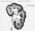
File:Keibel Mall 2 585.jpg ==Fig. 585. Frontal section through the kidney of a human fetus of the nineteenth week 175 mm in length==(1,280 × 1,144 (277 KB)) - 10:02, 13 November 2018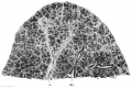
File:Keibel Mall 2 346.jpg ==Fig. 346. Section through the lower lobe of the right lung of a fetus of 100 mm vertex-breech length==(1,200 × 801 (224 KB)) - 16:50, 8 March 2014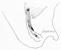
File:Lineback1920 fig03.jpg ==Fig. 3. Sagittal section of a human fetus 52 mm. CR. length==(800 × 660 (39 KB)) - 13:55, 18 November 2017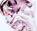
File:Fetal week 9 head lateral 01.jpg [[Category:Human Fetus]] [[Category:Musculoskeletal]] [[Category:Bone]] [[Category:Head]] [[Catego(700 × 600 (78 KB)) - 11:00, 16 May 2013
File:Streeter1917-fig08-09.jpg ==Figure 8 and 9. Lateral and Medial Views of left membranous labyrinth human fetus 130 mm== ...uction of the left membranous labyrinth and the periotic spaces in a human fetus, 130 mm. CR length (Carnegie Collection, No. 1018) enlarged 9 diameters. Th(1,200 × 853 (218 KB)) - 14:39, 17 September 2015File:2017 BGDA Lecture - Development of the Embryo-Fetus 2.pdf PDF version of 2017 lecture page: [[BGDA Lecture - Development of the Embryo/Fetus 2]](3.23 MB) - 08:44, 15 May 2017
File:Kollmann446.jpg Human fetus of 25 cm head steißlänge. Menschlicher Fetus von 25 cm Kopf steißlänge.(723 × 414 (38 KB)) - 20:42, 23 October 2011
File:Keibel Mall 2 177.jpg ==Fig. 177. Transition of the lens epithelium into the lens fibres, in a fetus at the beginning of the fourth month==(965 × 389 (48 KB)) - 11:42, 21 February 2014File:Human week 10 fetus 06.jpg ==Human Female Fetus - Midgut Herniation (10 week)== {{Human Female Fetus Week 10 gallery}}(1,200 × 900 (251 KB)) - 17:01, 25 May 2016
File:Bidloo1690 table56.jpg ==Fetus and Uterus==(698 × 1,000 (229 KB)) - 08:15, 8 November 2012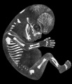
File:Mall1906 fig02.jpg [[Category:Human Fetus]][[Category:Week 10]](1,317 × 1,532 (205 KB)) - 14:37, 6 May 2018
File:Mall Meyer1921 fig19.jpg ...cle with the little nodule was somewhat larger than the one containing the fetus, although both chorionic vesicles were covered by approximately the same qu(808 × 875 (134 KB)) - 17:07, 24 November 2012
File:Placental artery 01.jpg [[Category:Human Fetus]](1,200 × 838 (371 KB)) - 07:47, 31 March 2012
File:Placental artery.jpg [[Category:Human Fetus]](600 × 509 (78 KB)) - 07:47, 31 March 2012
File:Human bilateral renal agenesis-hypoplasia-dysplasia.png ...s include maternal insulin dependent diabetes mellitus and male sex of the fetus. In the majority of cases, no specific etiology can be established, althoug A. t(2;6) case shows a left lateral photograph of this fetus with craniorachischisis and severely abnormal facial structures.(1,613 × 906 (2.75 MB)) - 10:24, 7 December 2012
File:Human- fetal week 10 sagittal plane A.jpg '''Human Fetus'''(500 × 573 (96 KB)) - 16:19, 27 April 2010
File:Mall1917 fig07.jpg Fig. 7 Group 7, giving two specimens of fetus compressus.(1,000 × 881 (100 KB)) - 20:33, 5 November 2013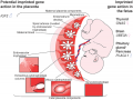
File:Placenta potential imprinted genes.png ...s only. Other genes may influence growth in utero via the placenta, or the fetus and placenta. The IUGR seen in SRS, and overgrowth in BWS are suggestive of(600 × 452 (416 KB)) - 10:19, 20 March 2018
File:Watson1918 plate03.jpg ...e section through the bla(lder, arnpullae, and seminal vesicles in a human fetus 276 mm. crown—rump length, estimated age 31 Weeks (series no. 7). This is '''24 to 27''' Transverse sections through the seminal apparatus in a human fetus at birth, crown-rump length 338 min. (series no. 8). This is the same speci(1,200 × 1,629 (320 KB)) - 09:39, 8 December 2016
File:Anson1948 fig04.jpg ==Fig. 4. Developmental stages 175 mm (20 week) fetus== Drawings (continued) of developmental stages from a 175 mm (20 week) fetus (Wisconsin series 104: (a) slide 17, section 5; (b) slide 16, section 2); x(1,280 × 1,541 (247 KB)) - 09:21, 15 October 2017
File:Kollmann054.jpg ...g. 54. Median section through the base of the skull of a 2.3 cm long human fetus beginning third month==(890 × 823 (60 KB)) - 14:28, 23 October 2011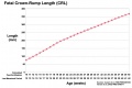
File:Fetal length change.jpg ...asurement used in embryology to more accurately stage the early embryo and fetus. Measured from the curvature at the top (crown) to the curvature at the bot [[Category:Human Fetus]] [[Category:Statistics]](972 × 648 (72 KB)) - 13:47, 26 May 2019
File:Statistics-Utah heart defects.jpg ...ategory:Genetic Abnormalities]] [[Category:Human Embryo]] [[Category:Human Fetus]](580 × 350 (35 KB)) - 12:36, 11 October 2009
File:Placental cord cross-section.jpg [[Category:Human Fetus]](525 × 525 (50 KB)) - 07:46, 31 March 2012
File:Streeter1917-fig08.jpg ==Figure 8 and 9. Lateral Views of left membranous labyrinth human fetus 130 mm== ...uction of the left membranous labyrinth and the periotic spaces in a human fetus, 130 mm. CR length (Carnegie Collection, No. 1018) enlarged 9 diameters. Th(610 × 853 (110 KB)) - 15:12, 17 September 2015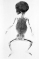
File:Fetal 5 month x-ray.jpg ==X-Ray of a Fetus at 5 Months==(1,500 × 2,298 (308 KB)) - 16:58, 23 May 2016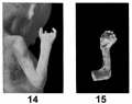
File:Mall1917 fig14-15.jpg '''Fig. 15''' Left hand which is club-shaped from a fetus compressus. No. 230, CR 57 mm., from the late Dr. J. P. West, Bellaire, Ohi(900 × 715 (51 KB)) - 07:13, 6 November 2013
File:Human- fetal week 10 lower body D.jpg '''Human Fetus'''(600 × 450 (91 KB)) - 15:36, 27 April 2010
File:Trisomy18male.jpg ...ategory:Genetic Abnormalities]] [[Category:Human Embryo]] [[Category:Human Fetus]](480 × 284 (10 KB)) - 09:53, 26 January 2010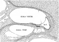
File:Streeter1917-fig01.jpg ==Fig. 1 Section through the second turn of the cochlea in a human fetus 130 mm CR length==(1,128 × 800 (298 KB)) - 13:21, 16 September 2015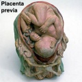
File:Galletti1770 placenta previa.jpg The fetus is shown in the normal vertex presentation, the placenta lies above the hea(450 × 450 (36 KB)) - 15:46, 19 July 2018
File:Kingsbury1932 plate02.jpg 11 Cat; 75-mm. fetus. Nearly median section of the tonsillar region, at the beginning of tonsill 12 Cat; 100—mm. fetus. Nearly median section. Early differentiation of the tonsil. Above are os b(1,280 × 1,981 (447 KB)) - 21:53, 28 March 2017
File:Kollmann755.jpg ==Fig. 755. Acoustic region in a human fetus of 22.5 cm 5th Month== bei einem menschlichen Fetus von 22,5 cm. 5. Monat. (Rekonstruktion.) Ansicht(814 × 457 (59 KB)) - 22:22, 24 October 2011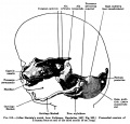
File:Keibel Mall 312.jpg Primordial cranium of a human fetus at the end of the third month (8 cm long).(704 × 677 (79 KB)) - 09:39, 25 February 2013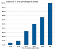
File:Drug-clearance-rates.png The drug clearance data below are only approximate calculated rates for the fetus and infant.(652 × 542 (14 KB)) - 21:26, 23 January 2019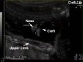
File:Cleft lip 100.jpg ...ng of the lip which may also include cleft palate. The movie above shows a fetus (at 18 weeks gestation, 20 weeks obstetric) which has a facial cleft.(585 × 438 (33 KB)) - 00:24, 18 May 2011
File:Keibel Mall 313.jpg Primordial cranium of a human fetus at the end of the third month (8 cm long).(702 × 652 (84 KB)) - 20:06, 9 February 2014
File:Lineback1920 fig02.jpg ==Fig. 2. Sagittal section of a human fetus 46 mm CR. length==(800 × 612 (34 KB)) - 23:19, 6 February 2017
File:Anson1948 fig12.jpg ==Fig. 12. Drawings of a reconstruction of the stapes in the 180 mm Fetus== Drawings of a reconstruction of the stapes in the 180 mm. (21 week) fetus (Wisconsin series 45 B); X 7: (a) the entire stapes, in superolateral view;(1,280 × 1,490 (289 KB)) - 20:52, 16 October 2017File:Human week 10 fetus 23.jpg ==Human Female Fetus - Pelvic Region (10 week)== {{Human Female Fetus Week 10 gallery}}(1,600 × 1,200 (393 KB)) - 17:21, 25 May 2016File:Human week 10 fetus 03.jpg ==Human Female Fetus - Pelvic Region (10 week)== {{Human Female Fetus Week 10 gallery}}(1,600 × 1,200 (370 KB)) - 17:21, 25 May 2016
File:Braune2 B1.jpg ==The Position of the Uterus and Fetus at Term (1872)== ...n through the maternal anatomy shows the relative size and position of the fetus during pregnancy.(1,200 × 3,263 (413 KB)) - 14:00, 30 October 2012File:Human week 10 fetus 26.jpg ==Human Female Fetus - Midgut Herniation (10 week)== {{Human Female Fetus Week 10 gallery}}(1,200 × 900 (262 KB)) - 17:03, 25 May 2016
File:Kingsbury1932 plate03.jpg 16 Cat; 100-mm. fetus. Nearly median section, caudal portion of tonsil, and fascia pharyngobasila 17 Cat; 83-mm. fetus. Nearly median section. Caudal portion of nasopharynx, cephalic aspect to t(1,280 × 1,958 (404 KB)) - 21:48, 28 March 2017
File:Human- fetal week 10 sagittal plane B.jpg '''Human Fetus'''(500 × 573 (99 KB)) - 15:49, 27 April 2010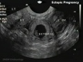
File:Ectopic 01.jpg [[Category:Ultrasound]] [[Category:Human Fetus]] [[Category:Abnormal Development]] [[Category:Ectopic Pregnancy]](655 × 491 (40 KB)) - 10:06, 19 January 2012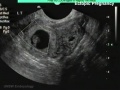
File:Ectopic 01 zoom.jpg [[Category:Ultrasound]] [[Category:Human Fetus]] [[Category:Abnormal Development]] [[Category:Ectopic Pregnancy]](655 × 491 (42 KB)) - 10:08, 19 January 2012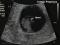
File:Ectopic 01 heart.jpg [[Category:Ultrasound]] [[Category:Human Fetus]] [[Category:Abnormal Development]] [[Category:Ectopic Pregnancy]](655 × 491 (34 KB)) - 10:09, 19 January 2012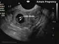
File:Ectopic 01 limbs.jpg [[Category:Ultrasound]] [[Category:Human Fetus]] [[Category:Abnormal Development]] [[Category:Ectopic Pregnancy]](655 × 491 (37 KB)) - 10:08, 19 January 2012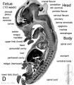
File:Human- fetal week 10 sagittal plane D.jpg ==Human Fetus Week 10==(500 × 573 (105 KB)) - 07:41, 1 May 2011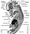
File:Human- fetal week 10 sagittal plane C.jpg '''Human Fetus'''(500 × 573 (98 KB)) - 15:48, 27 April 2010
File:Keibel Mall 314.jpg Visceral skeleton of the labyrinth of a human fetus at the end of the third month (8 cm long).(701 × 625 (89 KB)) - 09:30, 27 August 2012
File:Human- fetal week 10 urogenital A.jpg '''Human Fetus'''(600 × 450 (109 KB)) - 15:52, 27 April 2010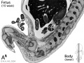
File:Human- fetal week 10 lower body A.jpg '''Human Fetus'''(600 × 450 (96 KB)) - 15:52, 27 April 2010
File:Fetal week 10 hard palate 01.jpg [[Category:Human Fetus]] [[Category:Musculoskeletal]] [[Category:Bone]] [[Category:Head]] [[Catego(800 × 532 (77 KB)) - 11:26, 16 May 2013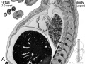
File:Human- fetal week 10 upper body A.jpg '''Human Fetus'''(600 × 450 (104 KB)) - 15:53, 27 April 2010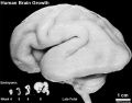
File:Human brain growth 01.jpg [[Category:Human Embryo]][[Category:Neural]][[Category:Human Fetus]](1,022 × 800 (119 KB)) - 09:30, 9 February 2016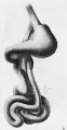
File:Bardeen1914-fig01.jpg ==Fig. 1. Ventral view of of Human Fetus 27 mm long==(439 × 853 (51 KB)) - 08:25, 29 April 2017
File:Watson1918 fig02.jpg .... 2 Transverse section through the right and left Wolffian ducts in a human fetus 80.3 mm crown-rump length==(1,378 × 841 (449 KB)) - 09:56, 8 December 2016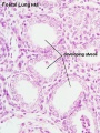
File:Fetal lung histology.jpg [[Category:Respiratory]] [[Category:Histology]] [[Category:Human Fetus]](450 × 600 (83 KB)) - 22:42, 28 February 2012
File:Wyburn1939-plate05.jpg ==Plate V. Human Fetus 60 mm==(1,727 × 2,500 (789 KB)) - 09:38, 15 September 2015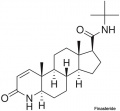
File:Finasteride.jpg ...a potential endocrine disruptor, exposed pregnant women can impact on male fetus genetial development.(500 × 463 (18 KB)) - 14:45, 25 March 2019
File:Hertig1946b fig15b.jpg ==Fig. 15B. Human 14 Week Fetus Yolk Sac==(800 × 508 (87 KB)) - 08:57, 8 August 2017
File:Human- fetal week 10 upper body D.jpg '''Human Fetus'''(600 × 450 (106 KB)) - 15:42, 27 April 2010
File:Boyd1950 fig10.jpg {{Online Editor}} - [[Fetal Development|Fetus]] 145mm is about 16 weeks ({{GA}} 18 weeks).(1,000 × 453 (114 KB)) - 08:29, 21 March 2017
File:Bardeen1914-fig02.jpg ==Fig. 2. Lateral view of Human Fetus 27 mm long==(624 × 861 (54 KB)) - 08:26, 29 April 2017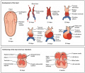
File:Progressive development of the Embryonic Heart.jpeg Development of the heart in the fetus and partitioning of the heart into four chambers(1,024 × 883 (154 KB)) - 11:30, 4 September 2018
File:Mall1917 fig10.jpg Figure 10 a and 10 b show an embryo and a fetus with hare lip.(1,000 × 507 (62 KB)) - 12:53, 22 April 2014
File:Human- fetal week 10 urogenital B.jpg '''Human Fetus'''(600 × 450 (109 KB)) - 15:49, 27 April 2010
File:Human- fetal week 10 lower body B.jpg '''Human Fetus'''(600 × 450 (93 KB)) - 15:51, 27 April 2010
File:Keibel Mall 2 123-125.jpg ==Figs. 123-125. The summits of three taste-buds from a human fetus of 21.3 cm==(1,020 × 520 (61 KB)) - 07:22, 3 March 2017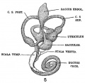
File:Streeter1917-fig05.jpg ==Fig. 4 Medial view of a model reconstructed from a human fetus 50 mm CR length==(556 × 544 (64 KB)) - 15:06, 17 September 2015
File:Kollmann687.jpg die Muscheln betrifft, bei dem Fetus komplizierter gebaut als bei dem Erwach-(618 × 398 (32 KB)) - 10:12, 21 October 2011
File:Human- fetal week 10 upper body B.jpg '''Human Fetus'''(600 × 450 (105 KB)) - 15:51, 27 April 2010
File:Kollmann766-769.jpg Fig. 767. Linkes Ohr eines 6 Monate alten menschlichen Fetus. Fig. 768. Ohrmuschel eines menschlichen Fetus vom 6. Monat.(694 × 311 (25 KB)) - 12:42, 21 October 2011
File:Human- fetal week 10 head A.jpg ==Human Fetus (week 10) Fetal==(600 × 544 (113 KB)) - 21:22, 29 May 2011
File:Streeter1922-fig07.jpg ==Fig. 7. Reconstruction of left auricular cartilage of a 50 mm fetus==(584 × 794 (54 KB)) - 13:49, 22 November 2017
File:Human- fetal week 10 lower body C.jpg '''Human Fetus'''(600 × 450 (94 KB)) - 15:47, 27 April 2010
File:Human- fetal week 10 head C.jpg '''Human Fetus'''(600 × 544 (118 KB)) - 15:47, 27 April 2010
File:Kollmann686.jpg von einem menschlichen Fetus des 4. Monats. Von der Seite und etwas von(833 × 606 (102 KB)) - 10:11, 21 October 2011
File:Fetal 10wk urogenital 1.jpg ==Human Fetus (week 10) Female==(800 × 600 (109 KB)) - 21:52, 8 July 2012
File:Kollmann615.jpg ==Fig. 615. Base of the brain of a human fetus from the beginning of 4th Month== The rhinencephalon has developed remarkably in the human fetus at this age. You can see the two olfactory Bulbis with short, almost withou(660 × 496 (35 KB)) - 18:09, 18 October 2011
File:Simkins1928 plate08.jpg 37 Detail of a seminiferous tubule of a 154—mm. fetus. X 750.(1,558 × 1,797 (220 KB)) - 16:57, 31 January 2018
File:Placental cord epithelium 01.jpg [[Category:Human Fetus]](1,200 × 805 (130 KB)) - 14:22, 20 November 2012
File:Human- fetal week 10 upper body C.jpg '''Human Fetus'''(600 × 450 (109 KB)) - 15:46, 27 April 2010
File:Fetal week 10 hard palate 03.jpg [[Category:Human Fetus]] [[Category:Musculoskeletal]] [[Category:Bone]] [[Category:Head]] [[Catego(600 × 450 (122 KB)) - 11:34, 16 May 2013File:Fetal heartbeat 01.mp3 ==Human Fetus (17 week) heart beat==(225 KB) - 18:16, 9 August 2010
File:Fetal head section 04.jpg [[Category:Human Fetus]] [[Category:Week 12]] [[Category:Tongue]] [[Category:Taste]](1,200 × 821 (188 KB)) - 17:52, 18 March 2012
File:Fetal lung histology 01.jpg [[Category:Respiratory]] [[Category:Histology]] [[Category:Human Fetus]](1,280 × 1,024 (339 KB)) - 22:43, 28 February 2012
File:Fetal 10wk urogenital 4.jpg ==Human Fetus (week 10) Female==(800 × 600 (105 KB)) - 17:58, 28 May 2011
File:Human- fetal week 10 urogenital D.jpg ==Human Fetus (10 week) Female==(600 × 450 (101 KB)) - 17:53, 28 May 2011
File:Human- fetal week 10 head D.jpg ==Human Fetus (week 10) Fetal==(600 × 544 (111 KB)) - 14:50, 23 April 2013
File:Human- fetal week 10 head B.jpg ==Human Fetus (week 10) Fetal==(600 × 544 (66 KB)) - 21:22, 29 May 2011
File:Mall Meyer1921 fig37.jpg The specimen consists of a rather poorly preserved young fetus, measuring 16.5 mm. CR. The ventral abdominal wall had been seriously injur(398 × 665 (57 KB)) - 07:11, 4 December 2012
File:Prentiss 286.jpg ==Fig. 286 Diagrammatic outline of the organs of circulation in the fetus of six months== Diagrammatic outline of the organs of circulation in the fetus of ail months (Allen Thomson). RA, Right atrium of the heart; RV, right ve(799 × 933 (121 KB)) - 02:59, 28 September 2012
File:Fetal 10wk urogenital 2.jpg ==Human Fetus (week 10) Female==(800 × 600 (110 KB)) - 21:17, 29 May 2011
File:Human- fetal week 10 head A1.jpg ==Human Fetus==(1,200 × 1,088 (159 KB)) - 06:58, 7 October 2010
File:Process of amniocentesis.jpeg ...luid, as well as the use of ultrasound to guide it and prevent harm to the fetus.(3,648 × 2,432 (2.14 MB)) - 10:25, 24 September 2010
File:Kollmann720.jpg sehen. Die Iris ist nach dem frischen Präparat eines menschlichen Fetus vom(775 × 579 (84 KB)) - 10:51, 21 October 2011
File:Fetal 10wk urogenital 3.jpg ==Human Fetus (week 10) Female==(800 × 600 (107 KB)) - 21:17, 29 May 2011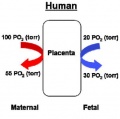
File:Placenta oxygen exchange levels.jpg ...gen tension on the maternal and fetal sides of the placenta. Note that the fetus has a fetal haemoglobin that is able to exchange oxygen and carbon dioxide(404 × 401 (18 KB)) - 12:31, 28 August 2014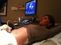
File:ZUltrasound Exam.JPG ...of a pregnant woman having an ultrasound examination to screen the growing fetus for any overt structural abnormalities. The gel applied to her abdomen allo(1,600 × 1,200 (846 KB)) - 14:28, 19 August 2014
File:Rubella virus.jpg ...enital rubella syndrome (CRS) with serious malformations of the developing fetus. The type and degree of abnormality relates to the time of maternal infecti(210 × 158 (5 KB)) - 07:34, 7 February 2016File:Week17 fetal heart rate.mp4 ==Human Fetus (17 week) heart beat==(398 KB) - 10:31, 29 April 2013
File:Streeter1917-fig03.jpg ...3 Detail of the section through the second turn of the cochlea in a human fetus 130 mm CR length==(1,000 × 420 (173 KB)) - 14:23, 17 September 2015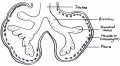
File:Lung historical image.PNG ...rther branching and mesenchymal differentation will for the alveoli in the fetus.(856 × 472 (326 KB)) - 14:53, 9 November 2014
File:Fetal valproate syndrome 01.jpg ...al Development]] [[Category:Environmental Abnormalities]] [[Category:Human Fetus]](544 × 408 (28 KB)) - 17:50, 24 November 2014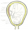
File:Arey1924 fig055.jpg ==Fig. 55. Section of the uterus, illustrating the relation of an advanced fetus to the placenta and membranes==(1,280 × 1,360 (244 KB)) - 17:47, 4 January 2017
File:Mall Meyer1921 plate05.jpg Figs. 42-50. Various forms of cyemata classed as fetus compressus: Nos. 1463 (X2), 1806 (X 1.5), 1301 (X8.5), 1513 (X0.58), 1495c .... Illustrating the absence of fundamental differences between the group of fetus compressus and grade 3 of the normal specimens. No. 12956 (fig. 52) patholo(924 × 1,200 (255 KB)) - 09:51, 6 December 2012
File:Mice model and limb development.gif ...the humerus (blue-stained area labeled H) is visible only in the wild-type fetus (C,D) and obscures the scapula in the ventral view (D).(270 × 440 (51 KB)) - 17:01, 15 October 2016
File:Tubal pregnancy historic.jpg :This historic drawing uses artistic license by depicting a fetus within the tube.(350 × 287 (12 KB)) - 10:44, 25 October 2016
File:LeeHalpert1932 plate03.jpg '''4''' {{Gallbladder}} of a 200 mm fetus in longitudinal section.(1,242 × 2,066 (251 KB)) - 14:15, 23 January 2019
File:Kollmann618.jpg ==Fig. 618. Brain of a human fetus of 6 months== Fig. 618. Gehirn eines menschlichen Fetus vom 6« Monat(940 × 592 (50 KB)) - 18:00, 18 October 2011
File:Kollmann717.jpg Schnitt durch das Auge eines viermonatKchen menschlichen Fetus in der Augenachse.(514 × 292 (29 KB)) - 10:50, 21 October 2011
File:Stages of development.png ...stages of development are where abnormalities may arise in the developing fetus; the stages are, 1. Proliferation or growth stage; 2. Migration stage; 3. O(912 × 597 (495 KB)) - 10:06, 26 October 2017
File:Gray0502.jpg The fetal blood is returned from the placenta to the fetus by the umbilical vein. This vein enters the abdomen at the umbilicus, and p ...t ventricle, and thence passes into the pulmonary artery. The lungs of the fetus being inactive, only a small quantity of the blood of the pulmonary artery(1,000 × 1,329 (215 KB)) - 21:34, 9 June 2014
File:Cullen1916 fig28.jpg ==Fig. 28. The Umbilical Region in a Fetus about Five Months Old Viewed from the Left==(1,000 × 882 (141 KB)) - 14:16, 28 October 2018
File:Fetal limb X-ray-01.jpg Normal fetus the limbs are properly positioned and no deformities are present.(685 × 931 (57 KB)) - 07:55, 25 May 2018
File:Barr body.JPG ...r body is able to be seen under the microscope, and therefore the sex of a fetus can be found through analysis of the amniotic fluid.(3,648 × 2,432 (2.01 MB)) - 11:52, 12 September 2010
File:Kollmann619.jpg ==Fig. 619. Base of the brain of a human fetus from the beginning of the 6th month== Fig. 619. Hirnbasis eines menschlichen t Fetus vom Anfang des 6. Monats.(750 × 639 (51 KB)) - 17:57, 18 October 2011
File:Infant rubella virus.jpg ...enital rubella syndrome (CRS) with serious malformations of the developing fetus. The type and degree of abnormality relates to the time of maternal infecti(564 × 374 (66 KB)) - 14:56, 1 February 2015
File:Gray0034.jpg [[Category:Fetal]] [[Category:Human Fetus]] [[Category:Placenta]](800 × 613 (98 KB)) - 11:09, 17 August 2017
File:Preterm fetal-infant growth chart.jpg ...ated growth chart allows a comparison of an infant's growth first with the fetus as early as 22 weeks and then with the term infant to 10 weeks. Comparison [[Category:Human Fetus]] [[Category:Birth]] [[Category:Preterm Birth]] [[Category:Statistics]] [[C(1,000 × 1,361 (267 KB)) - 12:23, 30 June 2011
File:Arey1924 fig232.jpg ...itudinal section through a developing hair from a five and one-half months fetus==(1,200 × 928 (173 KB)) - 12:59, 23 October 2016
File:Simkins1928 plate06.jpg 25 Cross-section of the ovary of a 90~mm. fetus. X 200.(1,587 × 2,088 (277 KB)) - 16:52, 31 January 2018
File:WHO - The global action report on preterm birth.jpg [[Category:Human Fetus]] [[Category:Birth]] [[Category:Preterm Birth]](150 × 194 (5 KB)) - 08:55, 26 May 2019
File:Fetal week 9 hard palate fusion 01.jpg [[Category:Human Fetus]] [[Category:Palate]][[Category:Musculoskeletal]] [[Category:Bone]] [[Categ(661 × 400 (51 KB)) - 11:01, 20 August 2018
File:Apoptotic trophoblast.jpg ...ophoblast and some erythroblast, will undergo apoptosis and the DNA of the fetus will be fragmented. Eventually, they will be able to distribute and migrate(1,932 × 1,002 (517 KB)) - 21:18, 23 July 2014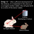
File:Crabbit 4.jpg ...nto the uterine wall of a surrogate mother rabbit where it develops into a fetus. The surrogate mother then gives birth to a clone of the donor rabbit. The(700 × 700 (76 KB)) - 18:06, 11 October 2009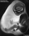
File:Fetal polyhydramnios MRI-01.jpg Fetus ({{GA}} week 33) normal anatomy in polyhydramnios ([[International Classifi(768 × 932 (60 KB)) - 10:50, 25 July 2016
File:Mall Meyer1921 fig212.jpg ==Fig. 212. Appearance of fetus before fixation==(400 × 564 (38 KB)) - 05:31, 4 December 2012
File:Newborn - vernix caseosa.jpg Vernix caseosa forms on the fetus by the developing epidermis shedding the periderm cell layer and then mixed(800 × 463 (73 KB)) - 18:40, 25 May 2016
File:Streeter1917-fig06.jpg ...uction of the left membranous labyrinth and the periotic spaces in a human fetus 85 mm CR length==(539 × 749 (93 KB)) - 14:49, 17 September 2015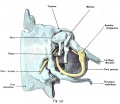
File:Kollmann757.jpg ==Fig. 757. Middle ear of a human fetus 8 cm CRL== Fig. 757. Mitteiohrraum^ Cavum tympani emes menschlichen Fetus(807 × 697 (98 KB)) - 22:19, 24 October 2011
File:Early placental structure.jpg '''(A)''' Structure and orientation of fetus and placenta in uterus at ~6 weeks of gestation. Fetal structures are repre ...xpression decreases as VE-cadherin expression rises in distal (relative to fetus) extravillous cytotrophoblasts.(792 × 367 (90 KB)) - 13:46, 18 March 2014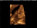
File:ZFetus yawning.jpg This is a 3D ultrasound image showing a fetus yawning.(500 × 383 (35 KB)) - 12:38, 4 February 2011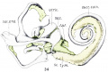
File:Bast1933 fig34.jpg ==Fig. 34. Human Fetus CRL 50 mm==(1,000 × 677 (73 KB)) - 13:38, 13 December 2018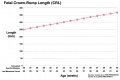
File:Third trimester Crown-Rump Length graph.jpg ...asurement used in embryology to more accurately stage the early embryo and fetus. Measured from the curvature at the top (crown) to the curvature at the bot(972 × 648 (59 KB)) - 10:06, 15 June 2016
File:Haemoglobin comparison oxygen saturation curve.png Remember that overall oxygen tension in the fetus is lower than that found in maternal arterial blood.(639 × 541 (15 KB)) - 01:28, 18 August 2010
File:Streeter1917-fig02.jpg ==Fig. 2. Section through the cochlea in a human fetus 130 mm. CR length==(1,000 × 847 (276 KB)) - 13:20, 16 September 2015
File:Streeter027.jpg ==Fig. 27. Median view of a model reconstructed from a human fetus 50 mm crown-rump length== Median view of a model reconstructed from a human fetus 50 mm. crown-rump length (Carnegie Collection, No. 84).(774 × 1,000 (51 KB)) - 17:31, 18 May 2015
File:Mall Meyer1921 fig132.jpg Fig. 133. Marked macerated and deformed fetus from the same case. X4.(840 × 838 (168 KB)) - 09:12, 6 December 2012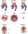
File:Aortic arch and ductus arteriosus.jpg ...red) from the placenta passes though the aorta. Oxygen-poor blood from the fetus (in blue) passes the DA entering the descending aorta (in purple).(600 × 720 (68 KB)) - 12:45, 20 October 2011
File:Kollmann620.jpg ==Fig. 620. human fetus from the end of 7th month== Fig. 620. Circumvolutiones pallii eines menschlichen Fetus vom Ende des(772 × 613 (50 KB)) - 17:57, 18 October 2011
File:Z3DCleft Lip Picture.jpg This is a 3D ultrasound image showing a fetus with a cleft lip. The 3D rendering enables the abnormality to be clearly vi(640 × 480 (32 KB)) - 14:27, 19 August 2014
File:Keibel Mall 2 583.jpg ==Fig. 583. Frontal section of the kidney of a human fetus of 160 mm==(1,280 × 1,044 (259 KB)) - 09:58, 13 November 2018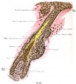
File:Kollmann666.jpg ==Fig. 666. Development of the wool hair (lanugo) in a human fetus of 5.5 months== bei einem menschlichen Fetus von 5 ^/2 Monaten. (Rückenhaut.) 460 mal vergr.(897 × 1,000 (140 KB)) - 06:01, 22 October 2011
File:Wallaby embryo 02.jpg ==Tammar Wallaby Embryo and Fetus==(1,138 × 986 (89 KB)) - 11:02, 29 July 2019
File:Placental villi 3.jpg [[Category:Human Fetus]] [[Category:Third Trimester]][[Category:Placenta]] [[Category:Histology]](1,280 × 1,024 (99 KB)) - 20:44, 8 October 2015
File:Placental villi 4.jpg [[Category:Human Fetus]][[Category:Third Trimester]] [[Category:Placenta]] [[Category:Histology]](1,280 × 1,024 (89 KB)) - 20:43, 8 October 2015
File:Simkins1928 plate02.jpg 7 Cross—section of the ovary of a 43-mm. fetus. X 200.(1,464 × 2,126 (223 KB)) - 16:43, 31 January 2018
File:Kollmann622.jpg ==Fig. 622. fetus from the end of 7th month== Fig. 622. Circumvolutiooes pallii eines menschlicheo Fetus vom Ende des(712 × 617 (46 KB)) - 17:48, 18 October 2011
File:Arey1924 fig231.jpg ==Fig. 231. Section through the facial integument of a three-months fetus==(1,200 × 748 (188 KB)) - 12:55, 23 October 2016
File:Fetal cardiac ultrasound 01.jpg ...standardization of fetal position in the STIC volume dataset to place the fetus in the exact dorsal supine position, navigating systematically in the volum(900 × 600 (88 KB)) - 11:58, 10 May 2015
File:Fetal temporomandibular joint 06.jpg Human fetus (300 mm GL; 32th week of development). SEM (920). Synovial lining cell (A(600 × 389 (50 KB)) - 11:49, 3 July 2015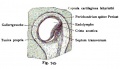
File:Kollmann745.jpg Fig. 745. Ampulle eines measchliclien Fetus(588 × 338 (43 KB)) - 12:32, 21 October 2011File:Dog fetus day 28.jpg ==Dog Fetus day 28==(400 × 375 (24 KB)) - 16:38, 25 September 2012File:Dog fetus day 30.jpg ==Dog Fetus day 28==(400 × 375 (32 KB)) - 16:39, 25 September 2012File:Dog fetus day 32.jpg ==Dog Fetus day 28==(400 × 375 (28 KB)) - 16:40, 25 September 2012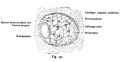
File:Kollmann747.jpg Menschlicher Fetus aus dem Anfang des 3. Monats.(753 × 388 (44 KB)) - 12:33, 21 October 2011
File:Mckay1955 plate03.jpg Fetus no. 5, 57 mm. Trophoblast. X 150(1,280 × 1,912 (367 KB)) - 16:04, 5 March 2017
File:Streeter026.jpg ==Fig. 26. Lateral view of a model reconstructed from a human fetus 50 mm crown-rump length== Lateral view of a model reconstructed from a human fetus 50 mm. crown-rump length (Carnegie Collection, No. 84).(774 × 1,000 (45 KB)) - 17:31, 18 May 2015
File:Kingsbury1932 fig1.jpg ...midiagrammatic) made from a. series through the pharyngeal tonsil of a cat fetus, 100-mm. length, cut in a plane parallel to the surface. Arteries cross-mar(1,000 × 1,047 (113 KB)) - 22:05, 28 March 2017
File:Abnormal81-92-neuron.png ...ategory:Genetic Abnormalities]] [[Category:Human Embryo]] [[Category:Human Fetus]] [[Category:Neural]](481 × 344 (9 KB)) - 17:09, 10 July 2012
File:Abnormal81-92-heart.png ...ategory:Genetic Abnormalities]] [[Category:Human Embryo]] [[Category:Human Fetus]](481 × 344 (6 KB)) - 23:16, 24 October 2013
File:Mall Meyer1921 plate23.jpg ...us (No. 1151), showing disproportion between the chorionic vesicle and the fetus, sparse development of the villi, and some hydatiform degeneration. Xl.5.(948 × 1,200 (279 KB)) - 09:45, 6 December 2012
File:MRI confirming renal agenesis.jpg ...k of gestation from a coronal frame from the dorsal to ventral side of the fetus showing from a-h.(600 × 1,301 (251 KB)) - 10:31, 18 December 2018
File:Macklin1914 plate03.jpg ==Plate 3. Wax plate reconstruction of the chondrocranium of a 40mm human fetus==(2,095 × 2,550 (597 KB)) - 22:22, 20 June 2016
File:Fetal temporomandibular joint 05.jpg Human fetus (233 mm GL; 24th week of development). SEM (198).(600 × 392 (71 KB)) - 10:13, 18 February 2015
File:Wallaby embryo 03.jpg ==Tammar Wallaby Embryo and Fetus==(1,000 × 986 (79 KB)) - 13:17, 23 May 2012
File:Kollmann760.jpg , / bei dem reifen Fetus einer Maus.(719 × 641 (85 KB)) - 12:41, 21 October 2011
File:Fetal cardiac state diagram 01.jpg * a - a cardiac state diagram CSD of septum from a healthy fetus at 36 weeks of gestation(1,200 × 321 (29 KB)) - 11:22, 28 August 2014
File:Figure 3. Fetal Lip and Primary Palate Three dimensional versus Two-dimensional US.gif ...al rendered frontal oblique US image shows a median cleft lip (arrow) in a fetus at 32 weeks gestational age.(354 × 440 (126 KB)) - 21:44, 8 October 2011
File:Kollmann752.jpg ==Fig. 752. Another embodiment of the auditory organs in a human fetus of 8 cm CRL== Fetus von 8 cm Kopfsteiftlänge.(751 × 415 (67 KB)) - 22:38, 24 October 2011
File:Placenta-maternal neuroendocrine links.jpg ...ology and postnatal maternalism to synchronize with the development of the fetus.(1,280 × 699 (82 KB)) - 09:06, 22 December 2017
File:Model male androsterone synthesis.jpg ==Proposed steroidogenic pathways in the second trimester human male fetus== ...rosterone synthesis and masculinization in the second trimester human male fetus.(740 × 518 (92 KB)) - 13:06, 1 June 2019
File:Human fetal temporal bone and mandible 01.jpg [[Category:Human Fetus]] [[Category:Musculoskeletal]] [[Category:Bone]] [[Category:Head]] [[Categ(1,200 × 805 (170 KB)) - 17:14, 24 July 2017
File:Jimenez-Castellanos1949 fig06.jpg Photomicrograph through a human fetus of 40 mm. at the middle level indicated in fig. 3. ac 15. (1) Suprarenal gl(724 × 527 (111 KB)) - 04:09, 17 August 2017
File:Cullen1916 fig33.jpg ==Fig. 33. A Diagrammatic Representation of the Umbilical Region of a Fetus at Term==(1,154 × 2,052 (422 KB)) - 22:14, 28 October 2018
File:Streeter028.jpg ...teral view of left membranous labyrinth and the periotic spaces in a human fetus 85 mm CRL== ...uction of the left membranous labyrinth and the periotic spaces in a human fetus 85 mm. crown-rump length (Carnegie Collection, No. 1400-30), enlarged 11.4(774 × 1,000 (69 KB)) - 17:32, 18 May 2015
File:Gray0027.jpg ...ing this sac is drawn ventrally to enclose (cover) the entire embryo, then fetus. The presence of this membane led to the description of reptiles, bird, and(500 × 500 (39 KB)) - 12:06, 25 February 2014
File:Abnormal AusData81-92Graph.png ...ategory:Genetic Abnormalities]] [[Category:Human Embryo]] [[Category:Human Fetus]](509 × 320 (7 KB)) - 10:42, 10 July 2012
File:Streeter014-19.jpg Section through the lateral semicircular canal in a human fetus 46 mm. crown-rump length (Carnegie Collection, No. 9.5, slide 72, section 1 Section thiough the posterior semicircular canal in a human fetus 50 mm. crown-rump length (Carnegie Collection, No. 184, shde 23). The .sect(640 × 800 (199 KB)) - 02:46, 15 February 2011
File:Canine embryo E35-38 image001.jpg ...rtilisation as bitches are ready for mating a few days prior to ovulation. Fetus located within the intact fetal membranes and placenta.(1,200 × 635 (199 KB)) - 14:27, 23 August 2016
File:Canine embryo E35-38 image002.jpg ...rtilisation as bitches are ready for mating a few days prior to ovulation. Fetus located within the intact fetal membranes and placenta. The placenta is vie(1,000 × 774 (210 KB)) - 15:58, 12 April 2011
File:Canine embryo E35-38 image003.jpg ...rtilisation as bitches are ready for mating a few days prior to ovulation. Fetus located within the intact fetal membranes and placenta. The placenta is vie(1,000 × 750 (192 KB)) - 14:32, 5 July 2018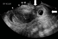
File:Cervical ectopic ultrasound.jpg [[Category:Ultrasound]] [[Category:Human Fetus]] [[Category:Abnormal Development]] [[Category:Ectopic Pregnancy]](800 × 541 (62 KB)) - 12:30, 1 June 2019
File:Streeter030.jpg ...teral view of left membranous labyrinth and the periotic spaces in a human fetus 130 mm CRL== ...uction of the left membranous labyrinth and the periotic spaces in a human fetus 130 mm. crown-rump length (Carnegie Collection, No. 1018), enlarged 11.4 di(774 × 1,000 (78 KB)) - 17:33, 18 May 2015
File:Abnormal AusData81-92.png ...ategory:Genetic Abnormalities]] [[Category:Human Embryo]] [[Category:Human Fetus]] [[Category:Graph]](523 × 358 (10 KB)) - 13:31, 3 April 2019
File:Mouse palatal rugae pattern E16.jpg Figure shows the mouse ruga pattern in the adult and the fetus shown by in situ hybridization against [[Developmental Signals - Sonic hedg In ED16.0 fetus, Shh gene expression pattern (as seen by whole-mount in situ hybridization)(569 × 700 (56 KB)) - 13:09, 5 February 2016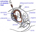
File:Amniocentesis.jpg ...ategory:Genetic Abnormalities]] [[Category:Human Embryo]] [[Category:Human Fetus]](284 × 250 (31 KB)) - 13:52, 28 April 2012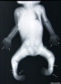
File:Fetal limb abnormalities X-ray-02.jpg Fetus with limb deformities including bowing of the digits, bowing and hypoplasia(685 × 931 (62 KB)) - 09:34, 1 November 2018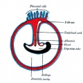
File:Gray0028.jpg ...(placental cord) The placental cord is the structure connecting the embryo/fetus to the placenta. It is initially extra-embryonic mesoderm forming the conne(500 × 500 (50 KB)) - 12:08, 25 February 2014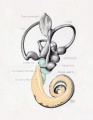
File:Streeter029.jpg ...edian view of left membranous labyrinth and the periotic spaces in a human fetus 85 mm CRL== ...uction of the left membranous labyrinth and the periotic spaces in a human fetus 85 mm. crown-rump length (Carnegie Collection, No. 1400-30), enlarged 11.4(774 × 1,000 (74 KB)) - 17:32, 18 May 2015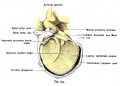
File:Kollmann754.jpg ==Fig. 754. Auditory meatus externus from a human fetus 22.5 cm (5th month)== Fig. 754. Äußerer Qehörgans, Meatus acusticus extemus von einem menschlichen Fetus von 22,5 cm.(802 × 577 (71 KB)) - 16:53, 12 July 2016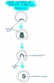
File:Cross section of genital tubercle male.jpg ...tudent drawn image of the cross sections of the genital tubercle in a male fetus, as it forms the tubular urethra within the penis.(576 × 886 (84 KB)) - 22:52, 8 November 2014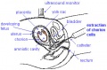
File:Cvs.jpg ...ategory:Genetic Abnormalities]] [[Category:Human Embryo]] [[Category:Human Fetus]](336 × 222 (28 KB)) - 14:46, 19 August 2014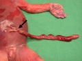
File:Placenta- umbilical cord torsion.jpg [[Category:Human]] [[Category:Human Fetus]] [[Category:Abnormal Development]] [[Category:Placenta]](397 × 298 (29 KB)) - 10:45, 27 August 2014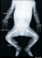
File:Fetal limb abnormalities X-ray-03.jpg Fetus with limb deformities (indicated by arrows), which include bowing of the ra(685 × 931 (57 KB)) - 12:33, 1 May 2016
File:Placenta and explant model.jpg (A) Structure and orientation of fetus and placenta in uterus at ~6 weeks of gestation. Fetal structures are repre ...xpression decreases as VE-cadherin expression rises in distal (relative to fetus) extravillous cytotrophoblasts.(800 × 682 (153 KB)) - 09:18, 11 May 2013
File:Mouse-E17.5 foot.jpg Skeletal preparations of E17.5 wild-type mouse fetus stained with alcian blue for cartilage and alizarin red for bone.(385 × 500 (29 KB)) - 10:20, 30 September 2010
File:Fetal head lateral.jpg [[Category:Human Fetus]] [[Category:Musculoskeletal]] [[Category:Bone]] [[Category:Cartilage]] [[C(632 × 447 (34 KB)) - 08:05, 13 November 2018
File:Neural-development.jpg [[Category:Neural]] [[Category:Human Fetus]] [[Category:Glia]] [[Category:Timeline]](800 × 535 (66 KB)) - 14:30, 27 May 2019
File:Complete hydatidiform mole 02.jpg ... × 15 × 10 cm) with many small vesicles, no normal gestational sac, and no fetus.(748 × 560 (60 KB)) - 14:32, 9 June 2015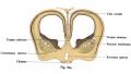
File:Kollmann624.jpg Fig. 624. Frootalschnitt durch das Gehirn efaies menschlichen Fetus vom(733 × 413 (52 KB)) - 17:17, 17 October 2011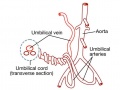
File:Placental cord vessels 01.jpg Vascular pattern in a normal fetus.(650 × 488 (51 KB)) - 11:45, 20 August 2018
File:Trisomy21arrow.gif ...ategory:Genetic Abnormalities]] [[Category:Human Embryo]] [[Category:Human Fetus]](200 × 175 (5 KB)) - 13:21, 5 August 2009
File:Streeter031.jpg ...edian view of left membranous labyrinth and the periotic spaces in a human fetus 130 mm CRL== ...uction of the left membranous labyrinth and the periotic spaces in a human fetus 130 mm. crown-rump length (Carnegie Collection, No. 1018), enlarged 11.4 di(774 × 1,000 (79 KB)) - 17:34, 18 May 2015
File:Mckay1956 plate01.jpg Fetus 33, 6mm, X 80 (green filter).(1,280 × 1,970 (377 KB)) - 15:11, 19 June 2017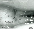
File:Fetal pineal gland 01.jpg ...rsal) view of the diencephalic-mesencephalic area of a 3.5-month-old human fetus.(700 × 603 (67 KB)) - 09:35, 4 September 2018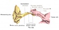
File:Kollmann753.jpg ==Fig. 753. Tubo-tympanic recess and external ear of a human fetus of 31 mm CRL== von einem menschlichen Fetus von 31 mm Scheitelsteißlänge. (Mitte des 3. Monats.)(734 × 380 (41 KB)) - 22:35, 24 October 2011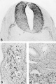
File:Mckay1955 plate02.jpg Fetus no. 5, 57mm. X 1.50(1,280 × 1,916 (464 KB)) - 16:06, 5 March 2017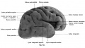
File:Kollmann621.jpg ==Fig. 621. Right hemisphere human Fetus from the end of the 7th month== Fetus vom Ende des 7. Monats.(976 × 550 (49 KB)) - 17:58, 18 October 2011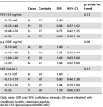
File:POPs and risk of hypospadias.jpg ...-DDE and HCB possess the potential to alter hormonal levels and affect the fetus, resulting hypospadias.(452 × 473 (224 KB)) - 22:59, 8 November 2014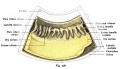
File:Kollmann726.jpg Segment aus dem vorderen Umfang des Bulbus eines menschlichen Fetus aus der(758 × 435 (68 KB)) - 10:54, 21 October 2011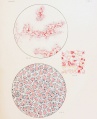
File:Wislocki1920 plate 3.jpg Fig. 12. Section through placenta of guinea-pig fetus measuring 40 mm., showing a vitally stained macrophage 26 hours after injec(975 × 1,200 (219 KB)) - 10:46, 16 June 2013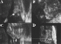
File:Figure 2. Fetal Lip and Primary Palate Three dimensional versus Two dimensional US.gif ...erse, and D, 3D rendered US images of a cleft lip (arrows) and palate in a fetus at 21 weeks gestational age. Image in D is viewed frontally. White line ind(440 × 317 (120 KB)) - 20:19, 8 October 2011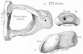
File:Anson1948 fig15.jpg (30 week) fetus (Wisconsin series 4) ; X 7: (a) the reconstruction entire, viewed from abov(1,280 × 829 (158 KB)) - 21:34, 16 October 2017
File:Ultrasound - fetal abdominal circumference.jpg [[Category:Ultrasound]] [[Category:Human Fetus]] [[Category:Second Trimester]](672 × 512 (23 KB)) - 09:41, 19 May 2015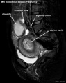
File:Abdominal ectopic pregnancy MRI.jpg ...sigmoid colon and was loosely adhered to the left abdominal sidewall. The fetus was localized at the right of the abdomen and was related to the placenta b(553 × 700 (45 KB)) - 12:27, 1 June 2019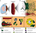
File:Human trophoblast invasion 01.jpg | valign=top width=300px|(A) The placenta connects the fetus to the uterine wall and establishes a vascular connection between mother an ...ssels into larger conduits to guarantee adequate blood flow to the growing fetus.(985 × 909 (223 KB)) - 15:24, 24 January 2017
File:Streeter004.jpg ==Fig. 4. Detail of the posterior canal in a human fetus 73 mm. long==(487 × 800 (92 KB)) - 02:48, 15 February 2011
File:Streeter1922-06-07.jpg ...jpg|'''Fig. 7.''']] Reconstruction of left auricular cartilage of a 50 mm. fetus (No. 84, Carnegie Collection). X 14. A model of the external form of the au(1,200 × 653 (107 KB)) - 11:00, 31 October 2015
File:Meyer1932history5 fig07.jpg ...opment. (This; to be sure. is a retained macerated, and probably abnormal, fetus.)(753 × 800 (140 KB)) - 21:15, 2 November 2015
File:CffDNA from apoptotic trophoblasts.jpg ...ophoblast and some erythroblast, will undergo apoptosis and the DNA of the fetus will be fragmented. Eventually, they will be able to distribute and migrate(3,028 × 1,696 (1,019 KB)) - 13:50, 22 September 2014
File:Kollmann685.jpg Frontalschnitt durch den Oberkiefer eines menschlichen Fetus von i8 cm Körper-(773 × 579 (126 KB)) - 10:11, 21 October 2011
File:Larsen's human embryology 5th ed.jpg ....au/lib/unsw/reader.action?docID=2074524&ppg=151 Fetal Development and the Fetus as Patient](391 × 499 (40 KB)) - 17:16, 22 March 2017
File:Placental villi 5.jpg [[Category:Human Fetus]][[Category:Third Trimester]][[Category:Placenta]] [[Category:Histology]](1,280 × 1,024 (238 KB)) - 20:44, 8 October 2015
File:Kollmann611.jpg Fig. 611. Qehirn eines menschlichen Fetus vom 3. Monat und von 42 mm(910 × 575 (55 KB)) - 17:00, 17 October 2011
File:Kollmann724.jpg liefert. Beim menschlichen Fetus von 12V8 cm vereinigen sich die beiden Venae(726 × 608 (96 KB)) - 10:53, 21 October 2011
File:Kollmann725.jpg in die äußere Ljamelle bei einem menschlichen Fetus vom Ende des 3. Monates(697 × 307 (33 KB)) - 10:53, 21 October 2011
File:Fetal temporomandibular joint 04.jpg | width=500px|Human fetus (175 mm GL; 18th week of development). Section of the posterior region of(600 × 390 (67 KB)) - 23:01, 21 March 2018
File:Simkins1928 plate07.jpg 29 Large and small cells in detail from the ovary of a 90-mm. fetus. X 750.(1,540 × 2,096 (239 KB)) - 16:54, 31 January 2018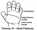
File:Trisomy21 hand.jpg ...ategory:Genetic Abnormalities]] [[Category:Human Embryo]] [[Category:Human Fetus]](360 × 295 (12 KB)) - 14:09, 21 October 2010
File:Thyroid uptake scans .jpg Graves' disease in mothers can cause thyrotoxic fetus - may have increased fetal motility and develop a range of abnormalities in(600 × 441 (34 KB)) - 14:57, 31 March 2012
File:Watson1918 fig01.jpg ...g a transverse section through the right and left Wolffian ducts in a human fetus 80.3 mm crown-rump length==(983 × 534 (264 KB)) - 09:49, 8 December 2016
File:Placental villi 6.jpg ...in villi blood vessels and in blood vessels within placental cord, embryo/fetus vessels and vitelline vessels.(1,000 × 750 (277 KB)) - 15:25, 17 March 2014
File:Meyer1920 fig18.jpg ...ave accompanied a living, 7-months fetus, having been expelled between the fetus and the placenta. Only a sniidl amount of clot, and what seems to be a smal(570 × 379 (26 KB)) - 07:19, 24 April 2012
File:Chromosome- triploidy.jpg ...dy can present with albuminuria, edema, or hypertension in the mother. The fetus may present with microcephaly and a {{placenta}} that is enlarged and fille(504 × 504 (227 KB)) - 07:31, 20 March 2019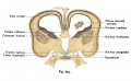
File:Kollmann625.jpg Fig. 625. Frontalschnitt durch das Hemisphärenhirn emes menschlichen Fetus(733 × 454 (62 KB)) - 17:18, 17 October 2011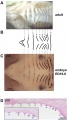
File:Mouse ruga pattern.jpg Figure shows the mouse ruga pattern in the adult and the fetus shown by in situ hybridization against [[Developmental Signals - Sonic hedg * '''C''' - In ED16.0 fetus, Shh gene expression pattern (as seen by whole-mount in situ hybridization)(600 × 1,059 (118 KB)) - 13:09, 5 February 2016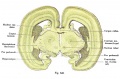
File:Kollmann628.jpg Fig. 628. Frootalschiiitt durch das Qehirn eines mensclilichen Fetus von(823 × 543 (126 KB)) - 11:18, 17 January 2020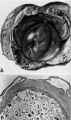
File:Hertig1946b fig15.jpg ==Fig. 15. Human Embryo 55 mm and older 14 Week Fetus==(800 × 1,348 (196 KB)) - 08:55, 8 August 2017
File:Mall Meyer1921 plate12.jpg Fig. 133. Marked macerated and deformed fetus from the same case. No. 1260 X4.(934 × 1,200 (252 KB)) - 15:21, 6 December 2012
File:Mall Meyer1921 fig145.jpg ==Fig. 145. Enlarged fetus from same, showing maceration effects==(603 × 461 (41 KB)) - 07:04, 4 December 2012
File:Harland Mossman.jpg ...w its efficiency in the exchange of nutrients and waste between mother and fetus. With W. J. Hamilton and J. D. Boyd he co-authored the most successful text(441 × 600 (49 KB)) - 15:14, 18 January 2020File:Fetus 35 week CT.jpg [[Category:Human Fetus]] [[Category:Computed Tomography]](700 × 874 (62 KB)) - 14:21, 24 August 2010
File:Kollmann714.jpg Frühe Stufe. Radiärschnitt durch die Iris eines 10,2 cm langen menschlichen Fetus(625 × 553 (54 KB)) - 10:48, 21 October 2011
File:Streeter1917 fig01.jpg ...the otic capsule is in a cartilaginous state. Ossification begins when the fetus has attained a crown-rump length of about 130 mm.(1,171 × 900 (131 KB)) - 08:25, 30 October 2015
File:Fetal head medial.jpg [[Category:Human Fetus]] [[Category:Musculoskeletal]] [[Category:Bone]] [[Category:Cartilage]] [[C(632 × 447 (34 KB)) - 08:09, 14 May 2014
File:LaryngealAtresia.jpg ...the fetal period or not. the caption only shows that it was present in the fetus. Reference should have been formatted with pubmed extension. I have now add(600 × 475 (51 KB)) - 10:38, 18 December 2018
File:Kollmann723.jpg und der Arteria hyaloidea bei einem menschlichen Fetus von lO cm Länge.(737 × 611 (81 KB)) - 10:52, 21 October 2011
File:Fetal temporomandibular joint 01.jpg | width=500px|Human fetus (55 mm GL; 10th week of development).(600 × 391 (107 KB)) - 23:03, 21 March 2018
File:Fetal temporomandibular joint 02.jpg | width=500px|Human fetus (95 mm GL; 12th week of development).(600 × 390 (109 KB)) - 23:02, 21 March 2018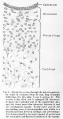
File:Streeter001.jpg .... 1. Detail of a section through the lateral semicircular canal in a human fetus 33 mm long==(423 × 800 (79 KB)) - 10:43, 30 July 2017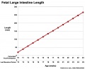
File:Fetal large Intestine length growth graph.jpg [[Category:Human]] [[Category:Human Fetus]] [[Category:Gastrointestinal Tract]] [[Category:Intestine]](800 × 653 (50 KB)) - 17:33, 21 April 2013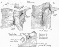
File:Anson1948 fig11.jpg ...struction of the fissular region of the otic capsule in a 180 mm. (21 week) fetus (Wisconsin series 45-b) with stapes removed; X 7: (a) viewed from a postero(1,220 × 981 (230 KB)) - 21:08, 16 October 2017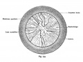
File:Kollmann719.jpg auf der vorderen Fläche der Linse aufliegend, injiziert; vom menschlichen Fetus(775 × 579 (87 KB)) - 17:28, 18 August 2012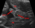
File:Ultrasound male.jpg Image shows an ultrasound of fetus with a penis and testes, indicating the male gender of the baby.(299 × 246 (29 KB)) - 23:06, 8 November 2014
File:Testis and ovary.jpg ...e and you have not included any detail related to hormonal function in the fetus? Minor, the figure is not very clear and should have been adjusted for brig(960 × 540 (37 KB)) - 11:08, 9 November 2014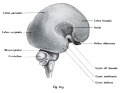
File:Kollmann613.jpg Fig. 613. Qehirn eines menschlichen Fetus vom Anfang des 4. Monates,(652 × 506 (31 KB)) - 17:01, 17 October 2011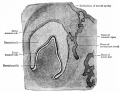
File:Bailey252.jpg ==Fig. 252. Section of developing tooth from a 3 months human fetus==(904 × 690 (166 KB)) - 12:25, 15 April 2011
File:Mouse-E17.5-foot-large.jpg Skeletal preparations of E17.5 wild-type mouse fetus stained with alcian blue for cartilage and alizarin red for bone.(600 × 779 (53 KB)) - 10:19, 30 September 2010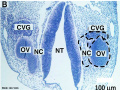
File:Human CS13 otic vesicle 01.jpg (B) Coronal section of a fetus with highlighted regions of interest (dashed black circles, CVG: cochlear-v(1,028 × 774 (112 KB)) - 12:34, 6 April 2018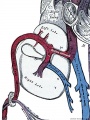
File:Fetal ductus venosus cartoon 01.jpg Fetal blood is returned from the placenta to the fetus by the placental or umbilical vein.(600 × 800 (129 KB)) - 13:15, 21 February 2015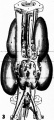
File:Jimenez-Castellanos1949 fig03.jpg ...econstruction of the juxtaaortic masses and the adjacent organs of a human fetus of 40 mm. :13 8.5. (1), (2), Vertebral body and arch; (3) aorta; (4) common(582 × 1,294 (133 KB)) - 04:08, 17 August 2017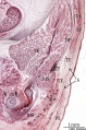
File:Human fetal neck 01.jpg Human fetus B503 (48 mm GL; 10 weeks of development). Frontal section. {{HE}}(549 × 827 (186 KB)) - 12:25, 21 September 2015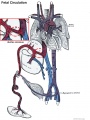
File:Fetal ductus venosus cartoon.jpg Fetal blood is returned from the placenta to the fetus by the placental or umbilical vein.(1,000 × 1,329 (255 KB)) - 12:11, 23 January 2016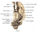
File:Kollmann612.jpg Fig. 612. Qehirn eines menschlicben Fetus vom 3. Monat und von 42 mm(666 × 583 (63 KB)) - 17:01, 17 October 2011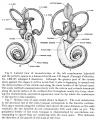
File:Streeter1917-fig06-07.jpg and the periotic spaces in a human fetus 85 mm. CR length (Carnegie Collection,(1,337 × 1,671 (639 KB)) - 14:49, 17 September 2015
File:ZDoppler Image of Fetal Aorta.jpg ...ur Doppler ultrasound image depicting a sagittal section of the chest of a fetus. The aortic arch and descending thoracic aorta can be seen extending from t(640 × 480 (53 KB)) - 12:36, 4 February 2011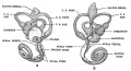
File:Streeter1917-fig04-05.jpg ...ces. This figure shows a lateral view of a model reconstructed from a human fetus 50 mm. CR length (Carnegie Collection, No. 84). The scala vestibuli is in t(1,000 × 544 (124 KB)) - 15:04, 17 September 2015
File:Anson1948 fig10.jpg ==Fig. 10. Otic capsule developmental stages 160 mm fetus==(1,191 × 1,236 (204 KB)) - 21:00, 16 October 2017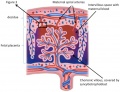
File:Placenta schematic.jpg ...c villous, intervillous space and maternal spiral arteries that supply the fetus.(784 × 600 (121 KB)) - 11:04, 9 November 2014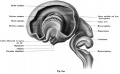
File:Kollmann610.jpg Fig. 610. Gehirn eines menschlichen Fetus vom 3. Monat und von 42 mm(898 × 555 (51 KB)) - 16:59, 17 October 2011
File:Fetal temporomandibular joint 03.jpg | width=500px|Human fetus (125 mm GL; 14th week of development). Sagittal section. Hematoxilyn-eosi(600 × 393 (48 KB)) - 10:03, 18 February 2015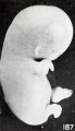
File:Mall Meyer1921 fig187.jpg ==Fig. 187. Slight swelling of fetus from brief maceration==(375 × 650 (31 KB)) - 05:42, 4 December 2012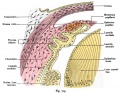
File:Kollmann715.jpg Fig. 715. Sphincter pupillae bei einem menschlichen Fetus von 19 cm Länge.(775 × 600 (128 KB)) - 10:49, 21 October 2011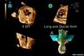
File:Fetal ultrasound ductal arch 01.jpg ...standardization of fetal position in the STIC volume dataset to place the fetus in the exact dorsal supine position, navigating systematically in the volum(800 × 533 (27 KB)) - 11:36, 28 August 2014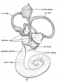
File:Streeter1917-fig09.jpg ==Figure 9. Medial Views of left membranous labyrinth human fetus 130 mm==(617 × 853 (109 KB)) - 15:12, 17 September 2015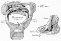
File:Anson1948 fig13.jpg Drawings of a reconstruction of the stapes in a 210 mm. (23 week) fetus (Wisconsin series 51) ; X 7: (a) reconstruction entire, seen from a supercl(1,239 × 841 (203 KB)) - 21:17, 16 October 2017
File:Kollmann627.jpg Fig. 627. Frontalschoitt durch das Qehiro eines meoschlicheo Fetus von(812 × 586 (122 KB)) - 17:19, 17 October 2011
File:Keibel Mall 274-278.jpg * Fig. 278 - Length of fetus 50 mm.(717 × 1,072 (123 KB)) - 00:51, 14 September 2012
File:Comparative genomic hybridization 01.jpg [[Category:Genetic Abnormalities]] [[Category:Human Embryo]] [[Category:Human Fetus]] [[Category:Comparative Genomic Hybridization]](453 × 536 (42 KB)) - 19:22, 6 September 2010


