Search results
From Embryology
Page title matches
File:Octopus bimaculatus embryonic development timeline.jpg ==Embryonic Development of Octopus bimaculatus== ...e table compares the embryonic development with the scale proposed for the development of L. pealii (Arnold 1965). *Observed in vivo.(1,280 × 1,394 (187 KB)) - 11:02, 21 May 2020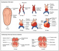
File:Progressive development of the Embryonic Heart.jpeg Development of the heart in the fetus and partitioning of the heart into four chambers ...gy, Connexions Web site. https://cnx.org/contents/FPtK1zmh@6.27:GxtDaPpg@3/Development-of-the-Heart/, Jun 19, 2013.(1,024 × 883 (154 KB)) - 11:30, 4 September 2018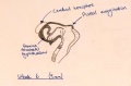
File:Week 6 embryonic development of CNS.jpg ==Week 6 embryonic development of CNS and emergence of pineal evagination.==(1,209 × 794 (165 KB)) - 12:43, 24 October 2014
File:Development of Trilaminar Embryonic Disc .png (1) Trilaminalar embryonic disc after gastrulation; lateral plate mesoderm resides between the endoder (4) By folding the lateral edges of the embryonic disc move inwards, creating the anterior intestinal portal. The persisting(1,921 × 5,223 (1.24 MB)) - 10:44, 13 October 2017


Page text matches
File:Hilfer1990 Fig16.jpg ==Figure 16. Townes and Holtfreter (1955) different embryonic layers in amphibians== ...o clumps and tended to take a position resembling that of normal embryonic development.(1,200 × 1,333 (247 KB)) - 10:43, 28 August 2014File:Octopus bimaculatus embryonic development timeline.jpg ==Embryonic Development of Octopus bimaculatus== ...e table compares the embryonic development with the scale proposed for the development of L. pealii (Arnold 1965). *Observed in vivo.(1,280 × 1,394 (187 KB)) - 11:02, 21 May 2020
File:HillH52 chorionic villi 04.jpg Human early placental villi development at 4-5 weeks. (Hill H52, x40) {{HE}} scale bar - 50 μm) * Embryonic mesenchyme(1,200 × 900 (254 KB)) - 13:34, 22 March 2014
File:HillH52 chorionic villi 05.jpg Human early placental villi development at 4-5 weeks. (Hill H52, x20) {{HE}} scale bar - 100 μm) * Embryonic mesenchyme(1,200 × 992 (371 KB)) - 13:54, 22 March 2014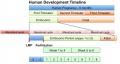
File:Human development timeline graph 02.jpg ==Human Development Timeline== |Simplified graphical view of human development timeline.(800 × 424 (61 KB)) - 09:37, 14 April 2016
File:Gray0032.gif ...mbryonic ectoderm that will contribute to embryonic and placental membrane development ...storic term for what we today call endoderm that will contribute to embryo development(500 × 417 (57 KB)) - 11:13, 22 May 2011
File:Bat embryo stage 19.mov ==Bat Embryonic Development (stage 19)== ...Cancer Center, who provided images and stage information on the embryonic development of the bat.(30 KB) - 01:12, 29 April 2011
File:Gray0032.jpg ...ough showing the trilaminar embryo suggests late week 2 to early week 3 of development. Uterine cavity is shown at bottom of image. ...mbryonic ectoderm that will contribute to embryonic and placental membrane development(800 × 667 (159 KB)) - 08:51, 21 April 2013
File:Bailey111.jpg ...the right are five individual cells showing stages of development from an embryonic cell to an adult fat cell.(916 × 567 (117 KB)) - 13:05, 18 January 2011
File:Stage13 and 22 thyroid development.jpg ==Thyroid Gland Embryonic Development Overview== Cross-sections of head and neck regions of embryonic stage 13 (week4-5) and stage 22 (week 8) are shown.(1,000 × 800 (283 KB)) - 11:49, 30 August 2011
File:Bat embryo stage 10 to 13.jpg ==Bat Embryonic Development (stage 10-13)== ...Cancer Center, who provided images and stage information on the embryonic development of the bat.(800 × 1,122 (66 KB)) - 13:21, 6 July 2012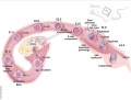
File:Mouse E0-E5.jpg ==Development of the Preimplantation Blastocyst in Mice== Cartoon shows an overview of mouse development from Embryonic Day 0 (E0) Through Day 5 (E5.0).(991 × 749 (90 KB)) - 10:08, 14 October 2016
File:HillH52 chorionic villi 08.jpg Human early placental villi development at 4-5 weeks. (Hill H52, x40) {{HE}} scale bar - 50 μm) ...phoblast shell enclosing mesenchyme (extra-embryonic mesoderm), containing embryonic blood vessels.(1,200 × 900 (229 KB)) - 14:59, 3 July 2014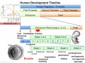
File:Human development timeline graph 01.jpg ==Human Development Timeline== This graph shows an overview of human development and details about embryonic development.(1,000 × 750 (141 KB)) - 08:49, 15 April 2014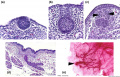
File:Mouse mammary development 01.jpg ==Embryonic Mouse Mammary Development== (a) [[:Category:Mouse E12.5|Embryonic day E12.5]] The epithelial cells have invaginated to form the initial bud,(1,200 × 773 (147 KB)) - 09:04, 20 March 2018
File:Kellicott 179.jpg From McMurrich (Development of the Human Body). ...ic ectoderm; the dotted line marks the line of the transition of the body (embryonic) ectoderm into that of the amnion.(893 × 800 (131 KB)) - 16:54, 23 December 2013
File:Human embryonic renal branching stage 22.jpg ==Embryonic Human Renal Urothelial Branching Development== ...he [[Kyoto Collection]] at Carnegie stage {{CS22}}. See also [[:File:Human embryonic renal branching 1.jpg|branching figure stages 14-22]].(500 × 756 (150 KB)) - 10:21, 18 January 2019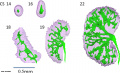
File:Human embryonic renal branching 1.jpg ==Embryonic Human Renal Urothelial Branching Development== ...{CS14}}, {{CS16}}, {{CS18}}, {{CS19}} and {{CS22}}. See also [[:File:Human embryonic renal branching stage 22.jpg|branching figure stage 22]].(1,280 × 779 (236 KB)) - 10:26, 18 January 2019- Error creating thumbnail: File with dimensions greater than 12.5 MP
File:Worm - embryonic cell lineage 01.jpg ==Worm - Embryonic Cell Lineage== Embryonic cell lineage developed by J .E. Sulston, E. Schierenberg, J. G. White, J. N(10,389 × 1,336 (598 KB)) - 11:25, 30 June 2012 
File:GladstoneHamilton1941 text-fig04.jpg ==Text-fig. 4. General .view of the embryonic disc at the anterior part of the primitive streak and groove== ...ion no. 57, the sections having been numbered from the anterior end of the embryonic disc. x 100.(1,280 × 633 (96 KB)) - 16:58, 26 February 2017
File:Mesoderm-cartoon4.jpg ==Mesoderm and Ectoderm Development cartoon== ...s a section through the trunk of the trilaminar embryo showing the further development of the 3 layers and the space (coelom) that forms in the mesoderm (only the(400 × 300 (20 KB)) - 10:09, 23 April 2020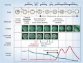
File:Model human blastocyst development.jpg ==Proposed Model for Human Embryo Development== ...gg (ESSP1) must be degraded as the transition from oocyte to embryo begins embryonic genome activation (EGA).(946 × 726 (84 KB)) - 14:47, 6 October 2015
File:Human Carnegie stage 1-23.jpg ==Human Embryonic Development== ..., showing human external appearance and growth during the first 8 weeks of development.(1,000 × 563 (98 KB)) - 11:32, 14 May 2017
File:Stage6 bf03.jpg ...howing development of the bilaminar (two layer) embryo and the early extra-embryonic coeloms (spaces). Extra-embryonic coelom(646 × 800 (65 KB)) - 14:48, 1 October 2018
File:Human- fetal week 10 sagittal planes.jpg ...he embryonic period (up to week 8) but still only 2 weeks into early fetal development.(600 × 250 (29 KB)) - 15:39, 27 April 2010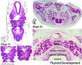
File:Stage13 and 22 thyroid development a.jpg ==Thyroid Gland Embryonic Development== ...e developing thyroid at the beginning of week 5 and at week 8 of embryonic development.(800 × 640 (94 KB)) - 08:50, 22 May 2012
File:Lung development overiview.png Summary of the lung formation from the embryonic stage to the fetal stage. Original Image is adapted from: Figure 1 from Embryonic Development of the Respiratory System page(717 × 549 (178 KB)) - 14:59, 9 November 2014
File:Bat embryo stage 12-17.jpg ==Bat Embryonic Development (stage 12-17)== :'''Links:''' [[Bat Development]] | [[:File:Bat embryo stage 10-13.jpg|stage 10 to 13]] | [[:File:Bat embry(548 × 767 (30 KB)) - 01:16, 29 April 2011
File:Bat embryo stage 18-24.jpg ==Bat Embryonic Development (stage 18-24)== :'''Links:''' [[Bat Development]] | [[:File:Bat embryo stage 10-13.jpg|stage 10 to 13]] | [[:File:Bat embry(518 × 734 (32 KB)) - 01:16, 29 April 2011
File:Thymic Epithelial Cell Development and Function.png ==Thymic Epithelial Cell Development and Function== '''Image shows Thymic Epithelial Cell Development and Function in fetal period'''(2,028 × 823 (3.17 MB)) - 11:37, 9 November 2014
File:Human- fetal week 10 heart ABCD.jpg ...he embryonic period (up to week 8) but still only 2 weeks into early fetal development.(600 × 450 (133 KB)) - 16:10, 27 April 2010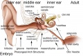
File:Adult hearing embryonic origins.jpg ==Adult Hearing Embryonic Origins== ...nd inner), each of which have their own separate components from different embryonic origins.(1,000 × 675 (80 KB)) - 14:26, 8 May 2018
File:Human- fetal week 10 cerebellum C.jpg ...he embryonic period (up to week 8) but still only 2 weeks into early fetal development.(347 × 284 (25 KB)) - 18:53, 20 September 2012
File:Human- fetal week 10 cerebellum D.jpg ...he embryonic period (up to week 8) but still only 2 weeks into early fetal development.(347 × 284 (23 KB)) - 18:53, 20 September 2012
File:Human- fetal week 10 cerebellum B.jpg ...he embryonic period (up to week 8) but still only 2 weeks into early fetal development.(347 × 284 (21 KB)) - 18:53, 20 September 2012
File:BGDA PracManual 2011 Practical 7.pdf ==BGDA Practical Manual 2011 Practical 7 - Embryonic Development==(203 KB) - 14:43, 22 April 2011
File:Mouse distal visceral endoderm 01.jpg ...MP signaling promotes the differentiation of primitive endoderm (PrE) into embryonic visceral endoderm (VE) until E4.5. (B) Embryonic visceral endoderm (VE) differentiates as a result of the concerted action o(959 × 1,280 (208 KB)) - 13:16, 3 May 2013
File:Placental membranes.jpg * '''amniotic sac''' - formed by the amniotic membrane (ectoderm and extra-embryonic mesoderm) completely surrounding the surrounding the embryo. * '''yolk sac''' - the yolk membrane (endoderm and extra-embryonic mesoderm) attached to the embryo at the umbilicus and continuous with the m(600 × 450 (99 KB)) - 07:46, 15 May 2014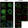
File:Antibody immunostaining of ligands and receptors in triploid human embryos.png ...mbryonic nuclei. Furthermore, growth factors were used to aid in embryonic development and promote blastocyst growth as well as IgG being applied to the negative ..., Shu Y, Cheng Y, Qiao J, et al. (2012) Promotion of Human Early Embryonic Development and Blastocyst Outgrowth In Vitro Using Autocrine/Paracrine Growth Factors.(3,439 × 3,444 (7.13 MB)) - 00:21, 20 August 2014
File:Zebrafish melanocyte development model.jpg ==Model for the parallel establishment of the zebrafish embryonic melanocyte lineage and the adult melanocyte stem cell lineage== ...and melanocytes during metamorphosis or regeneration, when inhibition from embryonic melanocytes (block arrow) is relieved. The MSC–derived melanocytes are se(484 × 277 (40 KB)) - 14:50, 20 February 2011
File:Aneuploidy model based on fragmentation.jpg ==Model for the Origin of Human Embryonic Aneuploidy Based on Fragmentation Timing== ...omal abnormalities and undergoes fragmentation as a survival mechanism. As development proceeds, these fragments either remain or are reabsorbed by the blastomere(668 × 790 (105 KB)) - 13:44, 1 April 2019
File:Bat embryo stage 10-13.jpg ==Bat Embryonic Development (stage 10-13)== :'''Links:''' [[Bat Development]] | [[:File:Bat embryo stage 10-13.jpg|stage 10 to 13]] | [[:File:Bat embry(547 × 767 (23 KB)) - 01:34, 29 April 2011
File:ThyroidDevelopment.png This image summarises the endodermal and mesodermal contribution to the development of the thyroid gland. The progenitor cells are from anterior endoderm and r ...ed to understand what has gone before, you have not included much in fetal development. There is no explanation of the molecular factors shown in the figure.(1,996 × 1,212 (49 KB)) - 11:46, 9 November 2014File:Human blastocyst day 3-6.mov ...asive imaging of human embryos before embryonic genome activation predicts development to the blastocyst stage. ...asive imaging of human embryos before embryonic genome activation predicts development to the blastocyst stage(4.26 MB) - 16:08, 15 October 2010
File:Embryonic upper limb - brachial and superficial brachial artery.jpg ==Embryonic upper limb - brachial and superficial brachial artery == ...development of the brachial artery and the superficial brachial artery in embryonic upper limb.(1,280 × 314 (122 KB)) - 14:25, 2 February 2020
File:Histology of human embryonic liver at 11 weeks.png ==Histology of human embryonic liver at 11 weeks== Paraffin-embedded sections of human embryonic liver at 11 weeks (9 weeks of gestation) stained for Hematoxylin and Eosin(1,988 × 1,642 (4.17 MB)) - 22:02, 8 November 2014
File:Stage9 bf2-primordial germ cell region.jpg # the relative size of the embryo and the associated extra-embryonic coeloms. # the shape of the early folded embryonic disc and rostro-caudal bendings.(814 × 1,000 (72 KB)) - 21:20, 17 May 2015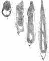
File:Bailey061.jpg ...ys and 22 hours after insemination (younger than B but further advanced in development), showing beginning of proamniotic cavity. ...astocyst 8 days after insemination (younger than B but further advanced in development), showing more advanced proamniotic cavity.(878 × 1,086 (184 KB)) - 15:02, 26 January 2011
File:1 Min Embryo - Human timeline.mp4 ...mplantation to 8 Weeks|BGDA Prac 6]] | [[Embryonic Development]] | [[Fetal Development]] | [[One Minute Embryology]] Lets look at an overview of human development(5.28 MB) - 10:40, 28 April 2016
File:Rugh 077.jpg ...r V. Hamburger and B. Mayer, unpublished. Redrawn from Spemann: "Embryonic Development and Induction," New Haven, Yale University Press.(861 × 800 (171 KB)) - 13:43, 12 April 2013
File:Human- fetal week 10 sagittal plane A.jpg ...he embryonic period (up to week 8) but still only 2 weeks into early fetal development.(500 × 573 (96 KB)) - 16:19, 27 April 2010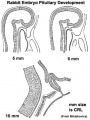
File:Pituitary rabbit development.jpg ==Rabbit Pituitary Development== Cartoon showing the changes in the embryonic rabbit pituitary.(374 × 500 (33 KB)) - 14:52, 27 May 2014
File:Stage19 em11.jpg ...EM images focussing on this developmentally important region and time for embryonic cleft lip and palate.(800 × 329 (46 KB)) - 10:11, 23 February 2014
File:Stage19 em01.jpg ...EM images focussing on this developmentally important region and time for embryonic cleft lip and palate.(800 × 329 (37 KB)) - 08:17, 23 February 2014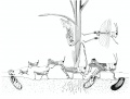
File:Grasshopper lifecycle.jpg # As soon as the eggs are laid, they begin embryonic development and reach an advanced stage in which they enter diapause and pass the winte # In spring the eggs complete embryonic development and hatch.(1,072 × 814 (122 KB)) - 13:40, 16 February 2016
File:Horseshoe.jpg ...]] within the lateral plate [[mesoderm]] that forms during week 3 of human development. ...:''' [[Renal_System_-_Abnormalities|Renal Abnormalities]] | [[Renal System Development]](400 × 400 (32 KB)) - 09:09, 13 October 2016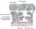
File:Gray0037.jpg ...evelopment contain core of mesoderm. Tertiary villi then have blood vessel development within this core. Extra-embryonic mesoderm grows into villi, covers the entire surface of chorionic sac.(500 × 412 (74 KB)) - 11:21, 9 June 2014
File:Gap junction 01.jpg * Also in embryonic development (see [[Blastocyst Development]])(800 × 562 (69 KB)) - 12:35, 25 March 2015
File:Endoderm cartoon.jpg ==Cartoon of endoderm development== ...3 images is from the animation [[Development_Animation_-_Endoderm|Endoderm Development]](587 × 262 (31 KB)) - 19:42, 11 June 2013
File:Human- fetal week 10 lower body D.jpg ...he embryonic period (up to week 8) but still only 2 weeks into early fetal development.(600 × 450 (91 KB)) - 15:36, 27 April 2010File:Stages 1-5 mouse.pdf Table 1: Mouse embryonic stages of development from fertilization to zona free blastocyst (stages 1 to 5).(29 KB) - 13:42, 31 March 2012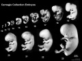
File:Human Carnegie stage 10-23.jpg ==Human Embryonic Development== ...tion, embryos compiled and scaled from original images. Note that weeks of development are form fertilisation (fertilization age), not gestational age {{GA}} that(1,024 × 768 (95 KB)) - 11:38, 16 February 2018
File:Elephant and calf.jpg * 50 days - embryonic vesicle earliest observation * 71 days - embryonic heartbeat and allantois visible as a single sacculation(538 × 404 (52 KB)) - 20:24, 1 November 2013
File:Morphological differences in early mouse embryonic development.png ...hological and lineage specification steps during the early mouse embryonic development== ...is formed as a layer separating epiblast from blastocoel (E4.5). After 4.5 embryonic days, the preimplantation embryo contains more than 100 cells.(2,061 × 886 (455 KB)) - 12:47, 18 August 2016
File:Hamilton1949 fig12.jpg ==Fig.12. Diagram to show the development of the secondary yolk sac, extra-embryonic coelom and remnant of the primary yolk sac==(1,312 × 1,265 (267 KB)) - 15:30, 15 May 2018
File:Hertig1956 fig82.jpg ...ssible to say what effect, if any, such an abnormality would have upon the development of the body stalk and the embryo. [[:Category:Carnegie Embryo 8299|Carnegie(1,000 × 796 (181 KB)) - 14:37, 25 February 2017
File:Human- fetal week 10 sagittal plane B.jpg ...he embryonic period (up to week 8) but still only 2 weeks into early fetal development.(500 × 573 (99 KB)) - 15:49, 27 April 2010
File:Bat - neural development 01.jpg ==Bat neural Development (stage 14)== :'''Links:''' [[Bat Development]] | [[Neural System Development]] | [[Carnegie stage 14]](733 × 498 (33 KB)) - 13:17, 6 July 2012
File:Human- fetal week 10 sagittal plane D.jpg ...he embryonic period (up to week 8) but still only 2 weeks into early fetal development.(500 × 573 (105 KB)) - 07:41, 1 May 2011
File:Cattle embryo staging 01.jpg ...al sections (H&E or CER1 stained) of, cattle embryos at the five stages of development between hatching and the start of {{gastrulation}}, based on data from sect ...ural) hypoblast; PS, primitive streak; RL, Rauber’s Layer (polar TB); VH, (embryonic) visceral hypoblast. All bars are 100 μm.(1,961 × 1,962 (708 KB)) - 13:45, 22 November 2018
File:Human- fetal week 10 sagittal plane C.jpg ...he embryonic period (up to week 8) but still only 2 weeks into early fetal development.(500 × 573 (98 KB)) - 15:48, 27 April 2010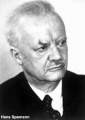
File:Hans Spemann.jpg A German embryologist who worked extensively on amphibian development and was the discoverer of the organiser region (or primitive node) the cont ...iology or Medicine "for his discovery of the organizer effect in embryonic development".(300 × 425 (18 KB)) - 10:48, 17 September 2014
File:Human- fetal week 10 cerebellum A.jpg ...he embryonic period (up to week 8) but still only 2 weeks into early fetal development.(347 × 284 (24 KB)) - 18:54, 20 September 2012
File:Human- fetal week 10 urogenital A.jpg ...he embryonic period (up to week 8) but still only 2 weeks into early fetal development.(600 × 450 (109 KB)) - 15:52, 27 April 2010
File:Human- fetal week 10 lower body A.jpg ...he embryonic period (up to week 8) but still only 2 weeks into early fetal development.(600 × 450 (96 KB)) - 15:52, 27 April 2010
File:Human- fetal week 10 upper body A.jpg ...he embryonic period (up to week 8) but still only 2 weeks into early fetal development.(600 × 450 (104 KB)) - 15:53, 27 April 2010
File:Human- fetal week 10 upper body D.jpg ...he embryonic period (up to week 8) but still only 2 weeks into early fetal development.(600 × 450 (106 KB)) - 15:42, 27 April 2010
File:Human embryonic-fetal tongue 01.jpg Photographs of mid-sagittal sections of an embryonic human tongue. (A, B) Tongue development (TD) stage 1. Co, copula; Ce, foramen cecum.(1,000 × 1,129 (490 KB)) - 19:02, 18 June 2018
File:Human- fetal week 10 urogenital B.jpg ...he embryonic period (up to week 8) but still only 2 weeks into early fetal development.(600 × 450 (109 KB)) - 15:49, 27 April 2010
File:Human- fetal week 10 lower body B.jpg ...he embryonic period (up to week 8) but still only 2 weeks into early fetal development.(600 × 450 (93 KB)) - 15:51, 27 April 2010
File:Human- fetal week 10 upper body B.jpg ...he embryonic period (up to week 8) but still only 2 weeks into early fetal development.(600 × 450 (105 KB)) - 15:51, 27 April 2010
File:Bat embryo stage 18 to 24.jpg ==Bat Embryonic Development (stage 18-24)== |+ '''Embryonic Bat Stages ''Carollia perspicillata'''''<ref name="PMID15861401"><pubmed>15(800 × 1,134 (104 KB)) - 12:22, 3 July 2012
File:Human- fetal week 10 head A.jpg ...he embryonic period (up to week 8) but still only 2 weeks into early fetal development.(600 × 544 (113 KB)) - 21:22, 29 May 2011
File:Mouse- preimplantation gene expression.jpg ==Characterization of global gene expression patterns during preimplantation development== ...in the characterization of gene expression patterns during preimplantation development as they have produced extensive catalogues of global stage specific gene ex(800 × 612 (106 KB)) - 09:59, 12 October 2010
File:Human- fetal week 10 lower body C.jpg ...he embryonic period (up to week 8) but still only 2 weeks into early fetal development.(600 × 450 (94 KB)) - 15:47, 27 April 2010
File:Human- fetal week 10 head C.jpg ...he embryonic period (up to week 8) but still only 2 weeks into early fetal development.(600 × 544 (118 KB)) - 15:47, 27 April 2010
File:Rugh 128.jpg ==Development of the olfactory organ of the frog== (Bottom) Schematic reconstruction of the embryonic olfactory organ.(826 × 800 (145 KB)) - 15:40, 14 April 2013
File:Fetal 10wk urogenital 1.jpg ...he embryonic period (up to week 8) but still only 2 weeks into early fetal development.(800 × 600 (109 KB)) - 21:52, 8 July 2012
File:Human embryonic tongue 01.jpg Photographs of mid-sagittal sections of an embryonic human tongue. (A, B) Tongue development (TD) stage 1. Co, copula; Ce, foramen cecum.(1,200 × 818 (443 KB)) - 14:12, 23 March 2016
File:Human- fetal week 10 upper body C.jpg ...he embryonic period (up to week 8) but still only 2 weeks into early fetal development.(600 × 450 (109 KB)) - 15:46, 27 April 2010
File:Human- fetal week 10 urogenital D.jpg ...he embryonic period (up to week 8) but still only 2 weeks into early fetal development.(600 × 450 (101 KB)) - 17:53, 28 May 2011
File:Fetal 10wk urogenital 4.jpg ...he embryonic period (up to week 8) but still only 2 weeks into early fetal development.(800 × 600 (105 KB)) - 17:58, 28 May 2011
File:Human- fetal week 10 head D.jpg ...he embryonic period (up to week 8) but still only 2 weeks into early fetal development.(600 × 544 (111 KB)) - 14:50, 23 April 2013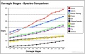
File:Carnegie stages species comparison.jpg * [[Monkey Development|Rhesus Monkey]]<ref>Hendrickx and Sawey '''Embryology of the rhesus monkey' ...Development|Mouse]]<ref>Theiler, K. '''The House Mouse: Atlas of Embryonic Development.''' 1989. New York: Springer-Verlag. ISBN 3 540 05940 7</ref>(800 × 514 (85 KB)) - 18:16, 3 June 2011
File:HeartILP001.jpg Embryo at approximately 18 days showing early angiogenesis and the development of the primordial heart tubes in the cardiogenic region. ...links to other resources. For example [[Carnegie_stage_7|Carnegie stage 7 embryonic disc]](1,507 × 898 (125 KB)) - 13:12, 8 September 2009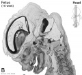
File:Human- fetal week 10 head B.jpg ...he embryonic period (up to week 8) but still only 2 weeks into early fetal development.(600 × 544 (66 KB)) - 21:22, 29 May 2011
File:Human- genital development critical periods.jpg ==Human Genital Development Critical Periods== This figure provides a broad general summary of key events in genital development in relation to critical periods.(1,000 × 494 (78 KB)) - 12:12, 15 May 2019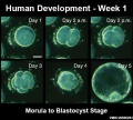
File:Human blastocyst day 1-5.jpg Human blastocyst development (in vitro) from day 1 to day 5. ...asive imaging of human embryos before embryonic genome activation predicts development to the blastocyst stage.(500 × 450 (46 KB)) - 13:50, 15 March 2014
File:Fetal 10wk urogenital 2.jpg ...he embryonic period (up to week 8) but still only 2 weeks into early fetal development.(800 × 600 (110 KB)) - 21:17, 29 May 2011
File:Human- fetal week 10 head A1.jpg ...he embryonic period (up to week 8) but still only 2 weeks into early fetal development.(1,200 × 1,088 (159 KB)) - 06:58, 7 October 2010
File:Placenta anchoring villi.jpg ...showing the junctional region between the maternal decidua (left) and the embryonic placental villi (right). {{HE}} ...ells differentiate in response to both steroid hormones (progesterone) and embryonic signals into large epitheliod decidual cells. This process is essential for(600 × 450 (167 KB)) - 09:32, 8 August 2016
File:Fetal 10wk urogenital 3.jpg ...he embryonic period (up to week 8) but still only 2 weeks into early fetal development.(800 × 600 (107 KB)) - 21:17, 29 May 2011
File:Early mouse development cartoon.jpg ...hological and lineage specification steps during the early mouse embryonic development=== * After 4.5 embryonic days, the preimplantation embryo contains more than 100 cells.(1,000 × 430 (81 KB)) - 09:39, 9 November 2011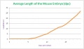
File:Average length of the mouse embryo.JPG ...llustrates the average length of the mouse embryo throughout its embryonic development. ...upon information in Dr Karl Theiler’s ‘The House Mouse; Atlas of Embryonic Development’, 1989(629 × 369 (23 KB)) - 11:43, 31 March 2012
File:Human- fetal week 10 urogenital C.jpg ...he embryonic period (up to week 8) but still only 2 weeks into early fetal development.(600 × 450 (105 KB)) - 17:42, 28 May 2011
File:Stage19- limb rotation.jpg ...ryo to show the direction in which the upper and lower limbs rotate during development. This distinctive embryonic rotation also establishes the future sensory pattern of [[:File:Dermatomes.(600 × 800 (27 KB)) - 07:45, 25 May 2018
File:Development.jpg Development journal issue 123 in 1996 ...e screens for developmental mutation in the zebrafish. Approximately 4,000 embryonic-lethal mutants were described.(600 × 478 (95 KB)) - 08:02, 15 October 2009
File:Melanocyte development cartoon.jpg ==Overview of Melanocyte Development== ...and acquire MITF, DCT and KIT expression. After colonising the developing embryonic hair follicles, some melanoblasts differentiate into melanocytes and produc(1,280 × 1,086 (176 KB)) - 22:43, 3 March 2015
File:Cleft palate.jpg ...showing cleft palate position. In humans, the timeline for lip and palate development occurs in two main phases. # A late embryonic - lip, maxillary and frontonasal prominence fusion.(653 × 776 (124 KB)) - 21:52, 15 September 2016
File:Bat embryo stage 12 to 17.jpg ==Bat Embryonic Development (stage 12-17)== |+ '''Embryonic Bat Stages ''Carollia perspicillata'''''<ref name="PMID15861401"><pubmed>15(800 × 1,120 (91 KB)) - 12:22, 3 July 2012
File:Inner cell mass cartoon.jpg ==The Three Germ Layers of Embryonic Development== During the third week of human embryonic development, the following layers form and will eventually differentiate and form speci(831 × 800 (78 KB)) - 14:29, 28 January 2013
File:Embryonic and fetal pituitary.jpg ==Embryonic and Fetal Pituitary== The links below will show additional images of pituitary development.(450 × 166 (14 KB)) - 14:59, 6 October 2015
File:Chicken skin timeline 01.jpg ==Chicken Skin Development Timeline== Three different processes in chicken embryo skin development based on morphogenesis.(1,547 × 1,709 (218 KB)) - 10:48, 6 March 2018
File:Mouse- embryo E9.5.jpg Original image file: Figure 1. Drp1 is required for embryonic mouse development. (+/+ embryo cropped from original full figure)(325 × 323 (10 KB)) - 12:46, 24 November 2010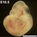
File:Mouse- embryo E10.5.jpg Original image file: Figure 1. Drp1 is required for embryonic mouse development. (+/+ embryo cropped from original full figure)(324 × 324 (14 KB)) - 12:46, 24 November 2010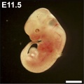
File:Mouse- embryo E11.5.jpg Original image file: Figure 1. Drp1 is required for embryonic mouse development. (+/+ embryo cropped from original full figure)(324 × 324 (14 KB)) - 12:45, 24 November 2010
File:Human blastocyst day 1-6.jpg Human blastocyst development (in vitro) from day 1 to day 6. ...asive imaging of human embryos before embryonic genome activation predicts development to the blastocyst stage.(708 × 338 (45 KB)) - 17:54, 11 December 2012
File:Stage20-23 limbs b.jpg ==Human Embryonic Limb Development== Carnegie stage 20 to 23 (week 8) extracts showing upper and lower limb development (Images are not to scale).(600 × 226 (13 KB)) - 09:15, 27 November 2015
File:Week4.jpg A Transverse section of the forebrain at Week 4 of Embryonic Development. The placodes can be seen as ventrolateral structures arising from the neur(521 × 712 (75 KB)) - 19:12, 3 October 2012
File:3–11 SS caudal foregut endoderm.png ...ntributed to the end of culture. Simple compass is provided indicating the embryonic axis. ...t template. As this is fetal I do not know why this is included in a fetal development project and it is not explained by the caption.(1,347 × 1,855 (315 KB)) - 22:10, 8 November 2014
File:Bilateral cleft palate.jpg ...showing cleft palate position. In humans, the timeline for lip and palate development occurs in two main phases. # A late embryonic - lip, maxillary and frontonasal prominence fusion.(214 × 300 (11 KB)) - 18:06, 18 May 2014
File:Rugh 069.jpg After W. Vogt, 1929b. From Spemann: "Embryonic Development and Induction," New Haven, Yale University Press.(1,200 × 603 (201 KB)) - 12:34, 12 April 2013
File:Edwin Conklin.jpg ...y. He was Chair of Biology for twenty-five years and studied the embryonic development of marine animals.(676 × 1,000 (93 KB)) - 13:16, 20 October 2016
File:Gray0025.jpg ...he allantois becomes expanded into a vesicle which projects into the extra-embryonic coelom. ...cting the embryonic disc to the chorion. Present mainly in week 3 of human development, this region will later form the placental cord region, becomes vascular, a(500 × 500 (29 KB)) - 12:10, 25 February 2014
File:Waddington1956 fig1.3.jpg ===Figure 1.3 The development of an (imaginary) embryonic field===(1,280 × 973 (80 KB)) - 14:02, 31 December 2019
File:Monozygotic twin embryos.jpg ...hese embryos are stage {{CS19}} in [[Week 7]] ({{GA}} week 9) of embryonic development. [[Category:Abnormal Development]][[Category:Twinning]](1,657 × 2,000 (511 KB)) - 12:13, 28 February 2019
File:Notch CNS simple diagram.png ...https://www.ncbi.nlm.nih.gov/pubmed/25815127 ''Neural differentiation from embryonic stem cells in vitro: An overview of the signaling pathways''] ...nceptually to the project page. Similar comment to the previous on cardiac development. You need to clearly show how Notch is involved in this process.(579 × 490 (50 KB)) - 11:40, 28 October 2016
File:Florian1930-text-fig03.jpg ==Text-fig 3. Schemes of the chief stages in the development of the human embryo== ...ve-mentioned structures. In this stage, approximately in the middle of the embryonic plate, there is present a distinct primordium of the primitive streak and,(407 × 425 (25 KB)) - 19:30, 23 September 2015
File:Stage7 intermediate-mesoderm.jpg ==Carnegie Stage 7 showing the intermediate mesoderm region of the embryonic disc== | [[Week 3]], [[Carnegie stage 7]] 15 - 17 days, 0.4 mm, embryonic disc, showing the epiblast viewed from the amniotic (dorsal) side.(690 × 800 (67 KB)) - 16:09, 17 August 2014
File:Bat icon.jpg ...rbee SD, Chen CH, Badwaik NK, Niswander L, Behringer RR, Rasweiler JJ 4th. Embryonic staging system for the short-tailed fruit bat, Carollia perspicillata, a mo [[Category:Animal Development]] [[Category:Bat]](100 × 120 (1 KB)) - 08:07, 14 September 2012
File:Mouse zygote pronuclei 02.jpg Embryonic gene expression (EGA, embryonic genome activation) is a rapid increase in the synthesis of transcripts. The ...s after hCG injection, which was used as the reference point for embryonic development (hours post-hCG i.e. hphCG).(1,000 × 501 (84 KB)) - 11:46, 29 December 2012
File:HillH202 Stage 17 bf04.jpg Chorionic sac has been open to observe the developing embryo and extra-embryonic coeloms (cavities). ...the individual functional units together of the fetal placenta. [[Placenta Development]](1,000 × 1,000 (138 KB)) - 17:58, 22 March 2014
File:Estradiol.jpg ...mones also regulate changes that occur each menstrual cycle. During female development the fetal adrenal gland synthesises DHEA, an oestrogen precursor, converted ...estis fluid at variable levels in different species. During male embryonic development exposure to high levels of estrogen can lead to genital abnormalities.(500 × 316 (20 KB)) - 11:40, 6 March 2019
File:Patten054.jpg ...ral body wall in the yolk-stalk region, results in the embryonic and extra-embryonic coelom retaining their open communication at this point for a long time aft In considering the early development of the heart (Chapter IX) the formation of the dorsal and ventral mesocardi(764 × 1,060 (179 KB)) - 09:11, 29 July 2011
File:Human embryology and developmental biology 5th.jpg Developmental Tables Carnegie Stages of Early Human Embryonic Development (Weeks 1-8) Major Developmental Events During the Fetal Period ===PART I Early Development and the Fetal-Maternal Relationship===(321 × 400 (25 KB)) - 10:57, 6 April 2016
File:Primary brain vesicles.jpg This image shows the primary brain vesicles on day 30 of embryonic development of CNS.(2,082 × 1,221 (473 KB)) - 12:45, 24 October 2014
File:Stage 22 image 186.jpg * Human embryonic pancreas and duodenum structure, [[Week 8]] [[Carnegie stage 22]]. ...Tract - Pancreas Development|Exocrine Pancreas]] | [[Endocrine - Pancreas Development|Endocrine Pancreas]] | [[Gastrointestinal_Tract_-_Intestine_Development|Int(1,000 × 658 (209 KB)) - 13:04, 17 April 2019
File:Human thyroid system and neural development.jpg ==Human Thyroid System Development== Timeline of human thyroid system and brain development from conception to birth. Estimation of neurogenesis adapted from Bayer et(1,032 × 728 (132 KB)) - 17:41, 25 May 2019
File:Human lung stages 01.jpg ==Overview of lung development== | Lung development is subdivided into 5 morphologically defined stages<br>(787 × 534 (138 KB)) - 13:29, 22 February 2019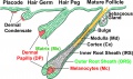
File:Hair development stages.jpg ==Hair Development Stages== ...pmental stages in hair follicle development. In mouse, this occurs between embryonic day 15 (~E15) and is complete at approximately postnatal day 4 (~P4).(1,000 × 590 (159 KB)) - 11:09, 23 March 2014
File:Theoria Generationis 1774.jpg ...spar Friedrich Wolff (1733-1794) of epigenesis, the gradual building up of embryonic structures, countered the existing theory of preformation. ...' [[Embryology History]] | [[Genital System Development]] | [[Renal System Development]](496 × 800 (84 KB)) - 08:37, 19 June 2019
File:Fgf signalling in palate development.PNG Fig 8. Regulation of Shh and Fgf18 expression during palate development. ...d Fgf18 beads (right side). (B) The patterns of GFP expression in ShhGFP/+ embryonic palatal explants treated with BSA beads (left side) and Fgf18 beads (right(1,634 × 1,942 (2.33 MB)) - 10:00, 31 August 2017
File:Gray0067.png ...espectively from the bodies, vertebral arches, and costal processes of the embryonic vertebræ. Note also the different shapes of vertebra at different column l ...s:''' [[Musculoskeletal_System_-_Axial_Skeleton_Development|Axial Skeleton Development]](350 × 463 (46 KB)) - 10:01, 7 October 2014
File:Anderson2016-fig48.jpg ...tage 20]], in the eighth week of development, just prior to closure of the embryonic interventricular foramen. The proximal cushions have fused, and are beginni(800 × 800 (203 KB)) - 21:53, 16 February 2017
File:Theiler stages 12-14 mouse.JPG ...y-nc-sa/3.0/legalcode. Figure 1: Illustration of Mouse embryonic stages of development from fertilization to zona free blastocyst (stages 1 to 5). E.Newton 2009(1,025 × 933 (94 KB)) - 10:43, 31 August 2009
File:Bailey081.jpg ==Fig. 81. Diagrams representing hypothetical stages in the development of the human embryo== ...ated, thus forming the amniotic cavity, while the remainder constitute the embryonic ectoderm; compare with Fig. 59.(782 × 755 (118 KB)) - 04:28, 13 April 2011
File:Parathyroid position in mouse embryo.jpg Mig12 expression analysis during embryonic development. (A) Whole mount in situ hybridization on E11.5 mouse embryo showing expres ...ed to understand what has gone before, you have not included much in fetal development of this gland and its function in the fetus.(500 × 641 (100 KB)) - 11:43, 9 November 2014
File:Anderson2016-fig40a.jpg ...ow cushions. There is an embryonic aortopulmonary foramen at this stage of development. ...e intrapericardial aorta and pulmonary trunk, at the same time closing the embryonic aortopulmonary foramen. By this stage, additional cushions, known as interc(800 × 800 (102 KB)) - 23:32, 16 February 2017
File:Anderson2016-fig40b.jpg ...ow cushions. There is an embryonic aortopulmonary foramen at this stage of development. ...e intrapericardial aorta and pulmonary trunk, at the same time closing the embryonic aortopulmonary foramen. By this stage, additional cushions, known as interc(800 × 800 (126 KB)) - 23:33, 16 February 2017
File:The Carnegie Staged Embryos cover.jpg ...ter fertilisation and explore for yourself the changes that occur in human development during this key period. ...ies of human embryos collected for basic research and medical education on development. I hope you enjoy learning about the amazing early events that begin to mak(459 × 600 (26 KB)) - 06:03, 23 March 2012
File:Hertig1956 fig66.jpg ...5 days of age on the basis of coital data, endometrial findings and ovular development. Abnormality of nulltinucleated blastomeres and some show variable degrees ...ng reaction of the blastomeres. The pale, larger cells may be of potential embryonic nature, whereas the darker irregular multinucleated ones may be of future t(633 × 501 (37 KB)) - 12:53, 25 February 2017
File:Human-critical periods of development.jpg ==Critical Periods of Human Development== ...eral, the effects for each system are more severe (major anomalies) in the embryonic period during organogenesis in the first trimester.(662 × 406 (53 KB)) - 12:11, 15 May 2019
File:Stage20-23 limbs.jpg ==Human Embryonic Limb Development (week 8)== Carnegie stage 20 to 23 ([[week 8]]) extracts showing upper and lower limb development (''Images are not to scale'').(1,000 × 376 (26 KB)) - 09:17, 27 November 2015
File:Hertig1956 plate16.jpg ...figure 82. Note endometrial defect. over the ovum, normal for this stage of development. [[:Category:Carnegie Embryo 8299|Carnegie 8299]], Sequence 4. X 22. ...ssible to say what effect, if any, such an abnormality would have upon the development of the body stalk and the embryo. [[:Category:Carnegie Embryo 8299|Carnegie(1,488 × 2,230 (590 KB)) - 15:41, 25 February 2017
File:Human fetal temporal bone and mandible 01.jpg ...the embryonic period (week 8, {{GA}} week 10) through the fetal period of development (to 9 months). :'''Links:''' [[Musculoskeletal_System_-_Skull_Development|Skull Development]](1,200 × 805 (170 KB)) - 17:14, 24 July 2017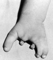
File:Clefthand-apical-defect.jpg ...normality is a deep median cleft of either the hand and/or foot due to the embryonic absence of the central rays. [[Category:Abnormal Development]] [[Category:Limb]] [[Category:Musculoskeletal]](500 × 568 (19 KB)) - 13:43, 15 August 2010
File:Melanoblast migration.png ==Directions of Melanoblast Migration in Embryonic Mouse Skin== ...- Melanocyte Development|Melanocyte Development]] | [[Integumentary System Development|Integumentary]](600 × 210 (40 KB)) - 14:00, 20 September 2016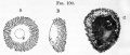
File:Foster109.jpg ==Fig. 109. The human ova during early stages of development== ...iew of an ovum figured by Keichert, supposed to be about thirteen days. e. embryonic area.(873 × 372 (70 KB)) - 06:12, 17 March 2012
File:Bailey422.jpg Showing the partial development of the dendrites of two cells of Purkinje. A, external limiting membrane; B, external (embryonic) granule layer; C, partly formed molecular (plexiform) layer; D, granular l(495 × 568 (95 KB)) - 08:44, 23 June 2015
File:Stage5 bf13.jpg ...es: implantation completed, inner cell mass, bilaminar embryo, trophoblast development * Embryonic disc - 0.132 x 0.1 mm(800 × 537 (142 KB)) - 15:13, 18 March 2014File:Week 6 embryonic development of CNS.jpg ==Week 6 embryonic development of CNS and emergence of pineal evagination.==(1,209 × 794 (165 KB)) - 12:43, 24 October 2014
File:Salamander- early development.jpg ==Salamander- early development== Early development of ''Ensatina eschscholtzii'', an amphibian with a large, yolky egg. Animal(1,200 × 887 (85 KB)) - 14:34, 30 September 2010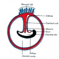
File:Gray0028.jpg ==Fig. 28. Diagram illustrating a later stage in the development of the umbilical cord== ...placental cords the placental blood vessels are initially paired, later in development only a single placental vein remains with a pair of placental arteries. Thi(500 × 500 (50 KB)) - 12:08, 25 February 2014
File:Aneuploidy model based on fragmentation 1.jpg Embryonic development was monitored by time-lapse imaging from the one- to four-cell stage follow :'''Links:''' [[Abnormal Development - Genetic]] | [[Trisomy 21]] | [[Zygote]] | [[Morula]](946 × 907 (154 KB)) - 09:58, 12 January 2015
File:FGF and FGFR expression patterns during endochondral and intramembranous bone development.jpeg ...and FGFR expression patterns during endochondral and intramembranous bone development= ...sification center. (G) Developmental progres- sion of intramembranous bone development. Cells and tissues are color-coded for expression domains of FGFs and FGFRs(800 × 1,215 (317 KB)) - 10:37, 30 April 2020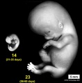
File:Stage14compare23.jpg ...32). These stages represent approximately the middle and at the end of the embryonic period. The embryo images have been scaled to the same magnification (5 mm ...stage 14]] | [[Carnegie stage 23]] | [[Week 5]] | [[Week 8]] | [[Embryonic Development]](593 × 601 (32 KB)) - 00:12, 11 December 2013
File:Model embryo to 32 cell stage icon.jpg ...constructed from original frames for the movie - simulation of embryonic development up to 32 cell stage.(696 × 256 (30 KB)) - 10:44, 11 November 2011
File:Mouse tongue Pax9 expression 01.jpg ==Expression patterns of Pax9 in different taste papillae of the embryonic mouse tongue== * '''B.''' Whole mount X-Gal staining of a Pax9+/LacZ mouse tongue at embryonic day 13.5 (E13.5). Note that expression is also seen in the mesenchyme adjac(1,200 × 687 (259 KB)) - 18:56, 18 June 2018
File:Stage8 sem1.jpg ...ay 18) SEM image showing the notochordal plate. This is an early embryonic development transient cellular structure and region lying above the primitive streak, t(828 × 1,000 (109 KB)) - 08:53, 23 August 2018
File:DickersonMcGurk1982 fig1.1.jpg ==Figure 1.1 Semidiagrammatic drawing of the development of the basic embryonic zones of the cortical plate==(1,280 × 689 (113 KB)) - 12:40, 2 July 2018
File:Mouse-Sp2 mRNA expression.png The white arrowheads denote the boundary between embryonic and extraembryonic tissue. ...) Is Essential for Mouse Development and Autonomous Proliferation of Mouse Embryonic Fibroblasts. PLoS ONE 5(3): e9587. doi:10.1371/journal.pone.0009587(600 × 503 (877 KB)) - 13:16, 30 July 2010
File:Lymphatic vasculature 02.jpg ==Main steps of mammalian lymphatic vascular development== # Lymphatic endothelial cells (LECs) are specified in embryonic veins.(1,280 × 454 (116 KB)) - 15:41, 12 February 2013
File:Limb patterning factors 03.jpg Shh expression in {{ME11.5}} mouse embryonic forelimb. [[Category:Limb]] [[Category:Musculoskeletal]] [[Category:Abnormal Development]] [[Category:Mouse]] [[Category:Mouse E11.5]](800 × 794 (36 KB)) - 22:38, 24 August 2020
File:Limb patterning factors 04.jpg Fgf8 expression in {{ME11.5}} mouse embryonic hindlimb. [[Category:Limb]] [[Category:Musculoskeletal]] [[Category:Abnormal Development]] [[Category:Mouse]] [[Category:Mouse E11.5]](800 × 794 (67 KB)) - 22:44, 24 August 2020
File:Day 19 Newborn Mouse.JPG :'''Links:''' [[Mouse Development]] | [[Mouse Stages]] | [[:Category:Mouse E19.0]] | [[2009_Group_Project_4|S ...ed upon Fig 285, in Dr Karl Theiler’s ‘The House Mouse; Atlas of Embryonic Development’, Springer - Verlag New York Inc, New York, 1989(448 × 518 (18 KB)) - 13:07, 25 June 2014
File:Mouse lung development 03.jpg ==Histological analyses of Mouse Lungs at Various Embryonic Stages== ...al mouse lung (low power)]] | [[Respiratory System Development]] | [[Mouse Development]](540 × 1,200 (349 KB)) - 11:27, 31 August 2017
File:Mouse cornea P0.jpg ==Mouse Corneal Development P0== ...Corneal Epithelial Cell Proliferation and Differentiation during Embryonic Development. PLoS ONE 10(1): e0117089. doi:10.1371/journal.pone.0117089(703 × 561 (101 KB)) - 11:52, 24 January 2015File:Chicken GIT peristalsis movie 01.mp4 Emergence and development of gut motility in the chicken embryo. ...se stages. Our work sets a baseline for further investigations of motility development in this important animal model.(6.19 MB) - 17:19, 1 August 2017
File:Mouse cornea E16.5.jpg ==Mouse Corneal Development E16.5== ...Corneal Epithelial Cell Proliferation and Differentiation during Embryonic Development. PLoS ONE 10(1): e0117089. doi:10.1371/journal.pone.0117089(701 × 562 (113 KB)) - 11:51, 24 January 2015
File:Limb patterning factors 06.jpg Msx2 expression in {{ME12.5}} wild-type mouse embryonic forelimb. [[Category:Limb]] [[Category:Musculoskeletal]] [[Category:Abnormal Development]] [[Category:Mouse]] [[Category:Mouse E12.5]](800 × 794 (41 KB)) - 22:46, 24 August 2020
File:Limb patterning factors 07.jpg Shh expression in {{ME12.5}} wild-type embryonic hindlimb autopod. [[Category:Limb]] [[Category:Musculoskeletal]] [[Category:Abnormal Development]] [[Category:Mouse]] [[Category:Mouse E12.5]](800 × 794 (44 KB)) - 22:46, 24 August 2020
File:External ear stages-14-23-adult.jpg ==Development of the External Ear== ...stage 14]]) through to [[week 8]] ([[Carnegie_stage_23|stage 23]]) showing development of the [[A#auricular hillocks|auricular hillocks]] (tubercles) that will fo(1,000 × 655 (42 KB)) - 09:59, 20 May 2014
File:Day 18 long whiskers.JPG :'''Links:''' [[Mouse Development]] | [[Mouse Stages]] | [[:Category:Mouse E18.0]] | [[2009_Group_Project_4|S ...ed upon Fig 268, in Dr Karl Theiler’s ‘The House Mouse; Atlas of Embryonic Development’, Springer - Verlag New York Inc, New York, 1989(462 × 514 (21 KB)) - 22:14, 15 November 2015
File:Day 6.5 Advanced Endometrial Reaction.JPG :'''Links:''' [[Mouse Development]] | [[Mouse Stages]] | [[:Category:Mouse E6.5]] | [[2009_Group_Project_4|St ...sed upon Fig 52, in Dr Karl Theiler’s ‘The House Mouse; Atlas of Embryonic Development’, Springer - Verlag New York Inc, New York, 1989(425 × 556 (17 KB)) - 13:12, 25 June 2014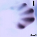
File:Limb patterning factors 05.jpg Sox9 expression in E12.5 wild-type mouse embryonic forelimb. [[Category:Limb]] [[Category:Musculoskeletal]] [[Category:Abnormal Development]] [[Category:Mouse]] [[Category:Mouse E12.5]](800 × 794 (37 KB)) - 22:45, 24 August 2020
File:Gray0026.jpg ==Fig. 26. Diagram showing stage of allantoic development with commencing constriction of the yolk-sac== Diagram showing later stage of allantoic development with commencing constriction of the yolk-sac.(500 × 500 (37 KB)) - 12:05, 25 February 2014
File:Gray0592.jpg ===Development of the Lymphatic Vessels=== ...begins as a series of sacs 108 at the points of junction of certain of the embryonic veins. These lymph-sacs are developed by the confluence of numerous venous(600 × 590 (55 KB)) - 12:49, 15 February 2013
File:Day 7 Amnion.JPG :'''Links:''' [[Mouse Development]] | [[Mouse Stages]] | [[:Category:Mouse E7.0]] | [[2009_Group_Project_4|St ...pon Fig. 64 (1), in Dr Karl Theiler’s ‘The House Mouse; Atlas of Embryonic Development’, Springer - Verlag New York Inc, New York, 1989(425 × 559 (18 KB)) - 13:12, 25 June 2014
File:Day 15 toes separate.JPG :'''Links:''' [[Mouse Development]] | [[Mouse Stages]] | [[:Category:Mouse E15.0]] | [[2009_Group_Project_4|S ...ed upon Fig 236, in Dr Karl Theiler’s ‘The House Mouse; Atlas of Embryonic Development’, Springer - Verlag New York Inc, New York, 1989(425 × 537 (22 KB)) - 13:05, 25 June 2014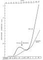
File:Yolk sac and amniotic cavity volume graph.jpg Change in human embryo extra-embryonic cavity volumes {{yolk sac}} and amniotic cavity between week 2 to 3 (stage :'''Links:''' [[Yolk Sac]] | [[Coelomic Cavity Development]] | [[Placenta - Membranes]](719 × 1,000 (50 KB)) - 00:42, 13 April 2018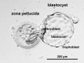
File:Human carnegie stage 3 label.jpg Human Blastocyst "hatching" from zona pellucida, in early [[Embryonic Development]] designated as [[Carnegie stage 3]]. ...ie stage 3 label.jpg|labeled image]] | [[Carnegie stage 3]] | [[Blastocyst Development]] | [[Week 1]](500 × 377 (26 KB)) - 13:39, 29 November 2012
File:Day 17 fingers and toes joined together.JPG :'''Links:''' [[Mouse Development]] | [[Mouse Stages]] | [[:Category:Mouse E17.0]] | [[2009_Group_Project_4|S ...ed upon Fig 257, in Dr Karl Theiler’s ‘The House Mouse; Atlas of Embryonic Development’, Springer - Verlag New York Inc, New York, 1989(425 × 554 (20 KB)) - 13:07, 25 June 2014
File:Ewart1897 02.jpg ...terine milk — from the uterus, and in fixing the embryo during its uterine development. The allantois (all.) never reaches the outer sac. It is vascular, and serv(555 × 600 (44 KB)) - 16:14, 3 May 2013
File:Stage5 bf04.jpg ...es: implantation completed, inner cell mass, bilaminar embryo, trophoblast development * Embryonic disc - 0.132 x 0.1 mm(1,410 × 794 (108 KB)) - 09:51, 6 April 2020
File:Bone signalling pathway.gif =Signals regulating growth plate development= ...feration and maturation through FGFR3 in the growth plate during embryonic development and postnatal bone growth. A number of WNTs expressed by growth plate chond(200 × 122 (12 KB)) - 15:10, 15 October 2016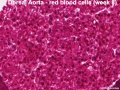
File:Stage 22 image 224.jpg ==Human Embryo Dorsal Aorta - Embryonic Red Blood Cells (Carnegie stage 22)== ...File:Stage 22 image 224.jpg|image RBCs]] | [[Cardiovascular System - Blood Development]] | [[Carnegie stage 22]](600 × 450 (99 KB)) - 12:02, 14 June 2016
File:Day 9 Formation and closure of anterior neuropore.JPG :'''Links:''' [[Mouse Development]] | [[Mouse Stages]] | [[2009_Group_Project_4|Student Project]] ...d upon Fig. 108, in Dr Karl Theiler’s ‘The House Mouse; Atlas of Embryonic Development’, Springer - Verlag New York Inc, New York, 1989(945 × 558 (54 KB)) - 12:56, 25 June 2014
File:Mouse oogenesis 02.jpg ...tion of secondary and antral follicles at 7 and 14 days post-partum (dpp), development of mature gametes as oocyte–granulosa cell complexes, oocyte meiosis and :Links: [[Oocyte Development]] | [[Ovary Development]] | [[Mouse Development]] | [[:File:Mouse oogenesis 01.jpg|full figure 1]](1,386 × 355 (40 KB)) - 10:20, 8 March 2018
File:Mouse - blastocoel formation.jpg ...ortant research directions aimed at alleviating culture-induced changes in embryonic programming. New levels of regulation are emerging and greater insight into(800 × 319 (47 KB)) - 10:05, 12 October 2010
File:Day 8 First Somites.JPG :'''Links:''' [[Mouse Development]] | [[Mouse Stages]] | [[:Category:Mouse E8.0]] | [[2009_Group_Project_4|St ...ed upon Fig. 82, in Dr Karl Theiler’s ‘The House Mouse; Atlas of Embryonic Development’, Springer - Verlag New York Inc, New York, 1989(1,087 × 590 (82 KB)) - 13:10, 25 June 2014
File:Human embryology and developmental biology 4th edn.jpg ...o major families of important developmental molecules. Clinical aspects of development are covered throughout in boxed sections of text. First-rate illustrations ...clear drawings, to help you to memorize and understand normal and abnormal development.(127 × 165 (37 KB)) - 01:04, 23 April 2016
File:FMR1 gene silencing.jpg ==The timing of FMR1 gene silencing during development== ...t (primed cells) of affected fetuses, during the developmental stages when embryonic stem cell lines are established.(1,280 × 1,124 (103 KB)) - 14:09, 5 April 2018
File:Day 13 Anterior Footplate Indented, marked pinna.JPG :'''Links:''' [[Mouse Development]] | [[Mouse Stages]] | [[:Category:Mouse E13.0]] | [[2009_Group_Project_4|S ...ed upon Fig 200, in Dr Karl Theiler’s ‘The House Mouse; Atlas of Embryonic Development’, Springer - Verlag New York Inc, New York, 1989(437 × 470 (18 KB)) - 16:28, 22 October 2014
File:Mouse cornea E12.5.jpg ==Mouse Corneal Development E12.5== ...Corneal Epithelial Cell Proliferation and Differentiation during Embryonic Development. PLoS ONE 10(1): e0117089. doi:10.1371/journal.pone.0117089(700 × 558 (123 KB)) - 11:50, 24 January 2015
File:Blyakher1955 fig03.jpg ==Fig. 3. Table of drawings from K. F. Wolff's dissertation illustrating the development of the chicken== 1 . The embryonic spot (n) from the non-incubated egg in its natural size? b, c - circular(1,028 × 1,253 (219 KB)) - 22:06, 10 May 2018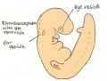
File:Day 9.5 Formation of posterior neuropore and forelimb bud.JPG :'''Links:''' [[Mouse Development]] | [[Mouse Stages]] | [[2009_Group_Project_4|Student Project]] ...d upon Fig. 116, in Dr Karl Theiler’s ‘The House Mouse; Atlas of Embryonic Development’, Springer - Verlag New York Inc, New York, 1989(752 × 568 (37 KB)) - 12:57, 25 June 2014
File:Model embryo to 32 cell stage 001.jpg First image in the simulation of embryonic development up to 32 cell stage.(696 × 256 (22 KB)) - 10:08, 11 November 2011
File:Model embryo to 32 cell stage 240.jpg Last image in the simulation of embryonic development up to 32 cell stage.(696 × 256 (49 KB)) - 10:09, 11 November 2011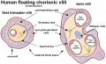
File:Human placental villi cartoon 01.jpg ...tween early ({{first trimester}}) and late ({{third trimester}}) placental development. ...ly shown in the term villi). Later {{Hofbauer cells}} may have a different embryonic origin.(1,084 × 663 (142 KB)) - 09:31, 26 March 2019
File:Stage10 sem10.jpg * note appearance of extra-embryonic mesoderm covering yolk sac (bottom) ...System Development]] | [[Cardiovascular System Development]] | [[Placenta Development]](1,000 × 740 (68 KB)) - 18:17, 28 May 2017
File:Regions of varying neural cell types in ventral neural tube.jpg Image depicting the neural tube (nt) during embryonic development, where the regions of different inter-neurons being V0, V1, V2, and V3, alo(600 × 537 (35 KB)) - 21:09, 28 October 2016
File:Boyden1931 fig02.jpg Ren. I and II, left renal veins representing persistence of primary embryonic renal veins (compare McClure and Butler, fig. 14); Y, remnant of intersupr [[Category:Renal]][[Category:Abnormal Development]][[Category:Historic Embryology]][[Catgeory:1930's]](773 × 860 (75 KB)) - 08:24, 16 September 2017
File:Lung human and mouse Sox expression.jpg ==Respiratory Development Human and Mouse Sox Expression== The lung originates from a region of the embryonic gut, the epithelium of which is called the anterior foregut endoderm (gray(1,280 × 692 (62 KB)) - 13:57, 23 February 2019
File:Patten1938 text-fig04.jpg ==Text-Fig. 4. Sectional plans of the embryonic heart in the frontal plane== Showing extent of growth of the various cardiac septa at several stages of development. These diagrams give specifically for the human embryo a more precise pict(1,000 × 1,432 (316 KB)) - 16:01, 27 February 2017
File:Day 7.5 Neural plate, presomite stage.JPG :'''Links:''' [[Mouse Development]] | [[Mouse Stages]] | [[:Category:Mouse E7.5]] | [[2009_Group_Project_4|St ...pon Fig. 64 (4), in Dr Karl Theiler’s ‘The House Mouse; Atlas of Embryonic Development’, Springer - Verlag New York Inc, New York, 1989(425 × 537 (23 KB)) - 13:11, 25 June 2014
File:CSt3.jpg Human Blastocyst "hatching" from zona pellucida, in early [[Embryonic Development]] designated as [[Carnegie stage 3]]. ...ie stage 3 label.jpg|labeled image]] | [[Carnegie stage 3]] | [[Blastocyst Development]] | [[Week 1]](500 × 377 (20 KB)) - 13:38, 29 November 2012
File:Anderson2016-fig10.jpg ...te, and is supplied through a ventricular septal defect, comparable to the embryonic interventricular communication shown in [[:File:Anderson2016-fig08b.jpg|Fig [[Category:Human]][[Category:Abnormal Development]](800 × 800 (109 KB)) - 10:25, 6 June 2017
File:Bailey073.jpg ...he double wall between the two would be the embryonic disk. The precocious development of the mesoderm, which as a loosely arranged tissue fills in all the space(705 × 501 (122 KB)) - 04:08, 13 April 2011
File:Mouse embryo vascular.png ==Surface Renderings of Embryonic Vascular Structures== ...Mouse Cephalic Plexus]] | [[Cardiovascular System Development]] | [[Mouse Development]](600 × 576 (518 KB)) - 13:34, 18 July 2019
File:Stage 13 image 097.jpg ===Gastrointestinal Development=== [[Gastrointestinal Tract Development]](1,000 × 720 (162 KB)) - 15:21, 27 April 2013
File:Mouse lung development 02.jpg ==Histological analyses of Mouse Lungs at Various Embryonic Stages== :'''Links:''' [[:File:Mouse lung development 01.jpg|Full image]](922 × 922 (239 KB)) - 15:08, 25 August 2011
File:Human embryonic shoulder girdle 04.jpg ==Human Embryonic Shoulder Girdle== Fawcett '''The Development and Ossification of the Human Clavicle.''' J Anat Physiol: 1913, 47(Pt 2);2(1,000 × 755 (71 KB)) - 13:46, 22 May 2017
File:Stage10 sem9a.jpg * note appearance of extra-embryonic mesoderm covering yolk sac (bottom) ...System Development]] | [[Cardiovascular System Development]] | [[Placenta Development]](592 × 800 (51 KB)) - 12:04, 17 March 2011
File:Stage10 sem9b.jpg * note appearance of extra-embryonic mesoderm covering yolk sac (bottom) ...System Development]] | [[Cardiovascular System Development]] | [[Placenta Development]](444 × 600 (32 KB)) - 12:04, 17 March 2011
File:Stage10 sem9c.jpg * note appearance of extra-embryonic mesoderm covering yolk sac (bottom) ...System Development]] | [[Cardiovascular System Development]] | [[Placenta Development]](296 × 400 (16 KB)) - 12:01, 17 March 2011
File:Stage10 sem10a.jpg * note appearance of extra-embryonic mesoderm covering yolk sac (bottom) ...System Development]] | [[Cardiovascular System Development]] | [[Placenta Development]](800 × 592 (48 KB)) - 12:04, 17 March 2011
File:Stage10 sem10b.jpg * note appearance of extra-embryonic mesoderm covering yolk sac (bottom) ...System Development]] | [[Cardiovascular System Development]] | [[Placenta Development]](600 × 444 (32 KB)) - 12:04, 17 March 2011
File:Stage10 sem10c.jpg * note appearance of extra-embryonic mesoderm covering yolk sac (bottom) ...System Development]] | [[Cardiovascular System Development]] | [[Placenta Development]](400 × 296 (16 KB)) - 12:04, 17 March 2011
File:Stage10 sem9.jpg * note appearance of extra-embryonic mesoderm covering yolk sac (bottom) ...System Development]] | [[Cardiovascular System Development]] | [[Placenta Development]](740 × 1,000 (72 KB)) - 12:03, 17 March 2011
File:Ewart1897 07.jpg ...consists of a vascular allantoic core, and a thin capsule derived from the embryonic sac. The villi are represented on too large a scale, but the embryo and yol ...ts oxygen, and also fluids containing all the ingredients required for the development and growth of the embryo. There is, however, no actual mixing of the matern(1,200 × 757 (167 KB)) - 15:48, 3 May 2013
File:Thyroid branching model cartoon.jpg Author proposed models of branching morphogenesis in thyroid development. ...nnects the embryonic thyroid from the pharyngeal endoderm. In late thyroid development, branching growth stimulated by Fgf10 derived from adjacent mesenchyme prom(860 × 1,280 (67 KB)) - 13:19, 9 March 2018
File:Day 14 Fingers Separate.JPG :'''Links:''' [[Mouse Development]] | [[Mouse Stages]] | [[:Category:Mouse E14]] | [[2009_Group_Project_4|Stu ...216 and Fig 217, in Dr Karl Theiler’s ‘The House Mouse; Atlas of Embryonic Development’, Springer - Verlag New York Inc, New York, 1989(517 × 541 (28 KB)) - 16:27, 22 October 2014
File:Mouse embryo meiotic to mitotic spindle 01.jpg ...essive transition from meiosis to mitosis throughout mouse preimplantation development that can be subdivided into three phases: # first three embryonic divisions - mechanism of acentrosomal spindle formation is largely shared w(800 × 684 (86 KB)) - 12:58, 3 May 2013
File:Eosin staining of mouse embryonic skin.png ==Image of Hematoxylin/ eosin staining of mouse embryonic skin== ...n describing what the figure is showing and how this relates to fetal skin development. No terms are described.(629 × 251 (283 KB)) - 23:53, 8 November 2014
File:Bat-embryonic stages 11-22.jpg ==Bat Embryonic Development== Images of the bat embryos of ''H. pratti'' at embryonic Stages 11-22.(600 × 703 (108 KB)) - 12:42, 3 July 2012
File:Day 8.5 Turning of embryo.JPG :'''Links:''' [[Mouse Development]] | [[Mouse Stages]] | [[:Category:Mouse E8.5]] | [[2009_Group_Project_4|St ...sed upon Fig 94, in Dr Karl Theiler’s ‘The House Mouse; Atlas of Embryonic Development’, Springer - Verlag New York Inc, New York, 1989(1,173 × 557 (65 KB)) - 13:09, 25 June 2014
File:P. maniculatus embryo E17.5-21.5.PNG Davis SW, Keisler JL (2016) Embryonic Development of the Deer Mouse, Peromyscus maniculatus. PLoS ONE 11(3): e0150598. https:(1,922 × 1,724 (4.8 MB)) - 17:00, 24 August 2017
File:Melanocyte Pathway.gif ...marked by asterisk). Following metamorphosis or melanocyte ablation of the embryonic pattern, zebrafish glial-pigment cell progenitors proliferate and migrate a(440 × 351 (33 KB)) - 15:57, 30 October 2018
File:Thomas Louis Jerome Auzoux.jpg ...ry in St. Aubin d'Ecrosville. These included models of embryonic and fetal development. Commercial model production continued after Dr. Auzoux's death and form pa(1,214 × 1,662 (906 KB)) - 08:49, 1 November 2018
File:Mouse cornea E13.5.jpg ==Mouse Corneal Development E13.5== ...Corneal Epithelial Cell Proliferation and Differentiation during Embryonic Development. PLoS ONE 10(1): e0117089. doi:10.1371/journal.pone.0117089(700 × 550 (103 KB)) - 11:50, 24 January 2015
File:Stage5 bf03.jpg ...es: implantation completed, inner cell mass, bilaminar embryo, trophoblast development * Embryonic disc - 0.132 x 0.1 mm(800 × 537 (132 KB)) - 09:30, 4 October 2018
File:Kollmann379.jpg The embryo at this stage of development of the embryonic disc(827 × 454 (45 KB)) - 12:02, 20 October 2011
File:Day 10 Closure of posterior neuropore, hind limb bud and tail bud.JPG :'''Links:''' [[Mouse Development]] | [[Mouse Stages]] | [[2009_Group_Project_4|Student Project]] ...29 and Fig. 134, in Dr Karl Theiler’s ‘The House Mouse; Atlas of Embryonic Development’, Springer - Verlag New York Inc, New York, 1989(1,089 × 555 (76 KB)) - 12:58, 25 June 2014
File:Congdon1922-plate01.jpg ==Plate 1. Human Embryonic Vascular Development== ...he arterial system of a 22-somite embryo. The first arch is at its maximum development and the dorsal and ventral outgrowths, which are to aid in the formation of(877 × 1,200 (145 KB)) - 21:21, 6 November 2018
File:Human liver week 9.jpg Paraffin-embedded sections of human embryonic liver at 9 weeks ({{GA}} 11 weeks.) :'''Links:''' [[Gastrointestinal_Tract_-_Liver_Development|Liver Development]](1,200 × 991 (425 KB)) - 13:19, 6 June 2014
File:Bailey110.jpg ...tery breaking up into capillary network' groups of fat cells developing in embryonic connective tissue. ...e there is also a small amount of fibrous tissue present. From the mode of development a small artery usually affords the blood supply for each lobule.(481 × 643 (110 KB)) - 10:37, 16 July 2012
File:Picture 1.JPG === '''Figure 1:''' Illustration of Mouse embryonic stages of development from fertilization to zona free blastocyst (stages 1 to 5). E.Newton 2009==(1,593 × 567 (109 KB)) - 01:25, 3 August 2011
File:Cervical ectopic ultrasound.jpg A gestational sac with a small embryonic pole with a fetal heartbeat of 122bpm located in the cervix below the scar [[Category:Ultrasound]] [[Category:Human Fetus]] [[Category:Abnormal Development]] [[Category:Ectopic Pregnancy]](800 × 541 (62 KB)) - 12:30, 1 June 2019
File:Mouse cornea development 01.jpg ==Mouse Corneal Development== ...Corneal Epithelial Cell Proliferation and Differentiation during Embryonic Development. PLoS ONE 10(1): e0117089. doi:10.1371/journal.pone.0117089(1,200 × 880 (325 KB)) - 11:48, 24 January 2015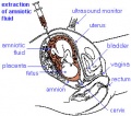
File:Amniocentesis.jpg Amniotic fluid is taken from the uterus, sent to a diagnostic laboratory and embryonic cells isolated from the amniotic fluid. [[Category:Abnormal Development]] [[Category:Genetic Abnormalities]] [[Category:Human Embryo]] [[Category:H(284 × 250 (31 KB)) - 13:52, 28 April 2012
File:Mammary gland luminal cell model.jpg ==Model for luminal cell hierarchy during mammary gland development== ...ressing (N1pos) multipotent stem cells exist only during embryonic mammary development, when they co-express myoepithelial cytokeratin (K5) and luminal cytokerati(1,200 × 1,680 (170 KB)) - 21:16, 14 March 2015
File:Cytomegalovirus.jpg Image shows human embryonic lung infected by cytomegalovirus demonstrated by an immunofluorescent techn [[Category:Virus]] [[Category:Abnormal Development]] [[Category:Environmental Abnormalities]](700 × 472 (49 KB)) - 10:07, 1 November 2011
File:Stage5 bf05.jpg ...es: implantation completed, inner cell mass, bilaminar embryo, trophoblast development * Embryonic disc - 0.132 x 0.1 mm(1,200 × 692 (256 KB)) - 17:56, 4 April 2015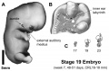
File:Stage 19 ear.jpg Stage 19 embryo (week 7) showing the ear development features. ...d shape of the inner ear labyrinth at weeks 6, 7, and 8; by the end of the embryonic period (week 8) it approximates the shape of the adult structure.(1,200 × 786 (116 KB)) - 19:01, 6 May 2011
File:Hertig1956 fig84.jpg ...may have resulted from failure of proper trophoblastic development at the embryonic pole. The polypoid protrusion may have caused mechanical pressure from the(734 × 713 (92 KB)) - 14:38, 25 February 2017
File:Day 12 Earliest signs of fingers.JPG :'''Links:''' [[Mouse Development]] | [[Mouse Stages]] | [[:Category:Mouse E12.0]] | [[2009_Group_Project_4|S ...83 and Fig. 184, in Dr Karl Theiler’s ‘The House Mouse; Atlas of Embryonic Development’, Springer - Verlag New York Inc, New York, 1989(510 × 466 (25 KB)) - 13:03, 25 June 2014
File:- compound mouse mutants demonstrate partially rescued upper and lower molars.png ...mutants showed rescue from bud to cap phase transition in embryonic dental development. Both upper and lower molars showed an enlargement in the tooth buds (H, K, ...ription of the genes shown in the figure or how they relate to fetal tooth development.(539 × 258 (289 KB)) - 23:56, 8 November 2014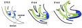
File:Mouse cloacal septation 01.jpg ...the ventral part of the cloaca (cl) extends rostrally to give rise to the embryonic urogenital sinus and the primordium of the bladder (bl). ...ointestinal Tract Development]] | [[Genital System Development]] | [[Mouse Development]](917 × 318 (47 KB)) - 09:02, 18 January 2013
File:Regulation of Nodal-Activin signalling during heart formation.png ...ling activity and gradients in mammals during the early stage of embryonic development provides clues for designing the initial stages of human pluripotent stem c(696 × 511 (238 KB)) - 12:26, 26 October 2017
File:Florian1930-text-fig02.jpg ==Text-fig 2. Schemes of the chief stages in the development of the human embryo== ...udally and dorsally. The yolk-sac correspondingly grows forwards below the embryonic plate almost up to the cranial end of the latter, and also grows in the cau(394 × 377 (20 KB)) - 19:30, 23 September 2015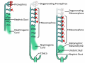
File:Stages of nephrogenesis.png ...eraction with metanephric mesenchyme <ref>Sawle. A. 2009. ''Development of embryonic nephrons.png''. Retrieved September 22, 2017, from(4,059 × 3,000 (1.8 MB)) - 14:30, 26 October 2017
File:Elizabeth M. Ramsey.jpg ...lacental circulation. She published extensively on placental and embryonic development and this paper uses both human and monkey placentas from the [[Carnegie Col(333 × 500 (27 KB)) - 07:20, 13 December 2018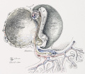
File:Cullen1916 fig02.jpg vitelline arteries and veins are clearly seen on the embryonic side of the yolk-sac. The amnion is now gradually as yet no umbilical cord. For the first stage of its development, See [[:File:Cullen1916 fig03.jpg|Fig. 3]]. folding of the exocoe1om. The a(1,280 × 1,110 (403 KB)) - 09:21, 28 October 2018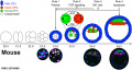
File:Mouse blastocyst development 01.jpg ==Schematic of Mouse Early Embryonic development== ...shows immunostaining of embryos at different stages during preimplantation development. Color coding is the same as in the panel above. The timing of the 4 differ(1,280 × 682 (149 KB)) - 16:05, 9 August 2017
File:Day 10.5 Deep Lens Indentation.JPG :'''Links:''' [[Mouse Development]] | [[Mouse Stages]] | [[:Category:Mouse E10.5]] | [[2009_Group_Project_4|S ...38 and Fig. 139, in Dr Karl Theiler’s ‘The House Mouse; Atlas of Embryonic Development’, Springer - Verlag New York Inc, New York, 1989(514 × 483 (28 KB)) - 13:00, 25 June 2014
File:Stage5 bf11b.jpg '''Facts:''' About 12 days, embryonic disc, 0.204 x 0.165 mm, chorionic cavity, 0.55 x 0.498 mm and chorion, 0.94 ...''' implantation completed, inner cell mass, bilaminar embryo, trophoblast development(464 × 600 (96 KB)) - 07:50, 25 September 2011
File:Stage5 bf22b.jpg '''Facts:''' About 12 days, embryonic disc, 0.204 x 0.165 mm, chorionic cavity, 0.55 x 0.498 mm and chorion, 0.94 ...''' implantation completed, inner cell mass, bilaminar embryo, trophoblast development(516 × 800 (95 KB)) - 10:49, 21 February 2017
File:Mouse eye cell proliferation E13.5.jpg ...Corneal Epithelial Cell Proliferation and Differentiation during Embryonic Development. PLoS ONE 10(1): e0117089. doi:10.1371/journal.pone.0117089(875 × 1,244 (227 KB)) - 12:59, 24 January 2015
File:Day 11.5 Lens Vesicle completely separated from surface.JPG :'''Links:''' [[Mouse Development]] | [[Mouse Stages]] | [[:Category:Mouse E11.5]] | [[2009_Group_Project_4|S ...64 and Fig. 165, in Dr Karl Theiler’s ‘The House Mouse; Atlas of Embryonic Development’, Springer - Verlag New York Inc, New York, 1989(511 × 484 (29 KB)) - 13:01, 25 June 2014
File:Stage5 bf22L.jpg '''Facts:''' About 12 days, embryonic disc, 0.204 x 0.165 mm, chorionic cavity, 0.55 x 0.498 mm and chorion, 0.94 ...''' implantation completed, inner cell mass, bilaminar embryo, trophoblast development(516 × 800 (110 KB)) - 13:39, 22 April 2012
File:Summary Figure of Notch in Cardiac Development.jpeg ...t. E, Notch signaling modulates coronary vessel morphogenesis in which the embryonic epicardium actively participates. F, Notch signaling regulates cardiac cond(800 × 289 (61 KB)) - 09:23, 16 September 2016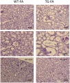
File:The kidney of a FA-injected mouse compared to a wildtype mouse.jpg ...nic line A homozygous mice (right)that have been injected with Gremlin, an embryonic gene that plays a role in nephrogenesis. These images show what can occur t ...ctor shown by their gene name in the figure and related it to fetal kidney development.(763 × 900 (324 KB)) - 21:40, 8 November 2014
File:Day 16 Reposition of umbilical hernia.JPG :'''Links:''' [[Mouse Development]] | [[Mouse Stages]] | [[:Category:Mouse E16.0]] | [[2009_Group_Project_4|S ...345 and Fig 246, in Dr Karl Theiler’s ‘The House Mouse; Atlas of Embryonic Development’, Springer - Verlag New York Inc, New York, 1989(640 × 532 (34 KB)) - 16:29, 22 October 2014
File:Day 11 Closure of lens vesicle.JPG :'''Links:''' [[Mouse Development]] | [[Mouse Stages]] | [[:Category:Mouse E11]] | [[2009_Group_Project_4|Stu ...51 and Fig. 152, in Dr Karl Theiler’s ‘The House Mouse; Atlas of Embryonic Development’, Springer - Verlag New York Inc, New York, 1989(560 × 528 (27 KB)) - 13:19, 18 August 2014File:Quail HH stage 2 fibronectin movement.mov :Links: [[Chicken Development]] | [[Gastrulation]] ...looked. We developed computational/optical methods that measure the extent embryonic cells move relative to the extracellular matrix. Our time-lapse data show t(3.07 MB) - 08:42, 5 February 2013File:Quail HH stage 2 fibronectin movement.mp4 :Links: [[Chicken Development]] | [[Gastrulation]] ...looked. We developed computational/optical methods that measure the extent embryonic cells move relative to the extracellular matrix. Our time-lapse data show t(2.68 MB) - 08:42, 5 February 2013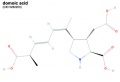
File:Domoic acid.jpg ...injection into maternal rat led to detectable levels in amniotic fluid and embryonic brain tissue within 1 hour. :Links: [[Abnormal Development - Biological Toxins|Biological Toxins]] | [[Abnormal_Development_-_Chemical(500 × 342 (15 KB)) - 10:35, 5 November 2011
File:Mouse tongue Pax9 expression 02.jpg ==Expression patterns of Pax9 in different taste papillae of the embryonic mouse tongue==(1,200 × 833 (299 KB)) - 18:55, 18 June 2018
File:Mouse E11.5 Bmp4 01.jpg ...li Activity Is Critical at Multiple Stages of Embryonic Mammary and Nipple Development. PLoS ONE 8(11): e79845. doi:10.1371/journal.pone.0079845(700 × 526 (41 KB)) - 10:36, 10 July 2014
File:Mouse E11.0 Bmp4 01.jpg ...li Activity Is Critical at Multiple Stages of Embryonic Mammary and Nipple Development. PLoS ONE 8(11): e79845. doi:10.1371/journal.pone.0079845(700 × 526 (38 KB)) - 13:16, 18 August 2014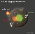
File:Mouse zygote pronuclei 01.jpg ...hydroxymethylcytosine (green) is a modified base form of cytosine found in embryonic stem cells, thought to have a role in regulating gene expression ...[[Zygote]] | [[Molecular_Development_-_Epigenetics|Epigenetics]] | [[Mouse Development]](800 × 784 (52 KB)) - 10:33, 31 July 2015
File:Stage22 ear.jpg ==Human Embryo (week 8, Carnegie stage 22) - Inner Ear Development== Stage 22 embryo (week 8) showing the embryo near the end of the embryonic period.(1,000 × 655 (80 KB)) - 17:32, 6 May 2011
File:Mouse-olfactory nerve pathway development.jpg ==Development of the olfactory nerve pathway== Miller et al. Neural Development 2010 5:20 doi:10.1186/1749-8104-5-20(600 × 1,764 (464 KB)) - 10:57, 31 July 2019
File:Anderson2016-fig12a.jpg ...and the crest of the muscular ventricular septum. This is the reorientated embryonic interventricular foramen. ...een the atrioventricular cushions, which have yet to fuse at this stage of development. This shows that there is a large defect, well described as an atrioventric(800 × 800 (120 KB)) - 18:11, 16 February 2017
File:Bat limb 02.jpg ...f the bat embryo ''Miniopterus schreibersii fuliginosus'' left forelimb at embryonic Stages 13-17. Original stage images have been scaled to approximately the s [[Category:Animal Development]][[Category:Bat]] [[Category:Embryo Stages]] [[Category:Limb]](1,200 × 430 (59 KB)) - 15:27, 25 June 2015
File:Quail HH stage 2 fibronectin movement.jpg ...]] | [[Media:Quail_HH_stage_2_fibronectin_movement.flv|Flash]] | [[Chicken Development]] | [[Gastrulation]] ...looked. We developed computational/optical methods that measure the extent embryonic cells move relative to the extracellular matrix. Our time-lapse data show t(729 × 256 (47 KB)) - 08:45, 5 February 2013
File:Mouse oogenesis 01.jpg ...tion of secondary and antral follicles at 7 and 14 days post-partum (dpp), development of mature gametes as oocyte–granulosa cell complexes, oocyte meiosis and :Links: [[Oocyte Development]] | [[Ovary Development]] | [[Mouse Development]](1,781 × 1,222 (173 KB)) - 10:14, 8 March 2018
File:Hertig1956 plate02.jpg ...Horizon Va, possessing solid syncytio- and cytotrophoblast without lacunar development, an amniotic cavity and/or an early amnion, a simple hilaminar germ disc an ...r less discrete multinucleated cells. The solid disc of trophoblast at the embryonic (implantation) pole is continuous with the membranous unaltered blastocyst(1,280 × 2,009 (482 KB)) - 10:57, 24 February 2017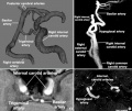
File:Trigeminal artery 01.jpg * '''A''' - The most common persistent embryonic carotid-vertebrobasilar anastomosis is the trigeminal artery. A persistent [[Category:Cardiovascular]][[Category:Abnormal Development]](947 × 800 (102 KB)) - 23:48, 8 September 2019
File:Hertig1956 fig04.jpg Normal preimplantation stages in human development; one segmenting egg found in the tube, Streeter Horizon II, and two free in ...unit in this and the two contiguous sections. Note that the nuclei of the embryonic blastomeres, although of equal size, are less vesicular and contain denser(745 × 562 (41 KB)) - 10:33, 24 February 2017
File:Cerebral blood supply development 01.jpg ==Cerebral Blood Supply Development== Embryonic stage(1,200 × 460 (67 KB)) - 09:09, 25 July 2019
File:Anderson2016-fig12b.jpg ...een the atrioventricular cushions, which have yet to fuse at this stage of development. This shows that there is a large defect, well described as an atrioventric ...and the crest of the muscular ventricular septum. This is the reorientated embryonic interventricular foramen.(800 × 800 (138 KB)) - 15:39, 24 May 2017
File:Mouse - forebrain Robo3 expression.jpg ...C, G, K) levels of the embryonic mouse forebrain during preplate stages of development (E11.5–13.5). ...ih.gov/omim/608630 OMIM - ROBO3] | [[Mouse Development]] | [[Neural System Development]](675 × 1,280 (289 KB)) - 12:19, 8 November 2010
File:Florian1930-text-fig05.jpg ==Text-fig 5. Schemes of the chief stages in the development of the human embryo== ...rther caudally. The primitive streak occupies the whole caudal half of the embryonic plate and the primordium of the head process has appeared. The end-knot of(659 × 444 (40 KB)) - 19:32, 23 September 2015
File:Zebrafish neural crest model.jpg ==Model for the role of H3.3-dependent histone replacement during CNC development== At the early embryonic blastula stage, cells have a broad potential with cis-regulatory elements f(600 × 480 (61 KB)) - 16:06, 14 October 2012
File:Hertig1956 plate01.jpg Normal preimplantation stages in human development; one segmenting egg found in the tube, Streeter Horizon II, and two free in ...unit in this and the two contiguous sections. Note that the nuclei of the embryonic blastomeres, although of equal size, are less vesicular and contain denser(1,280 × 1,904 (351 KB)) - 00:15, 7 June 2018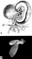
File:Hertig1946b fig09.jpg ...f of a 2.5 mm embryo representing a stage in the middle of the 4th week of development. The circulation is complete and the heart functions at this stage. Note th ...omphalo-mesenteric duct and its connection with yolk-sac and the primitive embryonic gut. Note the communication between the exocoelomic space around the yolk-s(800 × 1,481 (169 KB)) - 17:34, 7 August 2017
File:Liver hepatocyte from stem cell.png Genes specifically expressed at different steps during liver development are shown, in italics, under each step of lineage commitment. Cytokines use :"The protocol supports the stepwise differentiation of human embryonic stem cells (ESC) to cells with characteristics of primitive streak (PS)/mes(600 × 444 (96 KB)) - 17:56, 25 August 2010
File:Mouse Wnt signaling 01.jpg .... Expression of GFP mimics canonical Wnt/ß -catenin signaling activity. At embryonic day 10.5, as shown by live imaging, GFP expression is observed in the neura(600 × 450 (156 KB)) - 16:02, 6 October 2015
File:Hertig1956 fig11-12.jpg ...Horizon Va, possessing solid syncytioand cytotrophoblast without. lacunar development, an amniotic cavity and/or an early amnion, a simple hilaminar germ disc an ...s, hut interpreted as being slightly older than 8225 hecause of germ-disc. development. and greater trophoblastic maturity. Note greatest concentration of dark sy(1,280 × 655 (150 KB)) - 13:32, 31 October 2017
File:Patten 1953.jpg Recent contributions in two widely different aspects of human development have induced the author to prepare a second edition of this well known and ...es. Chapter 4 entitled Cleavage, Germ Layers arid the Establishment of the Embryonic Body, and Chapter 6 dealing with Fetal membranes and Placenta have been ext(781 × 1,262 (68 KB)) - 10:25, 8 March 2015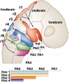
File:Hindbrain neural crest migration.jpg | width=500px|A schematic diagram of a chick head at embryonic day two ([[Hamburger Hamilton Stages]]), showing pathways of neural crest m :'''Links:''' [[Developmental Signals - Homeobox|Homeobox]] | [[Neural Crest Development]](450 × 545 (48 KB)) - 15:06, 16 July 2018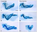
File:Chicken- wing cartilage.jpg ==Alcian-blue staining of the cartilaginous skeleton of embryonic chicken wings== ...Development]] | [[Musculoskeletal_System_-_Cartilage_Development|Cartilage Development]](1,200 × 1,022 (218 KB)) - 13:55, 22 September 2010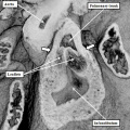
File:Anderson2016-fig42b.jpg ...onic interventricular communication, equivalent to CS23, or eight weeks of development, in the human.(800 × 800 (111 KB)) - 23:14, 16 February 2017
File:Mouse ventral body wall development 01.jpg ==Mouse Ventral Body Wall Development== The development of the ventral body wall and the pelvic girdle.(1,200 × 635 (130 KB)) - 15:59, 2 September 2011
File:Human embryonic shoulder girdle 02.jpg ==Human Embryonic Shoulder Girdle== Fawcett '''The Development and Ossification of the Human Clavicle.''' J Anat Physiol: 1913, 47(Pt 2);2(1,025 × 713 (109 KB)) - 08:44, 15 April 2012
File:Anderson2016-fig42a.jpg ...onic interventricular communication, equivalent to CS23, or eight weeks of development, in the human.(800 × 800 (90 KB)) - 17:55, 17 February 2017
File:Duodenal atresia 01.jpg ...erentiation of the fetus. Such defects can arise at any stage of embryonic development, vary greatly in type and severity, and are caused by a wide variety of det [[Category:Abnormal Development]][[Category:Duodenum]][[Category:Genetics]][[Category:Trisomy 21]][[Categor(750 × 592 (56 KB)) - 16:21, 16 April 2019
File:Mouse placenta 01.jpg ...lacental blood vessels Lateral View]] | [[Mouse Development]] | [[Placenta Development]] ...ch Institute who provided both images and information on (mouse) placental development. (Email: [mailto:s.dunwoodie@victorchang.unsw.edu.au Sally Dunwoodie])(429 × 463 (63 KB)) - 15:38, 2 June 2012
File:Human CS13-15 otic vesicle 01.jpg (A) FFPE slides containing embryonic tissues. :'''Links:''' [[Hearing - Inner Ear Development]] | [[Molecular Development - microRNA]] | [[Carnegie stage 13]] | [[Carnegie stage 14]] | [[Carnegie s(1,574 × 1,779 (364 KB)) - 12:31, 6 April 2018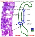
File:Stage14 respiratory tract.jpg ==Stage14 Respiratory Tract Development== ...gut and the early respiratory system at about stage 13 to 14 (week 4-5) of development.(406 × 431 (76 KB)) - 21:13, 24 February 2019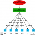
File:Molecular endocrine pancreas cells 01.jpg ==Molecular Development of Endocrine Pancreas Cells== ...both gastrin and ghrelin are frequently co-expressed with glucagon in the embryonic pancreas.(800 × 804 (71 KB)) - 17:36, 6 April 2018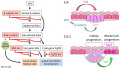
File:Integumentary touch dome model 01.jpg ...n of the sequential signaling mechanisms that drive TD MC specification in embryonic mouse skin. TD MC specification requires mesenchymal Wnt signaling which al ...ade of Wnt, Eda, and Shh Signaling Is Essential for Touch Dome Merkel Cell Development. PLoS Genet 12(7): e1006150. doi:10.1371/journal.pgen.1006150(1,280 × 724 (144 KB)) - 14:41, 12 August 2017
File:Mouse placenta 02.jpg ...placental blood vessels lateral View]] | [Mouse Development]] | [[Placenta Development]] ...ch Institute who provided both images and information on (mouse) placental development. (Email: [mailto:s.dunwoodie@victorchang.unsw.edu.au Sally Dunwoodie])(464 × 220 (21 KB)) - 16:00, 2 June 2012
File:Bat limb 01.jpg Images of the bat embryo ''Miniopterus schreibersii fuliginosus'' at embryonic Stages 13-17. [[Category:Animal Development]][[Category:Bat]] [[Category:Embryo Stages]] [[Category:Limb]](1,688 × 699 (149 KB)) - 15:26, 25 June 2015
File:Mouse pineal E15 to E21 neuroepithelium 01.jpg ...icroscopy of immunolabeled sagittal sections of rat pineal gland (PG) from embryonic day (E) 15 to E21. Both male and female rat embryos were used. ...Panels A3-G3 and A4-G4 show Pax6 and vimentin expression for each stage of development, respectively. Pineal organogenesis begins around E15 as an evagination of(1,280 × 2,123 (1.1 MB)) - 22:37, 20 May 2019
File:Stage18 em11.jpg Note at this embryonic stage of development the simple appearance of the lower lip and lower jaw (mandible) compared to(800 × 569 (99 KB)) - 08:53, 14 May 2014
File:Stage18 em01.jpg Note at this embryonic stage of development the simple appearance of the lower lip and lower jaw (mandible) compared to(800 × 569 (81 KB)) - 07:58, 23 February 2014
File:Zebrafish nephrogenesis signaling01.jpg ...cation factors impact etv4 in other embryonic locales to affect pronephros development. ...id|Retinoic acid]] | [[Developmental Signals - Notch|Notch]] | [[Zebrafish Development]](508 × 324 (29 KB)) - 22:48, 21 March 2018
File:Developmental gene expression context model.jpg ...n) and their hypothetical expression pattern at a given stage of embryonic development are shown. Embryos coloured blue show the activity of a given reporter gene(1,200 × 311 (48 KB)) - 16:52, 15 June 2015
File:Bailey089.jpg ...oximal and distal portion; the latter is the beginning of the foot. During development the fore-limb is always at a slightly more advanced stage than the hind-lim(482 × 690 (55 KB)) - 08:39, 13 April 2011
File:Bailey082.jpg ==Fig. 82. Diagrams representing stages of development of the human embryo== ...of von Spec's embryo (Fig. 77) . Only a part of the chorion is shown; the embryonic disk is slightly constricted from the yolk sac; note the belly stalk, compa(879 × 756 (137 KB)) - 04:28, 13 April 2011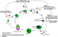
File:Model of Dazl germ cell function.jpg Green signifies Oct4ΔPE:GFP expression during the lifecycle of germ cell development. ...tive cells, and do not display stage specific genetic markers of germ cell development, or erasure of imprinting methylation marks at the H19 locus in males.(1,000 × 638 (81 KB)) - 11:47, 27 April 2020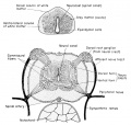
File:Rugh 119.jpg ==Development of the Spinal Cord of the Frog== ...are made up of the rapidly multiplying germinal neuroblasts (primitive or embryonic precursors of the neurons) and the supporting small and stellate cells know(842 × 800 (127 KB)) - 16:22, 30 April 2013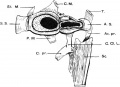
File:Human embryonic shoulder girdle 01.jpg ==Human Embryonic Shoulder Girdle== Fawcett '''The Development and Ossification of the Human Clavicle.''' J Anat Physiol: 1913, 47(Pt 2);2(1,000 × 726 (81 KB)) - 11:07, 5 January 2011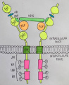
File:FGFR receptor subtype.jpeg ...ons and regulations of fibroblast growth factor signaling during embryonic development''](1,092 × 1,334 (130 KB)) - 17:09, 27 October 2016
File:Mouse lung development 01a.jpg ==Histological analyses of Mouse Lungs at Various Embryonic Stages== ...seen but sections from knockout embryos showed a delay in this sequence of development (panel A, l, p and t).(800 × 1,003 (495 KB)) - 13:01, 28 September 2015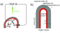
File:Signalling factors in lung branching cartoon.png Menshykau D, Kraemer C, Iber D (2012) Branch Mode Selection during Early Lung Development. PLoS Comput Biol 8(2): e1002377. doi:10.1371/journal.pcbi.1002377 ...is no further description to identify when this signalling process occurs (embryonic/fetal). There should also be an explanation of the factors in the figure.(1,201 × 657 (219 KB)) - 14:56, 9 November 2014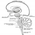
File:Gray0678.jpg ...e upper, greatly expanded part of the central nervous system. In its early embryonic condition it consists of three hollow vesicles, termed the hind-brain or rh Thus in the process of development the wall of the hind-brain undergoes modification to form the {{medulla obl(600 × 598 (35 KB)) - 13:18, 1 April 2019
File:Kollmann698.jpg ==Fig. 698. The embryonic bulbus two human embryos at different stages of development of the eye cup==(689 × 337 (45 KB)) - 18:58, 22 October 2012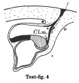
File:Florian1930-text-fig04.jpg ==Text-fig 4. Schemes of the chief stages in the development of the human embryo== ...ich, however, it has nothing to do, because it disappears during the later development by opening out into the yolk-sac cavity (Florian-Volker, 1929).(423 × 441 (31 KB)) - 19:31, 23 September 2015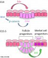
File:Integumentary touch dome model 03.jpg ...n of the sequential signaling mechanisms that drive TD MC specification in embryonic mouse skin. ...ade of Wnt, Eda, and Shh Signaling Is Essential for Touch Dome Merkel Cell Development. PLoS Genet 12(7): e1006150. doi:10.1371/journal.pgen.1006150(603 × 724 (73 KB)) - 14:42, 12 August 2017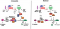
File:Hh signalling pathway.jpg |A signalling protein widely expressed in the tissue during embryonic development of vertebrates and some other species such as ''Drosophila'', and acts to p ...a'' hedgehog protein found in vertebrates, playing a key role in embryonic development of humans.(800 × 394 (53 KB)) - 11:23, 27 October 2016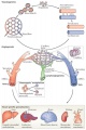
File:Vasculature development 01 cartoon.jpg ==Vasculature Development from Endothelial Progenitor Cells== ...e arterial (red) or venous (blue) fates and coalesce to generate the first embryonic blood vessels, the dorsal aorta and cardinal vein.(599 × 900 (106 KB)) - 13:17, 15 February 2013
File:Foster Balfour Sedgwick and Heap 1883.jpg ...|General Development]] | [[Book_-_The_Elements_of_Embryology_-_Mammalian_2|Embryonic Membranes and Yolk-Sac]] | [[Book_-_The_Elements_of_Embryology_-_Mammalian_(716 × 1,057 (89 KB)) - 21:36, 28 March 2012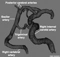
File:Trigeminal artery 02.jpg The most common persistent embryonic carotid-vertebrobasilar anastomosis is the trigeminal artery (TA), with a r [[Category:Cardiovascular]][[Category:Abnormal Development]](520 × 490 (35 KB)) - 23:49, 8 September 2019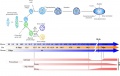
File:Adrenal and gonad steroidogenic factor 1 expression.jpg The key events of steroidogenic tissues development in mice are shown. Molecular players in male sex (SRY, SOX9, SF-1, Dax-1) a * '''E''' - embryonic day(1,000 × 636 (88 KB)) - 17:45, 6 April 2018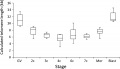
File:Early human telomere length.jpg ...uman oocyte GV and embryonic nuclei at different stages of preimplantation development, presented per embryo. ...t telomere length changes according to the stage of preimplantation embryo development."(1,800 × 1,034 (70 KB)) - 11:05, 25 October 2010
File:Mouse lung development 01.jpg ==Histological analyses of Mouse Lungs at Various Embryonic Stages== ...seen but sections from knockout embryos showed a delay in this sequence of development (panel A, l, p and t). At birth, lungs from knockout pups showed deficient(1,000 × 1,254 (791 KB)) - 13:00, 28 September 2015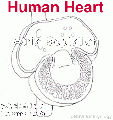
File:Heart1 atrium.gif ...ulmonary system. This channel will remain open through embryonic and fetal development until after birth. ...]] of the heart. In humans, this septation process begins in [[week 5]] of development with the septum primum, a membranous extension on the dorso-cranial wall gr(350 × 373 (243 KB)) - 11:42, 26 September 2015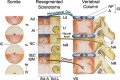
File:Vertebra development cartoon.jpg ...locations of the somites, resegmented sclerotomes, and vertebrae along the embryonic axis are approximately counter-registered. :'''Links:''' [[Musculoskeletal System - Axial Skeleton Development|Axial Skeleton]](800 × 533 (93 KB)) - 17:12, 20 April 2013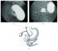
File:Duodenal atresia 02.jpg ...erentiation of the fetus. Such defects can arise at any stage of embryonic development, vary greatly in type and severity, and are caused by a wide variety of det [[Category:Abnormal Development]][[Category:Duodenum]][[Category:Genetics]][[Category:Trisomy 21]][[Categor(765 × 682 (68 KB)) - 16:21, 16 April 2019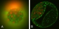
File:Bovine blastocyst KRT18 and MYL6 expression.jpg ...8, FN1 and MYL6 are differentially expressed during preimplantation embryo development and those genes can be considered as markers for bovine blastocyst formatio(800 × 393 (39 KB)) - 15:23, 14 October 2010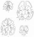
File:Bailey439.jpg ...zontal sections through human embryonic fore-brains at different stages of development==(932 × 1,016 (125 KB)) - 23:08, 22 February 2011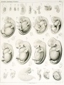
File:Keibel1897 plate01.jpg ...nd, as in the previous stage, evenly developed in circumference around the embryonic disc. ...are by such folds in not entirely inconsiderable proportion modified, the development processes nevertheless take place usually in the normal manner.(1,504 × 2,000 (365 KB)) - 16:14, 12 August 2017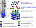
File:Spinal cord delta notch model.png Model of the embryonic rostro-caudal gradient of neurogenesis along the prospective spinal cord fr :'''Links:''' [[Chicken Development]] | [[Neural - Spinal Cord Development]](599 × 457 (171 KB)) - 11:54, 21 March 2018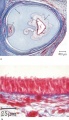
File:Proboscis histology.jpg ...that there is a defect in the definition of the median plane in embryonic development. It further can be concluded that the ethmoid plays an important role in the development of the bilateral symmetry of the face, and may play an important role in th(600 × 1,041 (166 KB)) - 13:30, 18 July 2019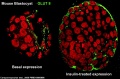
File:Mouse- blastocyst GLUT8 expression.jpg ...s. Presence of this transporter may be necessary for successful blastocyst development, fuel metabolism, and subsequent implantation. Moreover, the existence of a * mammalian development when embryonic fuel metabolism switches from the oxidation of lactate and pyruvate via the(789 × 519 (94 KB)) - 09:16, 3 December 2010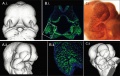
File:Mouse CT E10.5 head 01.jpg ...: procedural artifacts on assessments of embryonic craniofacial growth and development. Schmidt EJ, Parsons TE, Jamniczky HA, Gitelman J, Trpkov C, Boughner JC, L(1,000 × 636 (154 KB)) - 02:05, 16 April 2010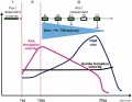
File:Hox 3 phase model.jpg ...o Hox genes are activated during phase II, allowing fast elongation of the embryonic axis. :Links: [[Developmental_Signals_-_Homeobox|Hox]] | [[Chicken Development]] | [[Presomitic Mesoderm Migration|Presomitic Mesoderm Migration Movie]](1,030 × 800 (81 KB)) - 16:12, 6 September 2015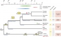
File:VIT Gene Evolution.jpg ...s that the emergence of lactation in the common mammalian ancestor and the development of placentation in eutherian and marsupial mammals allowed for the gradual(668 × 405 (76 KB)) - 18:34, 30 August 2010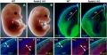
File:Mouse embryo cortex radial expansion.jpg ==RA Is Not Required for Radial Expansion of the Embryonic Cortex== (A–B) Raldh2−/− embryos at E14.5 exhibit relatively normal craniofacial development while they display stunted forelimbs (f).(1,200 × 624 (249 KB)) - 12:32, 31 August 2014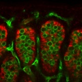
File:Mouse gonad sex determination 01.jpg The researchers started by examining Fgf9 expression during normal gonad development in the mouse. ...ce leads to sex reversal; in XX mice, loss of Wnt4 leads to partial testis development. Only when the protein products of Sry, Sox9, and Fgf9 are all expressed to(600 × 600 (81 KB)) - 12:54, 3 June 2019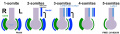
File:Mouse left-right axis 02.jpg ...ation]] | [[Gastrulation]] | [[:Category:Mouse E8.0|Mouse E8.0]] | [[Mouse Development]] ...ardiogenic plate begins to form and the foregut pocket is clearly visible. Embryonic age = 8 dpc (range 7.5-8.75 dpc) 1-7 somite pairs(1,313 × 374 (72 KB)) - 17:08, 30 April 2017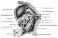
File:Bailey292.jpg ...cavity and the rest of the embryonic ccelom is soon partly cut off by the development of a transverse fold - the septum transversum. This septum is formed in clo(887 × 594 (94 KB)) - 13:51, 25 August 2011
File:Hertig1956 fig51.jpg ...in Horizon VIII. in this publication our primary interest is in the extra-embryonic structures of these ova. ...y angiogenesis is seen in the mesoblast of these villi t'oresha«lowing the development of the future fetal circulation which will be completed and functioning in(712 × 1,095 (109 KB)) - 22:17, 23 February 2017
File:Hertig1956 plate08.jpg ...lumen at the base of the ovum. This is a frequent finding at this stage of development and is due to leakage of blood from the lncunur spaces into the gland lumen ...f the ovum and the coagulated plasma (“Schlussconguluni” of Peters) at the embryonic pole. This maternal blood and plasma, leaking out from the intervillous spa(1,280 × 1,987 (497 KB)) - 12:33, 28 September 2018
File:Mouse ventral body wall development 02.jpg ==Mouse Ventral Body Wall Development==(1,000 × 1,222 (214 KB)) - 12:21, 11 April 2019
File:Bailey083.jpg ...diminution is caused by the relatively more rapid increase in size of the embryonic body and yolk sac. At this point it should be mentioned that the bending an ...as the cephalic flexure. This is the first of the flexures that appear as development proceeds. On the cephalic side of the yolk sac attachment is a protrusion w(924 × 774 (129 KB)) - 11:56, 28 May 2017
File:Gray0947.jpg ==Embryonic Head== ===Development===(600 × 398 (56 KB)) - 22:54, 21 August 2012
File:Duodenal atresia 03.jpg ...erentiation of the fetus. Such defects can arise at any stage of embryonic development, vary greatly in type and severity, and are caused by a wide variety of det(2,361 × 2,955 (425 KB)) - 16:39, 16 April 2019
File:Mouse left-right axis 01.jpg ...ation]] | [[Gastrulation]] | [[:Category:Mouse E8.0|Mouse E8.0]] | [[Mouse Development]] ...ardiogenic plate begins to form and the foregut pocket is clearly visible. Embryonic age = 8 dpc (range 7.5-8.75 dpc) 1-7 somite pairs(1,400 × 937 (172 KB)) - 12:36, 30 April 2017
File:Mouse model thymus development 01.jpg * '''Embryonic day 9.5 (E9.5''') - positioning :'''Links:''' [[Thymus Development]](1,200 × 634 (145 KB)) - 15:59, 1 March 2016
File:Mark Hill.jpg ...ublications/comprehensive-handbook-of-pediatric-audiology#contents Hearing Development: Embryology of the Ear]', in Tharpe AM;Seewald RC (ed.), [https://www.plura ...pter on human embryology (2015), written a new edition book chapter on the development of hearing (2016) and the integumentary system (2018).(300 × 400 (28 KB)) - 13:46, 1 December 2021
File:Keibel1910 plate01.jpg ==Plate 1. Development of the Salamander== ...of egg 10 days 10 hrs. old. Large circular blastopore; faint indication of embryonic anläge.(1,891 × 2,476 (444 KB)) - 10:16, 14 January 2015
File:Mouse head TFII-I expression 01.jpg ...regarding expression patterns at both an RNA and protein level throughout development are also comprehensively reviewed.(1,000 × 862 (298 KB)) - 12:09, 8 July 2011
File:Mouse- respiratory development 01.jpg ==Mouse Respiratory System Development== '''A.''' Initial events. From around embryonic day E8.5-9.5 the foregut endoderm (represented in grey) is specified into o(1,000 × 571 (125 KB)) - 06:47, 11 November 2010
File:Bailey355.jpg ==Fig. 355. Five stages in the development of a human hair== ...xceedingly fine and silky, develop in vast numbers over the surface of the embryonic body and are known collectively as the lanugo. This growth is lost (beginni(632 × 890 (109 KB)) - 13:29, 25 September 2012
File:Chicken embryo E-cad and P-cad gastrulation.png ...org/entry/602150 SLUG]) required for epithelial-mesenchymal transitions in development. ...lation]] is an important developmental process that establishes the 3 main embryonic layers (ectoderm, mesoderm and endoderm).(320 × 408 (253 KB)) - 13:28, 3 August 2017
File:Locy1895 plate26.jpg FIg. 2. Somewhat older embryo. The embryonic rim on the left side shows faintly Some of the metameric segments (not repr FIg. 18. specimen showing a large development of the central tongueislilte Process. Embryo slightly older than that in Fi(3,440 × 2,608 (439 KB)) - 15:19, 27 August 2018
File:Hertig1956 plate12.jpg ...5 days of age on the basis of coital data, endometrial findings and ovular development. They have, in common, the abnormality of nulltinucleated blastomeres. In a ...ng reaction of the blastomeres. The pale, larger cells may be of potential embryonic nature, whereas the darker irregular multinucleated ones may be of future t(1,280 × 1,894 (293 KB)) - 15:31, 25 February 2017
File:Hertig1956 plate04.jpg ...s the most distinctive gross feature of and first appears at this stage of development. It is due to flooding of the trophoblastic lacunae with maternal blood. ...he abembryonic pole is poorly differentiated, as contrasted to that at the embryonic or implantation pole. [[:Category:Carnegie Embryo 7700|Carnegie 7700]], Sec(1,280 × 1,904 (452 KB)) - 15:24, 25 February 2017
File:Hertig1956 plate09.jpg ...in Horizon VIII. in this publication our primary interest is in the extra-embryonic structures of these ova. ...y angiogenesis is seen in the mesoblast of these villi t'oresha«lowing the development of the future fetal circulation which will be completed and functioning in(1,280 × 1,922 (395 KB)) - 22:23, 23 February 2017
File:Keibel1910 plate02.jpg ==Plate 2. Development of the Salamander== Top view of egg 13 days 3 hrs. old. Small circular blastopore. Embryonic anläge triangulär in outline ; lateral boundaries indistinct. First appea(1,891 × 2,476 (360 KB)) - 10:30, 14 January 2015
File:Keibel1900 plate01.jpg ...ou can , if you compare this stage and slightly older, in which the actual embryonic plant and the first somites have occurred, little doubt . ...primitive streak . Since the formation is still set to such a low level of development that one sees nothing more than surveys and beads so that very reason appea(1,952 × 2,451 (519 KB)) - 21:05, 28 December 2013
File:National Museum of Health and Medicine.jpg ...in the 1970s and 1980s at the National Institute of Child Health and Human Development. ...collection consists principally of serial sections of normal human embryo development in the first eight weeks, as started by the Carnegie Institution of Washing(800 × 600 (201 KB)) - 11:52, 19 December 2018
File:Marcello Malpighi.jpg ...id regret, the \in timely death of many young soldiers and others from the development of such masses in the heart. ...he missed the micropyle which Grew had seen. Nevertheless, his work on the development of the ovum into the seed and of the ovary into the fruit and then of the g(652 × 869 (418 KB)) - 13:49, 20 February 2020


