Search results
From Embryology
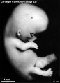
File:Stage22 bf3.jpg == Human Embryo Carnegie Stage 22== Carnegie collection Stage 22 Embryo No.8394(433 × 602 (27 KB)) - 13:22, 21 April 2012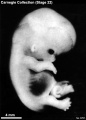
File:Stage22 bf6.jpg == Human Embryo Carnegie Stage 22== Carnegie collection Stage 22 Embryo No.6701(433 × 602 (28 KB)) - 10:42, 21 April 2012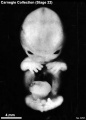
File:Stage22 bf7.jpg == Human Embryo Carnegie Stage 22== Carnegie collection Stage 22 Embryo No.6701(433 × 602 (26 KB)) - 13:20, 21 April 2012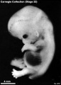
File:Stage22 bf8.jpg == Human Embryo Carnegie Stage 22== Carnegie collection Stage 22 Embryo No.6701(433 × 602 (27 KB)) - 13:20, 21 April 2012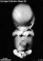
File:Stage22 bf4.jpg == Human Embryo Carnegie Stage 22== Carnegie collection Stage 22 Embryo No.8394(433 × 602 (25 KB)) - 13:21, 21 April 2012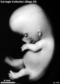
File:Stage22 bf5.jpg == Human Embryo Carnegie Stage 22== Carnegie collection Stage 22 Embryo No.8394(433 × 602 (26 KB)) - 13:22, 21 April 2012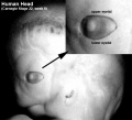
File:Stage22 eyelids.jpg ==Human Embryo Eyelids== Carnegie Stage 22(457 × 417 (27 KB)) - 10:56, 2 March 2011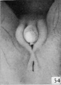
File:Spaulding-fig54.jpg ==Fig. 54. Carnegie Embryo No. 1476== ...e external genitalia in the human embryo.''']] Contributions to Embryology Carnegie Institution No.61 (1921). With four plates and two text-figures.(430 × 599 (27 KB)) - 22:42, 3 June 2015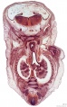
File:ME54 001.jpg ==Human Embryo - Stage 21== ME 54 [[Carnegie stage 21]], 22.5 mm, 8 weeks, female, frontal, {{HE}}(1,165 × 1,876 (464 KB)) - 15:36, 18 October 2016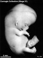
File:Stage21 bf2.jpg ==Human Embryo Carnegie Stage 21== Carnegie collection Stage 21 Embryo No. {{CE7592}}(413 × 555 (25 KB)) - 18:13, 29 September 2017
File:Stage21 bf3.jpg ==Human Embryo Carnegie Stage 21== Carnegie collection Stage 21 Embryo No. {{CE7592}}(413 × 555 (23 KB)) - 18:13, 29 September 2017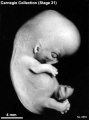
File:Stage21 bf5.jpg ==Human Embryo Carnegie Stage 21== Carnegie collection Stage 21 Embryo No. {{CE8553}}(413 × 555 (26 KB)) - 18:12, 29 September 2017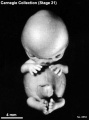
File:Stage21 bf6.jpg ==Human Embryo Carnegie Stage 21== Carnegie collection Stage 21 Embryo No. {{CE8553}}(413 × 555 (24 KB)) - 18:12, 29 September 2017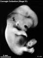
File:Stage21 bf8.jpg ==Human Embryo Carnegie Stage 21== Carnegie collection Stage 21 Embryo No. {{CE4090}}(413 × 555 (26 KB)) - 18:11, 29 September 2017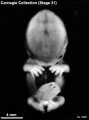
File:Stage21 bf9.jpg ==Human Embryo Carnegie Stage 21== Carnegie collection Stage 21 Embryo No. {{CE4090}}(413 × 555 (22 KB)) - 18:10, 29 September 2017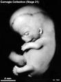
File:Stage21 bf4.jpg ==Human Embryo Carnegie Stage 21== Carnegie collection Stage 21 Embryo No. {{CE7592}}(413 × 555 (25 KB)) - 18:13, 29 September 2017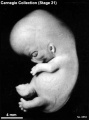
File:Stage21 bf7.jpg ==Human Embryo Carnegie Stage 21== Carnegie collection Stage 21 Embryo No. {{CE8553}}(413 × 555 (26 KB)) - 18:12, 29 September 2017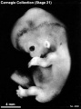
File:Stage21 bf10.jpg ==Human Embryo Carnegie Stage 21== Carnegie collection Stage 21 Embryo No. {{CE4090}}(413 × 555 (26 KB)) - 18:11, 29 September 2017
File:ME54 002.jpg ==Human Embryo - Stage 21== ME 54 [[Carnegie stage 21]], 22.5 mm, [[week 8]], female, frontal, {{HE}}(800 × 601 (157 KB)) - 22:56, 24 May 2016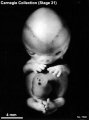
File:Stage21A.jpg ==Human Embryo Carnegie Stage 21== Carnegie collection Stage 22 Embryo No.7392(413 × 555 (23 KB)) - 08:11, 7 August 2011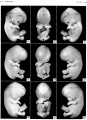
File:Streeter1957 stage 21 plate01.jpg Figs. 54-56. [[:Category:Carnegie Embryo 4090|No. 4090]]. Figs. 57-59. [[:Category:Carnegie Embryo 8553|No. 8553]].(1,500 × 2,077 (379 KB)) - 17:45, 6 November 2016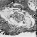
File:Hertig1956 fig17.jpg ...ther form the exocoelomic membrane. Carnegie Embryo {{CE8215}}, Section 12-54. X 300. [[Category:Carnegie Embryo 8215]](730 × 726 (100 KB)) - 13:36, 31 October 2017
File:Mall1906 fig02.jpg ==Fig.2. Embryo No. 284== Embryo {{CE284}}, 54 mm long, immediately beneath the squamo-zygomatic the alisphenoid may be se(1,317 × 1,532 (205 KB)) - 14:37, 6 May 2018
File:Stage22 bf1a.jpg == Human Embryo Carnegie Stage 22== Carnegie Stage 22(800 × 600 (18 KB)) - 05:41, 13 April 2011
File:Stage22 bf2.jpg == Human Embryo Carnegie Stage 22== Carnegie Stage 22(750 × 1,000 (36 KB)) - 10:32, 15 April 2011
File:Stage22 bf1b.jpg == Human Embryo Carnegie Stage 22== Carnegie Stage 22(600 × 450 (11 KB)) - 05:42, 13 April 2011
File:Stage22 bf2a.jpg == Human Embryo Carnegie Stage 22== Carnegie Stage 22(600 × 800 (36 KB)) - 10:33, 15 April 2011
File:Stage22 bf1c.jpg == Human Embryo Carnegie Stage 22== Carnegie Stage 22(400 × 300 (6 KB)) - 05:41, 13 April 2011
File:Stage22 bf2b.jpg == Human Embryo Carnegie Stage 22== Carnegie Stage 22(450 × 600 (22 KB)) - 10:33, 15 April 2011
File:Stage22 bf2c.jpg == Human Embryo Carnegie Stage 22== Carnegie Stage 22(300 × 400 (11 KB)) - 10:33, 15 April 2011
File:Stage22 bf1.jpg == Human Embryo Carnegie Stage 22== Carnegie Stage 22(1,000 × 750 (27 KB)) - 05:41, 13 April 2011
File:Streeter1915 fig07.jpg [[:Category:Carnegie Embryo 940|No. 940]] - [[Carnegie Collection]], [[Carnegie stage 17]] [[Week 6]] 42 - 44 days, 11 - 14 mm. ...jpg|Figure 7]] is a vertex view of a. human embryo 13.8 mm. long (No. 940, Carnegie Collection).(764 × 839 (93 KB)) - 08:12, 27 November 2016
File:Stage21 bf11.jpg ==Human Embryo Carnegie Stage 21== Facts: Week 8, 53 - 54 days, 22 - 24 mm(1,600 × 2,400 (387 KB)) - 15:40, 28 February 2014
File:Stage21 bf11a.jpg ==Human Embryo Carnegie Stage 21== Facts: Week 8, 53 - 54 days, 22 - 24 mm(800 × 1,200 (108 KB)) - 15:44, 28 February 2014
File:Stage21 bf11b.jpg ==Human Embryo Carnegie Stage 21== Facts: Week 8, 53 - 54 days, 22 - 24 mm(533 × 800 (56 KB)) - 15:45, 28 February 2014
File:ME54 003.jpg ==Human Embryo - Stage 21== ...This excerpt for the [[:File:ME54_001.jpg|coronal (frontal) section of the embryo]] cuts through the trachea, both lungs, pleural cavity and the rib cage.(800 × 601 (169 KB)) - 11:36, 31 August 2016
File:Streeter020-25.jpg ...mbryo''' Contributions to Embryology Carnegie Institution No.20 (1918) pp5-54, 4 text-figures and 4 plates.(636 × 800 (105 KB)) - 02:42, 15 February 2011
File:Streeter1915 fig08.jpg ...jpg|Figure 7]] is a vertex view of a. human embryo 13.8 mm. long (No. 940, Carnegie Collection). ...fig08.jpg|Figure 8]] is a vertex view of an embryo 20 mm. long (No. 349, Carnegie Collection).(836 × 843 (105 KB)) - 08:13, 27 November 2016
File:Streeter1915 fig09.jpg ...jpg|Figure 7]] is a vertex view of a. human embryo 13.8 mm. long (No. 940, Carnegie Collection). ...fig08.jpg|Figure 8]] is a vertex view of an embryo 20 mm. long (No. 349, Carnegie Collection).(907 × 840 (92 KB)) - 08:14, 27 November 2016
File:Gray0179.jpg Human embryo CRL 24 mm inner aspect * from CRL approximately [[Week 8]], [[Carnegie stage 22]] 54 - 56 days, 23 - 28 mm.(617 × 368 (47 KB)) - 18:34, 27 August 2012
File:Streeter005-13.jpg ...mbryo''' Contributions to Embryology Carnegie Institution No.20 (1918) pp5-54, 4 text-figures and 4 plates.(657 × 800 (178 KB)) - 21:56, 22 April 2012
File:Gray0178.jpg * Human embryo CRL 24 mm outer aspect. (From model by Low.) * from CRL approximately [[Week 8]], [[Carnegie stage 22]] 54 - 56 days, 23 - 28 mm.(617 × 368 (44 KB)) - 18:33, 27 August 2012
File:Streeter1921 fig07-09.jpg * Figure 7 is a vertex view of a human embryo 13.8 mm long (Carnegie Collection. No. 940). * Figure 8 is a vertex view of an embryo 20 mm long (Carnegie Collection, No. 349).(1,200 × 494 (92 KB)) - 11:19, 19 January 2017
File:Hertig1956 fig54.jpg ==Fig. 54 Reconstruction of half a 7-day ovum== ...rophy and dilation. See figures I1 and 12. [[:Category:Carnegie Embryo 8020|Carnegie 8020]]. X 260.(1,218 × 782 (185 KB)) - 13:24, 24 February 2017
File:Stage21 bf1.jpg ==Human Embryo Carnegie Stage 21== Facts: Week 8, 53 - 54 days, 22 - 24 mm(1,000 × 750 (35 KB)) - 05:43, 13 April 2011
File:Stage21 bf1a.jpg ==Human Embryo Carnegie Stage 21== Facts: Week 8, 53 - 54 days, 22 - 24 mm(800 × 600 (23 KB)) - 05:43, 13 April 2011
File:Stage21 bf1b.jpg ==Human Embryo Carnegie Stage 21== Facts: Week 8, 53 - 54 days, 22 - 24 mm(600 × 450 (14 KB)) - 05:43, 13 April 2011
File:Stage21 bf1c.jpg ==Human Embryo Carnegie Stage 21== Facts: Week 8, 53 - 54 days, 22 - 24 mm(400 × 300 (7 KB)) - 05:43, 13 April 2011
File:Streeter001.jpg (Carnegie Collection, No. {{CE145}}, slide 7, row 1, section 3) ...mbryo''' Contributions to Embryology Carnegie Institution No.20 (1918) pp5-54, 4 text-figures and 4 plates.(423 × 800 (79 KB)) - 10:43, 30 July 2017
File:Mall1906 fig04.jpg ==Fig.4. Head of Embryo No. 284== Mesial view of the head of embryo No.{{CE284}} (54 mm long). The centers of tke occipital bone surround the foramen magnum. Be(1,114 × 1,044 (137 KB)) - 14:35, 6 May 2018
File:Streeter004.jpg (Carnegie Collection, No. 1373, slide 9, row 3. section 1) The section is 10 microns ...mbryo''' Contributions to Embryology Carnegie Institution No.20 (1918) pp5-54, 4 text-figures and 4 plates.(487 × 800 (92 KB)) - 02:48, 15 February 2011
File:Hertig1956 plate11.jpg ...rophy and dilation. See figures I1 and 12. [[:Category:Carnegie Embryo 8020|Carnegie 8020]]. X 260. ...chorionic cavity. See figures 13 and 14. [[:Category:Carnegie Embryo 8155|Carnegie 8155]]. X 200.(1,280 × 1,909 (458 KB)) - 13:31, 24 February 2017
File:Streeter002-3.jpg (Carnegie Collection, No. 86), being the same as that shown in figure 11. The section (Carnegie Collection, No. 95, slide 72, section 1). This is the same canal as that sh(607 × 800 (106 KB)) - 12:41, 15 February 2011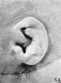
File:Streeter1922-fig54.jpg ==Fig. 54. Embryo No. 1716== Embryo No. {{CE1716}} 119 mm CRL(574 × 780 (88 KB)) - 13:52, 22 November 2017
File:Hertig1956 plate03.jpg ...hin the trophoblastic shell of the ovum. [[:Category:Carnegie Embryo 8215|Carnegie 8215]], Section 12-5-1-. X100. ...ger than that of the following specimens. [[:Category:Carnegie Embryo 8215|Carnegie 8215]], Sequence 3. X 22.(1,280 × 1,908 (581 KB)) - 15:22, 25 February 2017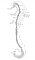
File:Bartelmez1923 fig02.jpg ==Fig. 2. A reconstruction of a four—somite embryo== ...reproduction. The plane of section is indicated by the position of section 54 which was shown as figure 1 in my 1922 paper.(1,295 × 2,189 (218 KB)) - 16:57, 24 October 2017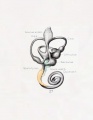
File:Streeter027.jpg ...view of a model reconstructed from a human fetus 50 mm. crown-rump length (Carnegie Collection, No. 84). ...mbryo''' Contributions to Embryology Carnegie Institution No.20 (1918) pp5-54, 4 text-figures and 4 plates.(774 × 1,000 (51 KB)) - 17:31, 18 May 2015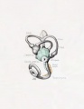
File:Streeter026.jpg ...view of a model reconstructed from a human fetus 50 mm. crown-rump length (Carnegie Collection, No. 84). ...mbryo''' Contributions to Embryology Carnegie Institution No.20 (1918) pp5-54, 4 text-figures and 4 plates.(774 × 1,000 (45 KB)) - 17:31, 18 May 2015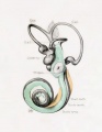
File:Streeter028.jpg ...byrinth and the periotic spaces in a human fetus 85 mm. crown-rump length (Carnegie Collection, No. 1400-30), enlarged 11.4 diameters. ...mbryo''' Contributions to Embryology Carnegie Institution No.20 (1918) pp5-54, 4 text-figures and 4 plates.(774 × 1,000 (69 KB)) - 17:32, 18 May 2015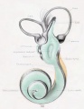
File:Streeter030.jpg ...yrinth and the periotic spaces in a human fetus 130 mm. crown-rump length (Carnegie Collection, No. 1018), enlarged 11.4 diameters. ...mbryo''' Contributions to Embryology Carnegie Institution No.20 (1918) pp5-54, 4 text-figures and 4 plates.(774 × 1,000 (78 KB)) - 17:33, 18 May 2015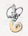
File:Streeter029.jpg ...byrinth and the periotic spaces in a human fetus 85 mm. crown-rump length (Carnegie Collection, No. 1400-30), enlarged 11.4 diameters. ...mbryo''' Contributions to Embryology Carnegie Institution No.20 (1918) pp5-54, 4 text-figures and 4 plates.(774 × 1,000 (74 KB)) - 17:32, 18 May 2015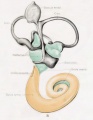
File:Streeter031.jpg ...yrinth and the periotic spaces in a human fetus 130 mm. crown-rump length (Carnegie Collection, No. 1018), enlarged 11.4 diameters. ...mbryo''' Contributions to Embryology Carnegie Institution No.20 (1918) pp5-54, 4 text-figures and 4 plates.(774 × 1,000 (79 KB)) - 17:34, 18 May 2015
File:Streeter1922-fig56.jpg ==Fig. 56. Embryo No. 1782, 135.6 mm CRL== ...phs are all shown at an enlargement of 4 diameters. Specimens are from the Carnegie Collection, and length given is crown-rump.(592 × 783 (92 KB)) - 08:32, 28 January 2013
File:Moffatt1957 plate02.jpg '''Fig 7''' - {{Fetal}} (no. {{CE3990}} 54 mm) The attachment of the developing disk to Meckel’s cartilage can be se ...Carnegie Embryo 3990]][[Category:Carnegie Embryo 6581]][[Category:Carnegie Embryo 1455]](1,500 × 1,891 (865 KB)) - 12:33, 28 October 2018
File:Streeter028-30.jpg ...view of a model reconstructed from a human fetus 50 mm. crown-rump length (Carnegie Collection, No. 84). The cistern and the scala vestibuli are shown in green ...byrinth and the periotic spaces in a human fetus 85 mm. crown-rump length (Carnegie Collection, No. 1400-30), enlarged 11.4 diameters. The cistern and the conn(748 × 1,000 (134 KB)) - 17:30, 18 May 2015
File:Streeter014-19.jpg ...the lateral semicircular canal in a human fetu.s 43 mm. crown-rump length (Carnegie Collection, No. 886, slide 42, section 3). The section is 100m thick and is ...the lateral semicircular canal in a human fetus 46 mm. crown-rump length (Carnegie Collection, No. 9.5, slide 72, section 1). The section i.s 100m thick and i(640 × 800 (199 KB)) - 02:46, 15 February 2011


