Search results
From Embryology
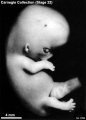
File:Stage22 bf3.jpg == Human Embryo Carnegie Stage 22== Carnegie collection Stage 22 Embryo No.8394(433 × 602 (27 KB)) - 13:22, 21 April 2012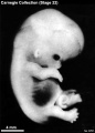
File:Stage22 bf6.jpg == Human Embryo Carnegie Stage 22== Carnegie collection Stage 22 Embryo No.6701(433 × 602 (28 KB)) - 10:42, 21 April 2012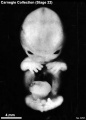
File:Stage22 bf7.jpg == Human Embryo Carnegie Stage 22== Carnegie collection Stage 22 Embryo No.6701(433 × 602 (26 KB)) - 13:20, 21 April 2012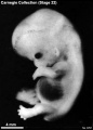
File:Stage22 bf8.jpg == Human Embryo Carnegie Stage 22== Carnegie collection Stage 22 Embryo No.6701(433 × 602 (27 KB)) - 13:20, 21 April 2012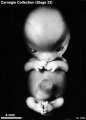
File:Stage22 bf4.jpg == Human Embryo Carnegie Stage 22== Carnegie collection Stage 22 Embryo No.8394(433 × 602 (25 KB)) - 13:21, 21 April 2012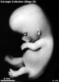
File:Stage22 bf5.jpg == Human Embryo Carnegie Stage 22== Carnegie collection Stage 22 Embryo No.8394(433 × 602 (26 KB)) - 13:22, 21 April 2012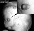
File:Stage22 eyelids.jpg ==Human Embryo Eyelids== Carnegie Stage 22(457 × 417 (27 KB)) - 10:56, 2 March 2011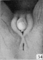
File:Spaulding-fig54.jpg ==Fig. 54. Carnegie Embryo No. 1476== ...e external genitalia in the human embryo.''']] Contributions to Embryology Carnegie Institution No.61 (1921). With four plates and two text-figures.(430 × 599 (27 KB)) - 22:42, 3 June 2015
File:ME54 001.jpg ==Human Embryo - Stage 21== ME 54 [[Carnegie stage 21]], 22.5 mm, 8 weeks, female, frontal, {{HE}}(1,165 × 1,876 (464 KB)) - 15:36, 18 October 2016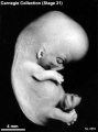
File:Stage21 bf5.jpg ==Human Embryo Carnegie Stage 21== Carnegie collection Stage 21 Embryo No. {{CE8553}}(413 × 555 (26 KB)) - 18:12, 29 September 2017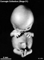
File:Stage21 bf6.jpg ==Human Embryo Carnegie Stage 21== Carnegie collection Stage 21 Embryo No. {{CE8553}}(413 × 555 (24 KB)) - 18:12, 29 September 2017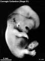
File:Stage21 bf8.jpg ==Human Embryo Carnegie Stage 21== Carnegie collection Stage 21 Embryo No. {{CE4090}}(413 × 555 (26 KB)) - 18:11, 29 September 2017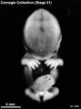
File:Stage21 bf9.jpg ==Human Embryo Carnegie Stage 21== Carnegie collection Stage 21 Embryo No. {{CE4090}}(413 × 555 (22 KB)) - 18:10, 29 September 2017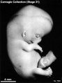
File:Stage21 bf2.jpg ==Human Embryo Carnegie Stage 21== Carnegie collection Stage 21 Embryo No. {{CE7592}}(413 × 555 (25 KB)) - 18:13, 29 September 2017
File:Stage21 bf3.jpg ==Human Embryo Carnegie Stage 21== Carnegie collection Stage 21 Embryo No. {{CE7592}}(413 × 555 (23 KB)) - 18:13, 29 September 2017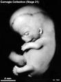
File:Stage21 bf4.jpg ==Human Embryo Carnegie Stage 21== Carnegie collection Stage 21 Embryo No. {{CE7592}}(413 × 555 (25 KB)) - 18:13, 29 September 2017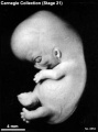
File:Stage21 bf7.jpg ==Human Embryo Carnegie Stage 21== Carnegie collection Stage 21 Embryo No. {{CE8553}}(413 × 555 (26 KB)) - 18:12, 29 September 2017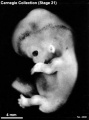
File:Stage21 bf10.jpg ==Human Embryo Carnegie Stage 21== Carnegie collection Stage 21 Embryo No. {{CE4090}}(413 × 555 (26 KB)) - 18:11, 29 September 2017
File:ME54 002.jpg ==Human Embryo - Stage 21== ME 54 [[Carnegie stage 21]], 22.5 mm, [[week 8]], female, frontal, {{HE}}(800 × 601 (157 KB)) - 22:56, 24 May 2016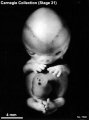
File:Stage21A.jpg ==Human Embryo Carnegie Stage 21== Carnegie collection Stage 22 Embryo No.7392(413 × 555 (23 KB)) - 08:11, 7 August 2011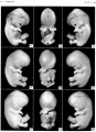
File:Streeter1957 stage 21 plate01.jpg Figs. 54-56. [[:Category:Carnegie Embryo 4090|No. 4090]]. Figs. 57-59. [[:Category:Carnegie Embryo 8553|No. 8553]].(1,500 × 2,077 (379 KB)) - 17:45, 6 November 2016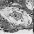
File:Hertig1956 fig17.jpg ...ther form the exocoelomic membrane. Carnegie Embryo {{CE8215}}, Section 12-54. X 300. [[Category:Carnegie Embryo 8215]](730 × 726 (100 KB)) - 13:36, 31 October 2017
File:Mall1906 fig02.jpg ==Fig.2. Embryo No. 284== Embryo {{CE284}}, 54 mm long, immediately beneath the squamo-zygomatic the alisphenoid may be se(1,317 × 1,532 (205 KB)) - 14:37, 6 May 2018
File:Stage22 bf1c.jpg == Human Embryo Carnegie Stage 22== Carnegie Stage 22(400 × 300 (6 KB)) - 05:41, 13 April 2011
File:Stage22 bf2b.jpg == Human Embryo Carnegie Stage 22== Carnegie Stage 22(450 × 600 (22 KB)) - 10:33, 15 April 2011
File:Stage22 bf2c.jpg == Human Embryo Carnegie Stage 22== Carnegie Stage 22(300 × 400 (11 KB)) - 10:33, 15 April 2011
File:Stage22 bf1.jpg == Human Embryo Carnegie Stage 22== Carnegie Stage 22(1,000 × 750 (27 KB)) - 05:41, 13 April 2011
File:Stage22 bf1a.jpg == Human Embryo Carnegie Stage 22== Carnegie Stage 22(800 × 600 (18 KB)) - 05:41, 13 April 2011
File:Stage22 bf2.jpg == Human Embryo Carnegie Stage 22== Carnegie Stage 22(750 × 1,000 (36 KB)) - 10:32, 15 April 2011
File:Stage22 bf1b.jpg == Human Embryo Carnegie Stage 22== Carnegie Stage 22(600 × 450 (11 KB)) - 05:42, 13 April 2011
File:Stage22 bf2a.jpg == Human Embryo Carnegie Stage 22== Carnegie Stage 22(600 × 800 (36 KB)) - 10:33, 15 April 2011
File:Streeter1915 fig07.jpg [[:Category:Carnegie Embryo 940|No. 940]] - [[Carnegie Collection]], [[Carnegie stage 17]] [[Week 6]] 42 - 44 days, 11 - 14 mm. ...jpg|Figure 7]] is a vertex view of a. human embryo 13.8 mm. long (No. 940, Carnegie Collection).(764 × 839 (93 KB)) - 08:12, 27 November 2016
File:Stage21 bf11.jpg ==Human Embryo Carnegie Stage 21== Facts: Week 8, 53 - 54 days, 22 - 24 mm(1,600 × 2,400 (387 KB)) - 15:40, 28 February 2014
File:Stage21 bf11a.jpg ==Human Embryo Carnegie Stage 21== Facts: Week 8, 53 - 54 days, 22 - 24 mm(800 × 1,200 (108 KB)) - 15:44, 28 February 2014
File:Stage21 bf11b.jpg ==Human Embryo Carnegie Stage 21== Facts: Week 8, 53 - 54 days, 22 - 24 mm(533 × 800 (56 KB)) - 15:45, 28 February 2014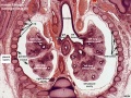
File:ME54 003.jpg ==Human Embryo - Stage 21== ...This excerpt for the [[:File:ME54_001.jpg|coronal (frontal) section of the embryo]] cuts through the trachea, both lungs, pleural cavity and the rib cage.(800 × 601 (169 KB)) - 11:36, 31 August 2016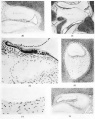
File:Streeter020-25.jpg ...mbryo''' Contributions to Embryology Carnegie Institution No.20 (1918) pp5-54, 4 text-figures and 4 plates.(636 × 800 (105 KB)) - 02:42, 15 February 2011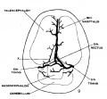
File:Streeter1915 fig09.jpg ...jpg|Figure 7]] is a vertex view of a. human embryo 13.8 mm. long (No. 940, Carnegie Collection). ...fig08.jpg|Figure 8]] is a vertex view of an embryo 20 mm. long (No. 349, Carnegie Collection).(907 × 840 (92 KB)) - 08:14, 27 November 2016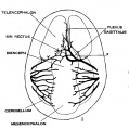
File:Streeter1915 fig08.jpg ...jpg|Figure 7]] is a vertex view of a. human embryo 13.8 mm. long (No. 940, Carnegie Collection). ...fig08.jpg|Figure 8]] is a vertex view of an embryo 20 mm. long (No. 349, Carnegie Collection).(836 × 843 (105 KB)) - 08:13, 27 November 2016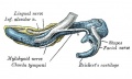
File:Gray0179.jpg Human embryo CRL 24 mm inner aspect * from CRL approximately [[Week 8]], [[Carnegie stage 22]] 54 - 56 days, 23 - 28 mm.(617 × 368 (47 KB)) - 18:34, 27 August 2012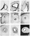
File:Streeter005-13.jpg ...mbryo''' Contributions to Embryology Carnegie Institution No.20 (1918) pp5-54, 4 text-figures and 4 plates.(657 × 800 (178 KB)) - 21:56, 22 April 2012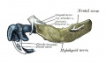
File:Gray0178.jpg * Human embryo CRL 24 mm outer aspect. (From model by Low.) * from CRL approximately [[Week 8]], [[Carnegie stage 22]] 54 - 56 days, 23 - 28 mm.(617 × 368 (44 KB)) - 18:33, 27 August 2012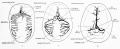
File:Streeter1921 fig07-09.jpg * Figure 7 is a vertex view of a human embryo 13.8 mm long (Carnegie Collection. No. 940). * Figure 8 is a vertex view of an embryo 20 mm long (Carnegie Collection, No. 349).(1,200 × 494 (92 KB)) - 11:19, 19 January 2017
File:Hertig1956 fig54.jpg ==Fig. 54 Reconstruction of half a 7-day ovum== ...rophy and dilation. See figures I1 and 12. [[:Category:Carnegie Embryo 8020|Carnegie 8020]]. X 260.(1,218 × 782 (185 KB)) - 13:24, 24 February 2017
File:Stage21 bf1.jpg ==Human Embryo Carnegie Stage 21== Facts: Week 8, 53 - 54 days, 22 - 24 mm(1,000 × 750 (35 KB)) - 05:43, 13 April 2011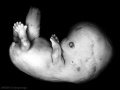
File:Stage21 bf1a.jpg ==Human Embryo Carnegie Stage 21== Facts: Week 8, 53 - 54 days, 22 - 24 mm(800 × 600 (23 KB)) - 05:43, 13 April 2011
File:Stage21 bf1b.jpg ==Human Embryo Carnegie Stage 21== Facts: Week 8, 53 - 54 days, 22 - 24 mm(600 × 450 (14 KB)) - 05:43, 13 April 2011
File:Stage21 bf1c.jpg ==Human Embryo Carnegie Stage 21== Facts: Week 8, 53 - 54 days, 22 - 24 mm(400 × 300 (7 KB)) - 05:43, 13 April 2011
File:Streeter001.jpg (Carnegie Collection, No. {{CE145}}, slide 7, row 1, section 3) ...mbryo''' Contributions to Embryology Carnegie Institution No.20 (1918) pp5-54, 4 text-figures and 4 plates.(423 × 800 (79 KB)) - 10:43, 30 July 2017
File:Mall1906 fig04.jpg ==Fig.4. Head of Embryo No. 284== Mesial view of the head of embryo No.{{CE284}} (54 mm long). The centers of tke occipital bone surround the foramen magnum. Be(1,114 × 1,044 (137 KB)) - 14:35, 6 May 2018


