Search results
From Embryology
Page title matches
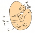
File:Day 10.5 Deep Lens Indentation.JPG Day 10.5: Deep lens indentation occurs. Tel = cerebral hemisphere, L = lens invagination, Hl = Hindlimb bud, Aa = forelimb bud, So = Somite 4, O = otic(514 × 483 (28 KB)) - 13:00, 25 June 2014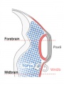
File:Lens-neural crest signaling 01.jpg ==Wnt mediates lens repression by neural crest cells and Transforming growth factor-β== # NCCs (blue) secrete TGF-βs, which signal to the non-lens ectoderm and dorsal optic vesicle.(300 × 400 (24 KB)) - 17:25, 31 August 2011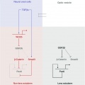
File:Lens-neural crest signaling 02.jpg ==Wnt mediates lens repression by neural crest cells and Transforming growth factor-β== Proposed molecular model to explain TGF-β- and Wnt-mediated lens restriction. Broken lines: interactions inferred from the literature.(521 × 522 (22 KB)) - 14:10, 29 April 2011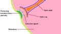
File:Formation of the lens 1.jpg ...ing the vital role of induction from the optic cup in the formation of the lens.(1,152 × 648 (102 KB)) - 17:42, 26 September 2012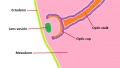
File:Formation of the lens 2.jpg ...erm, forming the lens vesicle. The combined structure of the optic cup and lens vesicle can now be referred to as the optic globe.(1,152 × 648 (94 KB)) - 17:17, 27 September 2012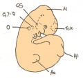
File:Day 11 Closure of lens vesicle.JPG Day 11: Closure of the lens vesicle occurs.(560 × 528 (27 KB)) - 13:19, 18 August 2014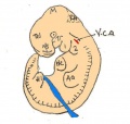
File:Day 11.5 Lens Vesicle completely separated from surface.JPG Day 11.5: the lens vesicle is completely separated from the surface.(511 × 484 (29 KB)) - 13:01, 25 June 2014


Page text matches
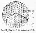
File:Hertwig268.jpg ==Fig. 268. Diagram of the arrangement of the lens-fibres== ...rior surface of the lens and their termination at the anterior star of the lens ; If", continuation of the same fibres Co the posterior star on the posteri(406 × 375 (39 KB)) - 21:48, 28 March 2012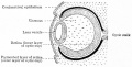
File:Bailey463.jpg ==Fig. 463. Diagram of developing lens and optic cup== ...ium. The mesodermal tissue between the latter and the anterior wall of the lens vesicle is the anlage of the substantia propria cornea.(679 × 345 (52 KB)) - 06:43, 31 August 2011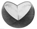
File:Keibel Mall 2 176.jpg ==Fig. 176. A lens at end of the third month formation of the lens sutures== ...the third month). The right upper quadrant has been removed. One sees the lens epithelium, its continuity with the lena fibres at the equator.(992 × 800 (134 KB)) - 11:34, 21 February 2014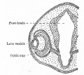
File:Bailey462.jpg ==Fig. 462. Showing somewhat later stage in development of optic cup and lens than is shown in Fig. 461== ...m, but about the eighth week a thin layer of mesoderm grows in between the lens vesicle and the surface ectoderm, completely separating them (Fig. 463).(463 × 411 (45 KB)) - 06:43, 31 August 2011
File:Gray0887.jpg ==Lens Epithelium== ...the margin of the lens, showing the transition of the epithelium into the lens fibers. (Babuchin.)(221 × 800 (67 KB)) - 22:32, 19 August 2012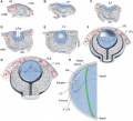
File:DevelopmentEye-PMC43029243.jpg '''Stages of lens formation in mouse embryos''' ...nchyme; 1° and 2° LFs, primary and secondary lens fibers; PLE, prospective lens ectoderm; RPE, retinal pigmented epithelium; SE, surface ectoderm.(654 × 594 (142 KB)) - 10:06, 19 November 2017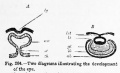
File:Hertwig264.jpg ...e between-brain (zh), is invaginated as a result of the development of the lens-pit (Ig). ...cup with double walls, an inner (ib) and an outer (ab) ; 1st, stalk of the lens ; gl, vitreous body.(653 × 395 (36 KB)) - 21:50, 28 March 2012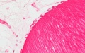
File:Stage 22 image 207.jpg ==Developing Lens - Human Embryo Carnegie stage 22== * lens (fibres)(1,200 × 753 (208 KB)) - 11:41, 14 June 2016File:Formation of the lens 2.jpg ...erm, forming the lens vesicle. The combined structure of the optic cup and lens vesicle can now be referred to as the optic globe.(1,152 × 648 (94 KB)) - 17:17, 27 September 2012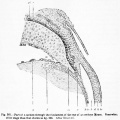
File:Hertwig267.jpg ...the eye ; I', transition of the lens epithelium into the lens-fibres ; Ic, lens-epithelium ; k, anterior chamber of the eye ; d, Descemet's membrane ; //,(700 × 701 (83 KB)) - 21:48, 28 March 2012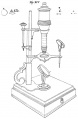
File:Meyer1932history7 fig02.jpg (8.) The Best lens, (b) Cuffs lens No. 4, (c) an English lens No. 0. The latter was used by Ledermuller and he says that it magnified eig(655 × 1,000 (85 KB)) - 21:28, 2 November 2015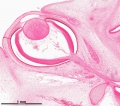
File:Stage 22 image 008-eye.jpg ...ages/Stage22/08-eye/Stage22-08-eye.html?zoom=4&lat=-2030&lon=2572&layers=B Lens and Cornea] ...ages/Stage22/08-eye/Stage22-08-eye.html?zoom=5&lat=-2242&lon=2982&layers=B Lens - anterior](1,200 × 1,059 (555 KB)) - 09:26, 17 December 2016
File:Bailey465.jpg ==Fig. 465. Successive stages in the development of the lens in the rabbit embryo== ...ngle layer of cuboidal cells, the anlage of the anterior epithelium of the lens (Figs. 463, 465, g, h, i).(806 × 931 (142 KB)) - 06:49, 31 August 2011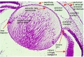
File:Stage 22 image 155.jpg ==Human Embryo Stage 22 Developing Lens== ...- Developing Retina]] | [[:File:Stage 22 image 155.jpg|Image - Developing Lens]](1,000 × 672 (239 KB)) - 07:46, 31 August 2011
File:Keibel Mall 2 177.jpg ==Fig. 177. Transition of the lens epithelium into the lens fibres, in a fetus at the beginning of the fourth month==(965 × 389 (48 KB)) - 11:42, 21 February 2014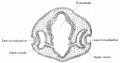
File:Bailey461.jpg ...epressed against the outet surface of the optic vesicle forming a distinct lens invagination (Fig. 461).(751 × 394 (54 KB)) - 06:42, 31 August 2011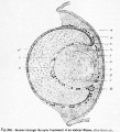
File:Hertwig266.jpg ...ch, choroidea ; If, lens-fibres ; le, lens-epithelium ; I' ', zone of the lens-fibre nuclei ; It, fundament of the cornea ; he, external corneal epitheliu(600 × 663 (83 KB)) - 21:48, 28 March 2012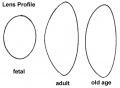
File:Gray0886.jpg ==Lens Profile== Profile views of the lens at different periods of life. 1. In the fetus. 2. In adult life. 3. In old(500 × 368 (17 KB)) - 22:33, 19 August 2012
File:McMurrich1914 fig277.jpg ...layer of optic cup; sf, spaces of Fontana; si, suspensory ligament of the lens; v, vitreous humor. - (Angelucci.)(1,000 × 715 (280 KB)) - 22:36, 31 January 2017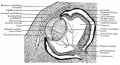
File:Bailey467.jpg ...d, the liquor Morgagni, may remain between the anterior epithelium and the lens fibers. ...the ''membrana pupillaris''. After the earlier and more rapid formation of lens fibers ceases, the hyaloid artery begins (about the seventh month) to under(946 × 515 (159 KB)) - 06:51, 31 August 2011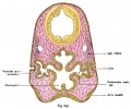
File:Kollmann697.jpg ...h increases the glass body and later allowed to develop. Law is made the lens pouch only in passing, left through the middle.(731 × 612 (94 KB)) - 19:34, 22 October 2012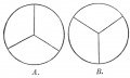
File:Gray0885.jpg ==Lens== ...and arrangement of the radiating lines on the front and back of the fetal lens.(667 × 400 (20 KB)) - 22:29, 19 August 2012File:Lens-neural crest signaling 01.jpg ==Wnt mediates lens repression by neural crest cells and Transforming growth factor-β== # NCCs (blue) secrete TGF-βs, which signal to the non-lens ectoderm and dorsal optic vesicle.(300 × 400 (24 KB)) - 17:25, 31 August 2011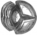
File:Gray0884.jpg ==Lens Structure== The crystalline lens, hardened and divided. (Enlarged.)(419 × 400 (58 KB)) - 15:12, 19 August 2012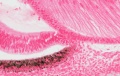
File:Stage 22 image 211.jpg ==Developing Lens and Iris - Human Embryo Carnegie stage 22== * lens(1,200 × 760 (242 KB)) - 11:41, 14 June 2016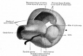
File:Bailey464.jpg ==Fig. 464. Model showing lens and formation of optic cup== ...oted the fact that typical optic cup formation may occur in cases where no lens is developed. The optic cup when first formed is not a complete cup, for th(869 × 592 (67 KB)) - 06:44, 31 August 2011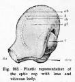
File:Hertwig265.jpg ==Fig. 265. Plastic representation of the optic cup with lens and vitreous body== ...its lower surface.) aus, Optic [choroid] fissure ; yl, vitreous body ; I, lens.(460 × 500 (36 KB)) - 21:49, 28 March 2012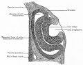
File:Bailey466.jpg ==Fig. 466. Section through optic cup and lens invagination of chick of fifty-four hours' incubation== Between the lens anlage and the pigmented layer of the retina is the broad inner layer of th(859 × 683 (139 KB)) - 15:54, 1 February 2011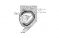
File:Streeter1957 fig06-19.jpg ...reous humor of the eye and forms a network on the posterior surface of the lens. X60.(1,280 × 834 (120 KB)) - 08:03, 18 April 2018File:Lens-neural crest signaling 02.jpg ==Wnt mediates lens repression by neural crest cells and Transforming growth factor-β== Proposed molecular model to explain TGF-β- and Wnt-mediated lens restriction. Broken lines: interactions inferred from the literature.(521 × 522 (22 KB)) - 14:10, 29 April 2011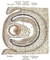
File:Gray0867.jpg ...enters the cup through the choroidal fissure and around the equator of the lens becomes intimately united with this reticular tissue, and contributes to fo(495 × 600 (131 KB)) - 08:59, 19 August 2012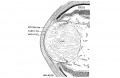
File:Streeter1957 fig06-23.jpg ...reous humor of the eye and forms a network on the posterior surface of the lens. X60. [[Category:Week 8]][[Category:Vision]][[Category:Lens]][[Category:Carnegie Embryo 4570]](1,280 × 834 (161 KB)) - 10:27, 9 May 2018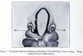
File:Brown006.jpg ...into the secondary optic vesicle (see A, Fig. 7), and eventually forms the lens, which will be described later. As the lens vesicle passes into the secondary optic vesicle, some of the mesoblastic ce(800 × 543 (63 KB)) - 07:12, 31 August 2011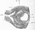
File:Foster128.jpg # the absence at this stage of mesoblast between the lens and the epiblast ; the interval between the two has however been made too g # the arteria centralis retinae forming the vascular capsule of the lens and continuous with vascular structures round the edges of the optic cup.(998 × 859 (223 KB)) - 07:05, 17 March 2012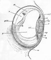
File:Foster051.jpg ...e elongated cells are shewn at ril, now forms nearly the whole mass of the lens, the front wall being reduced to a layer of flattened cells el. ...with the mesoblast m, and appears to be the rudiment of the capsule of the lens and suspensory ligament.(722 × 847 (112 KB)) - 10:24, 11 January 2011
File:Stage 22 image 212.jpg ==Developing Lens and Iris - Human Embryo Carnegie stage 22== * lens(1,200 × 753 (269 KB)) - 11:41, 14 June 2016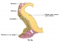
File:Kollmann695.jpg ==Figure 695 The lens unit with a rabbit embryo== ...of capillaries. In the whole extent of the lateral surface considered, the lens plate has a recess. It therefore describes it as an Linsengrübchen (foveol(734 × 519 (45 KB)) - 10:53, 18 May 2014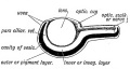
File:Keith1902 fig146.jpg ==Fig. 146. Diagrammatic Section of the Optic Cup and Lens==(900 × 480 (68 KB)) - 09:22, 8 January 2014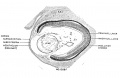
File:Streeter1957 fig06-21.jpg ...reous humor of the eye and forms a network on the posterior surface of the lens. X60.(1,280 × 834 (185 KB)) - 21:14, 15 March 2017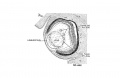
File:Streeter1957 fig06-20.jpg ...reous humor of the eye and forms a network on the posterior surface of the lens. X60.(1,280 × 834 (133 KB)) - 21:14, 15 March 2017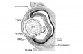
File:Streeter1957 fig06-22.jpg ...reous humor of the eye and forms a network on the posterior surface of the lens. X60.(1,280 × 834 (181 KB)) - 21:13, 15 March 2017
File:Optic cup at carnegie stage 17 .jpg Lens Cavity appears as a small moon shaped slit within the developing lens as the primary fibers slowly fill up cavity. The retina is beginning to dif(640 × 597 (297 KB)) - 21:02, 4 October 2012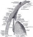
File:Gray0883.jpg ...ont, forming a deep concavity, the hyaloid fossa, for the reception of the lens. It is transparent, of the consistence of thin jelly, and is composed of an The Crystalline Lens (''lens crystallina'') — The crystalline lens, enclosed in its capsule, is situated immediately behind the iris, in front(616 × 700 (107 KB)) - 15:01, 23 August 2017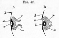
File:Foster047.jpg ...gun to close in. Owing to this involution, which forms the rudiment of the lens, the optic vesicle is doubled in, its front portion r being pushed against ...osterior wall u and an anterior wall r. In the hollow of this cup lies the lens , now completely detached from the superficial epiblast x. Its cavity is st(484 × 315 (22 KB)) - 10:04, 11 January 2011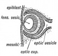
File:Keith1902 fig143.jpg ==Fig. 143. The manner in which the Lens Vesicle is severed from the Epiblast==(475 × 456 (48 KB)) - 09:21, 8 January 2014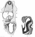
File:Keibel Mall 2 166.jpg ...ium (His); Tr.a., truncus arteriosus; Uk., lower jaw; Z., cell mass in the lens pit.(969 × 1,000 (144 KB)) - 10:32, 21 February 2014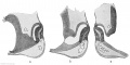
File:Keibel Mall 2 167.jpg ...ells occur between the distal layer of the optic cup and the anlage of the lens,(1,000 × 496 (86 KB)) - 10:32, 21 February 2014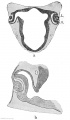
File:Keibel Mall 2 168.jpg ...which is being constricted off from the epidermis. In the interior of the lens vesicle there is a mass of degenerated cells, shown more distinctly in Fig.(583 × 1,000 (65 KB)) - 10:31, 21 February 2014
File:Stage15 bf1.jpg Ectoderm: sensory placodes, lens pit, otocyst, nasal pit, primary/secondary vesicles, fourth ventricle of br Head: 1st, 2nd and 3rd pharyngeal arch, forebrain, site of lens placode, site of otic placode, stomodeum(1,000 × 750 (25 KB)) - 17:22, 13 May 2018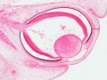
File:Stage 22 image 208.jpg * lens ...vessels present during development located in the vitreous and back of the lens.(1,200 × 903 (368 KB)) - 19:14, 13 September 2017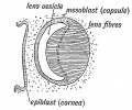
File:Keith1902 fig144.jpg ==Fig. 144. The Formation of the Lens Fibres from the Epithelium on the posterior Wall of the Vesicle==(602 × 503 (67 KB)) - 09:21, 8 January 2014
File:Stage15 bf1b.jpg Ectoderm: sensory placodes, lens pit, otocyst, nasal pit, primary/secondary vesicles, fourth ventricle of br Head: 1st, 2nd and 3rd pharyngeal arch, forebrain, site of lens placode, site of otic placode, stomodeum(600 × 450 (11 KB)) - 05:03, 13 April 2011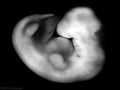
File:Stage15 bf1c.jpg Ectoderm: sensory placodes, lens pit, otocyst, nasal pit, primary/secondary vesicles, fourth ventricle of br Head: 1st, 2nd and 3rd pharyngeal arch, forebrain, site of lens placode, site of otic placode, stomodeum(400 × 300 (6 KB)) - 05:04, 13 April 2011
File:Stage15 bf1a.jpg Ectoderm: sensory placodes, lens pit, otocyst, nasal pit, primary/secondary vesicles, fourth ventricle of br Head: 1st, 2nd and 3rd pharyngeal arch, forebrain, site of lens placode, site of otic placode, stomodeum(800 × 600 (17 KB)) - 05:03, 13 April 2011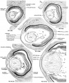
File:Streeter1957 fig06.jpg ...reous humor of the eye and forms a network on the posterior surface of the lens. X60.(1,280 × 1,541 (622 KB)) - 13:41, 22 May 2017
File:Foster049.jpg I the lens, I' the cavity of the lens, lying in the hollow of the optic cup. r the anterior, u the posterior wall(360 × 444 (29 KB)) - 10:13, 11 January 2011
File:Stage15 bf3.jpg Ectoderm: sensory placodes, lens pit, otocyst, nasal pit, primary/secondary vesicles, fourth ventricle of br Head: 1st, 2nd and 3rd pharyngeal arch, forebrain, site of {{lens}} {{placode}}, site of otic placode, stomodeum(448 × 600 (29 KB)) - 09:23, 2 October 2020
File:Stage15 bf4.jpg Ectoderm: sensory placodes, lens pit, otocyst, nasal pit, primary/secondary vesicles, fourth ventricle of br Head: 1st, 2nd and 3rd pharyngeal arch, forebrain, site of {{lens}} {{placode}}, site of otic placode, stomodeum(448 × 600 (22 KB)) - 09:23, 2 October 2020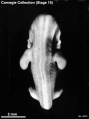
File:Stage15 bf5.jpg Ectoderm: sensory placodes, lens pit, otocyst, nasal pit, primary/secondary vesicles, fourth ventricle of br Head: 1st, 2nd and 3rd pharyngeal arch, forebrain, site of {{lens}} {{placode}}, site of otic placode, stomodeum(448 × 600 (23 KB)) - 09:24, 2 October 2020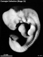
File:Stage15 bf6.jpg Ectoderm: sensory placodes, lens pit, otocyst, nasal pit, primary/secondary vesicles, fourth ventricle of br Head: 1st, 2nd and 3rd pharyngeal arch, forebrain, site of {{lens}} {{placode}}, site of otic placode, stomodeum(448 × 600 (28 KB)) - 09:22, 2 October 2020
File:Comparison of phenotypes with PAX6 gene mutation in different animals.png ...able and modifiable phenotype that consists of decreased eye size, reduced lens size, and malformation of the retina. Drosophila ey (a PAX6 ortholog) mutat(558 × 368 (291 KB)) - 22:12, 23 September 2012
File:Rugh 122.jpg ==Development of the optic cup and lens in Siredon pisciformis==(643 × 1,000 (187 KB)) - 10:56, 17 April 2013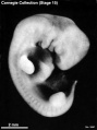
File:Stage15 bf7.jpg {{Ectoderm}}: sensory placodes, lens pit, otocyst, nasal pit, primary/secondary vesicles, fourth ventricle of br {{Head}}: 1st, 2nd and 3rd pharyngeal arch, forebrain, site of lens placode, site of otic placode, stomodeum(448 × 600 (28 KB)) - 17:42, 5 October 2020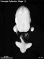
File:Stage15 bf8.jpg {{Ectoderm}}: sensory placodes, lens pit, otocyst, nasal pit, primary/secondary vesicles, fourth ventricle of br {{Head}}: 1st, 2nd and 3rd pharyngeal arch, forebrain, site of lens placode, site of otic placode, stomodeum(448 × 600 (22 KB)) - 17:43, 5 October 2020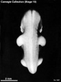
File:Stage15 bf9.jpg {{Ectoderm}}: sensory placodes, lens pit, otocyst, nasal pit, primary/secondary vesicles, fourth ventricle of br {{Head}}: 1st, 2nd and 3rd pharyngeal arch, forebrain, site of lens placode, site of otic placode, stomodeum(448 × 600 (22 KB)) - 17:44, 5 October 2020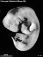
File:Stage15 bf10.jpg {{Ectoderm}}: sensory placodes, lens pit, otocyst, nasal pit, primary/secondary vesicles, fourth ventricle of br {{Head}}: 1st, 2nd and 3rd pharyngeal arch, forebrain, site of lens placode, site of otic placode, stomodeum(448 × 600 (28 KB)) - 17:44, 5 October 2020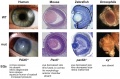
File:Pax6 eye phenotypes.jpg ...bsence of iris), corneal opacity (aniridia-related keratopathy), cataract (lens clouding), glaucoma, and long-term retinal degeneration. ...rsistent epithelial cells that remains attached between the cornea and the lens.(1,000 × 662 (152 KB)) - 12:58, 28 September 2015File:Day 10.5 Deep Lens Indentation.JPG Day 10.5: Deep lens indentation occurs. Tel = cerebral hemisphere, L = lens invagination, Hl = Hindlimb bud, Aa = forelimb bud, So = Somite 4, O = otic(514 × 483 (28 KB)) - 13:00, 25 June 2014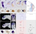
File:Eye-neural crest signaling.jpg ==Wnt mediates lens repression by neural crest cells and Transforming growth factor-β== ...for NCC-specific HNK1 (brown). Arrowheads indicate gene expression in non-lens ectoderm.(946 × 886 (436 KB)) - 14:10, 29 April 2011
File:Stage14 sem1a.jpg Features: midbrain, nasal placode, lens pit, 1,2,3 pharyngeal arches, fourth ventricle of brain, 1st pharyngeal gro Identify: midbrain region, nasal placode, lens pit, 1st, 2nd and 3rd pharyngeal arches, 1st pharyngeal groove, maxillary a(800 × 655 (66 KB)) - 20:42, 25 October 2013
File:Stage14 sem1b.jpg Features: midbrain, nasal placode, lens pit, 1,2,3 pharyngeal arches, fourth ventricle of brain, 1st pharyngeal gro Identify: midbrain region, nasal placode, lens pit, 1st, 2nd and 3rd pharyngeal arches, 1st pharyngeal groove, maxillary a(600 × 491 (41 KB)) - 20:42, 25 October 2013
File:Stage14 sem1c.jpg Features: midbrain, nasal placode, lens pit, 1,2,3 pharyngeal arches, fourth ventricle of brain, 1st pharyngeal gro Identify: midbrain region, nasal placode, lens pit, 1st, 2nd and 3rd pharyngeal arches, 1st pharyngeal groove, maxillary a(400 × 327 (23 KB)) - 09:37, 3 June 2014
File:Stage14 sem1.jpg Features: midbrain, nasal placode, lens pit, 1,2,3 pharyngeal arches, fourth ventricle of brain, 1st pharyngeal gro Identify: midbrain region, nasal placode, lens pit, 1st, 2nd and 3rd pharyngeal arches, 1st pharyngeal groove, maxillary a(1,000 × 819 (94 KB)) - 20:41, 25 October 2013
File:Stage14 bf1.jpg Features: midbrain, nasal placode, lens pit, 1,2,3 pharyngeal arches, fourth ventricle of brain, 1st pharyngeal gro Identify: midbrain region, nasal placode, lens pit, 1st, 2nd and 3rd pharyngeal arches, 1st pharyngeal groove, maxillary a(1,000 × 743 (27 KB)) - 09:38, 3 June 2014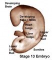
File:Stage13 surface bulges.jpg Features: site of midbrain, lens placode, site of nasal placode, 1,2,3 pharyngeal arches, L. ventricular pro Identify: site of midbrain, lens placode, site of nasal placode, 1st, 2nd and 3rd pharyngeal arches, L. vent(337 × 389 (14 KB)) - 06:08, 12 August 2011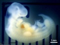
File:Stage14 bf1a.jpg Features: midbrain, nasal placode, lens pit, 1,2,3 pharyngeal arches, fourth ventricle of brain, 1st pharyngeal gro Identify: midbrain region, nasal placode, lens pit, 1st, 2nd and 3rd pharyngeal arches, 1st pharyngeal groove, maxillary a(800 × 594 (20 KB)) - 20:48, 25 October 2013
File:Stage14 bf1b.jpg Features: midbrain, nasal placode, lens pit, 1,2,3 pharyngeal arches, fourth ventricle of brain, 1st pharyngeal gro Identify: midbrain region, nasal placode, lens pit, 1st, 2nd and 3rd pharyngeal arches, 1st pharyngeal groove, maxillary a(600 × 446 (13 KB)) - 20:48, 25 October 2013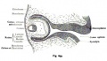
File:Kollmann699.jpg ...k bubble has become longer and is enclosed together with the optic cup and lens of the mesoderm, from which the cornea, sclera and choroid shape gradually.(722 × 418 (64 KB)) - 18:56, 22 October 2012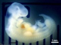
File:Stage14 bf1c.jpg Features: midbrain, nasal placode, lens pit, 1,2,3 pharyngeal arches, fourth ventricle of brain, 1st pharyngeal gro Identify: midbrain region, nasal placode, lens pit, 1st, 2nd and 3rd pharyngeal arches, 1st pharyngeal groove, maxillary a(400 × 297 (8 KB)) - 20:49, 25 October 2013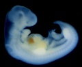
File:Stage14 human.jpg Features: midbrain, nasal placode, lens pit, 1,2,3 pharyngeal arches, fourth ventricle of brain, 1st pharyngeal gro Identify: midbrain region, nasal placode, lens pit, 1st, 2nd and 3rd pharyngeal arches, 1st pharyngeal groove, maxillary a(646 × 530 (32 KB)) - 11:01, 2 June 2015File:Formation of the lens 1.jpg ...ing the vital role of induction from the optic cup in the formation of the lens.(1,152 × 648 (102 KB)) - 17:42, 26 September 2012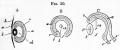
File:Foster050.jpg ...of the eye as to shave off a small portion of the posterior surface of the lens I, but so far in front as not to be carried at all through the stalk. Lette ...of the line z, 2, the wall of the optic cup would have extended up to the lens below as well as above. Letters as above.(792 × 327 (43 KB)) - 10:17, 11 January 2011
File:Stage12 bf5.jpg Features: day 26, 27 somites, forebrain, site of lens placode, site of otic placode , stomodeum, 1st pharyngeal arch, 2nd pharyng Identify: forebrain, site of lens placode, site of otic placode, stomodeum, 1st pharyngeal arch, 2nd pharynge(1,000 × 750 (59 KB)) - 05:56, 13 April 2011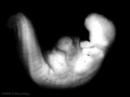
File:Stage12 bf5a.jpg Features: day 26, 27 somites, forebrain, site of lens placode, site of otic placode , stomodeum, 1st pharyngeal arch, 2nd pharyng Identify: forebrain, site of lens placode, site of otic placode, stomodeum, 1st pharyngeal arch, 2nd pharynge(800 × 600 (39 KB)) - 05:56, 13 April 2011
File:Stage12 bf5b.jpg Features: day 26, 27 somites, forebrain, site of lens placode, site of otic placode , stomodeum, 1st pharyngeal arch, 2nd pharyng Identify: forebrain, site of lens placode, site of otic placode, stomodeum, 1st pharyngeal arch, 2nd pharynge(600 × 450 (23 KB)) - 05:56, 13 April 2011
File:Stage12 bf5c.jpg Features: day 26, 27 somites, forebrain, site of lens placode, site of otic placode , stomodeum, 1st pharyngeal arch, 2nd pharyng Identify: forebrain, site of lens placode, site of otic placode, stomodeum, 1st pharyngeal arch, 2nd pharynge(400 × 300 (6 KB)) - 05:56, 13 April 2011
File:Stage12 sem6b.jpg * Features: day 26, 27 somites, forebrain, site of lens placode, site of otic placode , stomodeum, 1st pharyngeal arch, 2nd pharyng * Identify: forebrain, site of lens placode, site of otic placode, stomodeum, 1st pharyngeal arch, 2nd pharynge(800 × 796 (74 KB)) - 22:48, 4 September 2014
File:Stage12 sem6c.jpg * Features: day 26, 27 somites, forebrain, site of lens placode, site of otic placode , stomodeum, 1st pharyngeal arch, 2nd pharyng * Identify: forebrain, site of lens placode, site of otic placode, stomodeum, 1st pharyngeal arch, 2nd pharynge(600 × 597 (49 KB)) - 22:49, 4 September 2014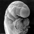
File:Stage12 sem6a.jpg * Features: day 26, 27 somites, forebrain, site of lens placode, site of otic placode , stomodeum, 1st pharyngeal arch, 2nd pharyng * Identify: forebrain, site of lens placode, site of otic placode, stomodeum, 1st pharyngeal arch, 2nd pharynge(1,000 × 995 (100 KB)) - 22:48, 4 September 2014
File:Stage12 sem6.jpg * Features: day 26, 27 somites, forebrain, site of lens placode, site of otic placode , stomodeum, 1st pharyngeal arch, 2nd pharyng * Identify: forebrain, site of lens placode, site of otic placode, stomodeum, 1st pharyngeal arch, 2nd pharynge(1,620 × 1,612 (190 KB)) - 22:47, 4 September 2014
File:Stage12 bf1.jpg Features: day 26, 27 somites, forebrain, site of lens placode, site of otic placode , stomodeum, 1st pharyngeal arch, 2nd pharyng Identify: forebrain, site of lens placode, site of otic placode, stomodeum, 1st pharyngeal arch, 2nd pharynge(482 × 1,000 (20 KB)) - 12:51, 15 April 2011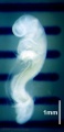
File:Stage12 bf1a.jpg Features: day 26, 27 somites, forebrain, site of lens placode, site of otic placode , stomodeum, 1st pharyngeal arch, 2nd pharyng Identify: forebrain, site of lens placode, site of otic placode, stomodeum, 1st pharyngeal arch, 2nd pharynge(386 × 800 (15 KB)) - 12:53, 15 April 2011
File:Stage12 bf1b.jpg Features: day 26, 27 somites, forebrain, site of lens placode, site of otic placode , stomodeum, 1st pharyngeal arch, 2nd pharyng Identify: forebrain, site of lens placode, site of otic placode, stomodeum, 1st pharyngeal arch, 2nd pharynge(290 × 600 (11 KB)) - 12:50, 15 April 2011
File:Stage12 bf1c.jpg Features: day 26, 27 somites, forebrain, site of lens placode, site of otic placode , stomodeum, 1st pharyngeal arch, 2nd pharyng Identify: forebrain, site of lens placode, site of otic placode, stomodeum, 1st pharyngeal arch, 2nd pharynge(193 × 400 (6 KB)) - 12:53, 15 April 2011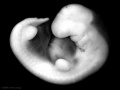
File:Stage14 bf2.jpg Features: midbrain, nasal placode, lens pit, 1,2,3 pharyngeal arches, fourth ventricle of brain, 1st pharyngeal gro Identify: midbrain region, nasal placode, lens pit, 1st, 2nd and 3rd pharyngeal arches, 1st pharyngeal groove, maxillary a(1,000 × 750 (71 KB)) - 12:54, 10 April 2014
File:Stage12 bf2.jpg Features: day 26, 27 somites, forebrain, site of lens placode, site of otic placode , stomodeum, 1st pharyngeal arch, 2nd pharyng Identify: forebrain, site of lens placode, site of otic placode, stomodeum, 1st pharyngeal arch, 2nd pharynge(471 × 1,000 (17 KB)) - 12:19, 7 September 2009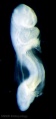
File:Stage12 bf2a.jpg Features: day 26, 27 somites, forebrain, site of lens placode, site of otic placode , stomodeum, 1st pharyngeal arch, 2nd pharyng Identify: forebrain, site of lens placode, site of otic placode, stomodeum, 1st pharyngeal arch, 2nd pharynge(377 × 800 (12 KB)) - 12:20, 7 September 2009
File:Stage12 bf2b.jpg Features: day 26, 27 somites, forebrain, site of lens placode, site of otic placode , stomodeum, 1st pharyngeal arch, 2nd pharyng Identify: forebrain, site of lens placode, site of otic placode, stomodeum, 1st pharyngeal arch, 2nd pharynge(283 × 600 (9 KB)) - 18:38, 17 August 2011
File:Stage12 bf2c.jpg Features: day 26, 27 somites, forebrain, site of lens placode, site of otic placode , stomodeum, 1st pharyngeal arch, 2nd pharyng Identify: forebrain, site of lens placode, site of otic placode, stomodeum, 1st pharyngeal arch, 2nd pharynge(189 × 400 (5 KB)) - 12:20, 7 September 2009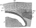
File:McMurrich1914 fig278.jpg ac, Anterior chamber of the eye; co, cornea; ec, ectoderm; I, lens; mc, ciliary muscle; p, pigment layer of the optic cup; r, retinal layer; t(800 × 617 (134 KB)) - 22:28, 31 January 2017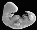
File:Stage14 SEM.jpg Features: midbrain, nasal placode, lens pit, 1,2,3 pharyngeal arches, fourth ventricle of brain, 1st pharyngeal gro Identify: midbrain region, nasal placode, lens pit, 1st, 2nd and 3rd pharyngeal arches, 1st pharyngeal groove, maxillary a(646 × 530 (36 KB)) - 22:47, 3 August 2009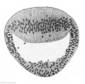
File:Keibel Mall 2 174.jpg ==Fig. 174. Lens of a human embryo of from 30 to 31 days==(607 × 600 (56 KB)) - 11:38, 21 February 2014
File:Stage14 bf2l.jpg Features: midbrain, nasal placode, lens pit, 1,2,3 pharyngeal arches, fourth ventricle of brain, 1st pharyngeal gro Identify: midbrain region, nasal placode, lens pit, 1st, 2nd and 3rd pharyngeal arches, 1st pharyngeal groove, maxillary a(1,000 × 834 (66 KB)) - 20:51, 25 October 2013
File:Stage14 bf2al.jpg Features: midbrain, nasal placode, lens pit, 1,2,3 pharyngeal arches, fourth ventricle of brain, 1st pharyngeal gro Identify: midbrain region, nasal placode, lens pit, 1st, 2nd and 3rd pharyngeal arches, 1st pharyngeal groove, maxillary a(800 × 667 (48 KB)) - 11:25, 4 September 2009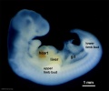
File:Stage14 bf2bl.jpg Features: midbrain, nasal placode, lens pit, 1,2,3 pharyngeal arches, fourth ventricle of brain, 1st pharyngeal gro Identify: midbrain region, nasal placode, lens pit, 1st, 2nd and 3rd pharyngeal arches, 1st pharyngeal groove, maxillary a(600 × 500 (31 KB)) - 11:25, 4 September 2009
File:Stage13 bf2.jpg Features: site of midbrain, lens placode, site of nasal placode, 1,2,3 pharyngeal arches, L. ventricular pro Identify: site of midbrain, lens placode, site of nasal placode, 1st, 2nd and 3rd pharyngeal arches, L. vent(1,000 × 750 (61 KB)) - 04:58, 13 April 2011
File:Stage13 bf2a.jpg Features: site of midbrain, lens placode, site of nasal placode, 1,2,3 pharyngeal arches, L. ventricular pro Identify: site of midbrain, lens placode, site of nasal placode, 1st, 2nd and 3rd pharyngeal arches, L. vent(800 × 600 (40 KB)) - 04:58, 13 April 2011
File:Stage13 bf2b.jpg Features: site of midbrain, lens placode, site of nasal placode, 1,2,3 pharyngeal arches, L. ventricular pro Identify: site of midbrain, lens placode, site of nasal placode, 1st, 2nd and 3rd pharyngeal arches, L. vent(600 × 450 (24 KB)) - 04:58, 13 April 2011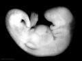
File:Stage13 bf2c.jpg Features: site of midbrain, lens placode, site of nasal placode, 1,2,3 pharyngeal arches, L. ventricular pro Identify: site of midbrain, lens placode, site of nasal placode, 1st, 2nd and 3rd pharyngeal arches, L. vent(400 × 300 (12 KB)) - 04:55, 13 April 2011
File:Stage12 sem7.jpg Features: day 26, 27 somites, forebrain, site of lens placode, site of otic placode , stomodeum, 1st pharyngeal arch, 2nd pharyng Identify: forebrain, site of lens placode, site of otic placode, stomodeum, 1st pharyngeal arch, 2nd pharynge(2,520 × 2,715 (479 KB)) - 14:16, 14 May 2011
File:Stage12 sem7a.jpg Features: day 26, 27 somites, forebrain, site of lens placode, site of otic placode , stomodeum, 1st pharyngeal arch, 2nd pharyng Identify: forebrain, site of lens placode, site of otic placode, stomodeum, 1st pharyngeal arch, 2nd pharynge(1,000 × 1,077 (139 KB)) - 14:18, 14 May 2011
File:Stage12 sem7b.jpg Features: day 26, 27 somites, forebrain, site of lens placode, site of otic placode , stomodeum, 1st pharyngeal arch, 2nd pharyng Identify: forebrain, site of lens placode, site of otic placode, stomodeum, 1st pharyngeal arch, 2nd pharynge(800 × 862 (102 KB)) - 14:18, 14 May 2011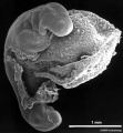
File:Stage12 sem7c.jpg Features: day 26, 27 somites, forebrain, site of lens placode, site of otic placode , stomodeum, 1st pharyngeal arch, 2nd pharyng Identify: forebrain, site of lens placode, site of otic placode, stomodeum, 1st pharyngeal arch, 2nd pharynge(600 × 647 (68 KB)) - 14:18, 14 May 2011
File:Stage12 sem1.jpg Features: day 26, 27 somites, forebrain, site of lens placode, site of otic placode , stomodeum, 1st pharyngeal arch, 2nd pharyng Identify: forebrain, site of lens placode, site of otic placode, stomodeum, 1st pharyngeal arch, 2nd pharynge(472 × 1,000 (69 KB)) - 15:06, 14 May 2011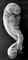
File:Stage12 sem1a.jpg Features: day 26, 27 somites, forebrain, site of lens placode, site of otic placode , stomodeum, 1st pharyngeal arch, 2nd pharyng Identify: forebrain, site of lens placode, site of otic placode, stomodeum, 1st pharyngeal arch, 2nd pharynge(378 × 800 (24 KB)) - 15:07, 14 May 2011
File:Stage12 sem1b.jpg Features: day 26, 27 somites, forebrain, site of lens placode, site of otic placode , stomodeum, 1st pharyngeal arch, 2nd pharyng Identify: forebrain, site of lens placode, site of otic placode, stomodeum, 1st pharyngeal arch, 2nd pharynge(284 × 600 (15 KB)) - 15:07, 14 May 2011
File:Stage12 sem1c.jpg Features: day 26, 27 somites, forebrain, site of lens placode, site of otic placode , stomodeum, 1st pharyngeal arch, 2nd pharyng Identify: forebrain, site of lens placode, site of otic placode, stomodeum, 1st pharyngeal arch, 2nd pharynge(189 × 400 (8 KB)) - 15:07, 14 May 2011
File:Stage14 bf2a.jpg Features: midbrain, nasal placode, lens pit, 1,2,3 pharyngeal arches, fourth ventricle of brain, 1st pharyngeal gro Identify: midbrain region, nasal placode, lens pit, 1st, 2nd and 3rd pharyngeal arches, 1st pharyngeal groove, maxillary a(800 × 600 (45 KB)) - 05:00, 13 April 2011
File:Stage14 bf2b.jpg Features: midbrain, nasal placode, lens pit, 1,2,3 pharyngeal arches, fourth ventricle of brain, 1st pharyngeal gro Identify: midbrain region, nasal placode, lens pit, 1st, 2nd and 3rd pharyngeal arches, 1st pharyngeal groove, maxillary a(600 × 450 (26 KB)) - 05:01, 13 April 2011
File:Stage14 bf2c.jpg Features: midbrain, nasal placode, lens pit, 1,2,3 pharyngeal arches, fourth ventricle of brain, 1st pharyngeal gro Identify: midbrain region, nasal placode, lens pit, 1st, 2nd and 3rd pharyngeal arches, 1st pharyngeal groove, maxillary a(400 × 300 (13 KB)) - 05:00, 13 April 2011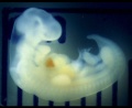
File:Stage14 human scale.jpg Features: midbrain, nasal placode, lens pit, 1,2,3 pharyngeal arches, fourth ventricle of brain, 1st pharyngeal gro Identify: midbrain region, nasal placode, lens pit, 1st, 2nd and 3rd pharyngeal arches, 1st pharyngeal groove, maxillary a(646 × 530 (40 KB)) - 22:42, 3 August 2009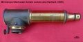
File:Hartnack camera lucida lens.jpg ==Microscope Oberhauser Camera Lucida Lens==(715 × 367 (58 KB)) - 11:30, 25 March 2015
File:Stage14 bf2cl.jpg Features: midbrain, nasal placode, lens pit, 1,2,3 pharyngeal arches, fourth ventricle of brain, 1st pharyngeal gro Identify: midbrain region, nasal placode, lens pit, 1st, 2nd and 3rd pharyngeal arches, 1st pharyngeal groove, maxillary a(400 × 333 (17 KB)) - 14:11, 21 October 2010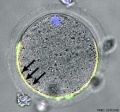
File:Mouse oocyte cortical granule ovastacin 01.jpg ...e contrast (DIC) after staining with rabbit anti-ovastacin (red) antibody, Lens culinaris agglutinin (green) a marker of cortical granules, and the nuclear(500 × 465 (54 KB)) - 18:33, 11 August 2018
File:Stage14 bf3.jpg Features: midbrain, nasal placode, lens pit, 1,2,3 pharyngeal arches, fourth ventricle of brain, 1st pharyngeal gro Identify: midbrain region, nasal placode, lens pit, 1st, 2nd and 3rd pharyngeal arches, 1st pharyngeal groove, maxillary a(1,368 × 2,000 (159 KB)) - 17:43, 30 May 2011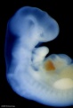
File:Stage14 bf3a.jpg Features: midbrain, nasal placode, lens pit, 1,2,3 pharyngeal arches, fourth ventricle of brain, 1st pharyngeal gro Identify: midbrain region, nasal placode, lens pit, 1st, 2nd and 3rd pharyngeal arches, 1st pharyngeal groove, maxillary a(684 × 1,000 (61 KB)) - 17:43, 30 May 2011
File:Stage14 bf3b.jpg Features: midbrain, nasal placode, lens pit, 1,2,3 pharyngeal arches, fourth ventricle of brain, 1st pharyngeal gro Identify: midbrain region, nasal placode, lens pit, 1st, 2nd and 3rd pharyngeal arches, 1st pharyngeal groove, maxillary a(547 × 800 (43 KB)) - 17:43, 30 May 2011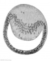
File:Keibel Mall 2 175.jpg ==Fig. 175. Section through the lens of a human embryo of 12.5 mm greatest length==(496 × 597 (40 KB)) - 11:35, 21 February 2014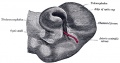
File:Gray0865.jpg ...h each other at the cup margin, which ultimately overlaps the front of the lens and reaches as far forward as the future aperture of the pupil. The invagin(759 × 400 (70 KB)) - 08:52, 19 August 2012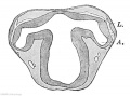
File:Keibel Mall 2 163.jpg ...el and Elze, Fig. 12h.) X30. A., anlageof the optic cup; L., anlage of the lens.(675 × 500 (51 KB)) - 10:33, 21 February 2014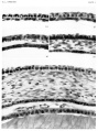
File:Streeter1957 plate01.jpg ...the space between the mesothelium (Descement's) and the epithelium of the lens.(1,500 × 2,009 (486 KB)) - 17:37, 6 November 2016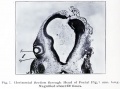
File:Brown007.jpg * In Figs. 7 and 8 the lens vesicle will be seen to have been entirely separated from the surface.(800 × 594 (78 KB)) - 13:30, 14 February 2011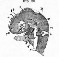
File:Foster038.jpg ...the lens, the shaded portion being the expression of its cavity. Below the lens between the two limbs of the horseshoe is the choroidal fissure.(432 × 416 (46 KB)) - 13:14, 29 January 2016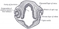
File:Gray0863.jpg ...h each other at the cup margin, which ultimately overlaps the front of the lens and reaches as far forward as the future aperture of the pupil. The invagin(813 × 400 (67 KB)) - 15:06, 23 August 2017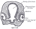
File:Gray0864.jpg ...h each other at the cup margin, which ultimately overlaps the front of the lens and reaches as far forward as the future aperture of the pupil. The invagin(600 × 495 (81 KB)) - 15:06, 23 August 2017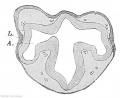
File:Keibel Mall 2 164.jpg ...l and Elze, Fig. llf.) X30. A., anlage of the optic cup; L., anlage of the lens.(610 × 500 (44 KB)) - 10:33, 21 February 2014
File:Kollmann698.jpg ...(van Bambecke) are the edges of the lens pit already grown and there is a lens vesicle is formed, which still related to the remaining ectoderm.(689 × 337 (45 KB)) - 18:58, 22 October 2012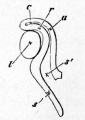
File:Foster048.jpg ...E EYE AND THE OPTIC NERVE AT AN EARLY STAGE (from Lieberkuhn), to shew the lens '''I''' occupying the whole hollow of the optic cup, the inclination of the(209 × 294 (12 KB)) - 13:16, 11 January 2011
File:Meyer1932history8 fig04.jpg (a) The lens holder, (b) the different lenses used, (c) glass object carrier, (d) locati(743 × 1,000 (137 KB)) - 21:39, 2 November 2015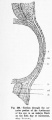
File:Hertwig269.jpg Ce, Corneal epithelium ; h, lens-epithelium ; h, structureless sheet of the corneal fundament ; li, embryoni(346 × 800 (47 KB)) - 21:41, 28 March 2012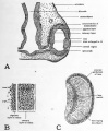
File:Patten045.jpg '''C''' drawing to show cellular organization of the lens.(800 × 968 (156 KB)) - 08:32, 29 July 2011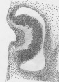
File:Retinal Disc to form Optic Cup at Carnegie stage 14.jpg Lens Pit can also be seen forming. Contour forming slowly.(572 × 800 (400 KB)) - 21:11, 4 October 2012
File:Meyer1932history7 fig01.jpg (I) and (II), front and back respectively. (c) Lens. (g) adjusting screw, (h) stage, (i) rotating object carrier, (k) handle of(800 × 665 (48 KB)) - 21:23, 2 November 2015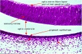
File:Stage 22 image 154.jpg ...- Developing Retina]] | [[:File:Stage 22 image 155.jpg|Image - Developing Lens]](1,000 × 665 (205 KB)) - 07:45, 31 August 2011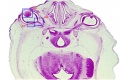
File:Stage 22 image 152.jpg ...- Developing Retina]] | [[:File:Stage 22 image 155.jpg|Image - Developing Lens]](1,000 × 670 (127 KB)) - 07:45, 31 August 2011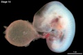
File:Stage14 bf28.jpg * Head: 1st, 2nd and 3rd pharyngeal arch, forebrain, site of lens placode, site of otic placode, stomodeum(1,280 × 848 (115 KB)) - 22:07, 14 June 2016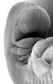
File:Stage14 sem2.jpg Features: midbrain, nasal placode, lens pit, 1,2,3 pharyngeal arches, 1st pharyngeal groove, heart prominence, cerv(1,261 × 2,000 (234 KB)) - 16:39, 9 June 2011
File:Stage14 sem2a.jpg Features: midbrain, nasal placode, lens pit, 1,2,3 pharyngeal arches, 1st pharyngeal groove, heart prominence, cerv(631 × 1,000 (83 KB)) - 16:39, 9 June 2011
File:Stage14 sem2b.jpg Features: midbrain, nasal placode, lens pit, 1,2,3 pharyngeal arches, 1st pharyngeal groove, heart prominence, cerv(505 × 800 (58 KB)) - 16:39, 9 June 2011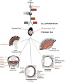
File:Eye evolution.jpg ...nism, we propose intercalary evolution of progressively more genes such as lens genes into the eye developmental pathway.(447 × 563 (106 KB)) - 09:35, 31 August 2017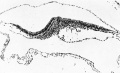
File:GladstoneHamilton1941 text-fig05.jpg ...ned on their distal aspect and sides by endothelioid cells with rounded or lens-like nuclei; on the central side, however, the mesothelial layer is often d(1,280 × 774 (152 KB)) - 17:02, 26 February 2017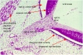
File:Stage 22 image 153.jpg ...- Developing Retina]] | [[:File:Stage 22 image 155.jpg|Image - Developing Lens]](1,000 × 662 (191 KB)) - 07:45, 31 August 2011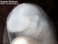
File:Stage13 bf7.jpg * Head: 1st, 2nd and 3rd pharyngeal arch, forebrain, site of lens placode, site of otic placode, stomodeum(600 × 451 (25 KB)) - 08:34, 28 August 2013
File:Stage13 bf9.jpg * Head: 1st, 2nd and 3rd pharyngeal arch, forebrain, site of lens placode, site of otic placode, stomodeum(800 × 600 (43 KB)) - 08:34, 28 August 2013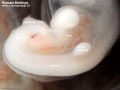
File:Stage13 bf10.jpg * Head: 1st, 2nd and 3rd pharyngeal arch, forebrain, site of lens placode, site of otic placode, stomodeum(800 × 600 (45 KB)) - 08:36, 28 August 2013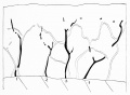
File:Keibel Mall 2 514.jpg ...ixture (acid fuchsin and picric acid), made by the use of an oil-immersion lens (Zeiss obj. 2 mm. and ocular 6).(1,280 × 941 (101 KB)) - 09:29, 22 December 2018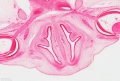
File:Stage 22 image 209.jpg * lens(1,200 × 808 (305 KB)) - 10:56, 14 August 2017
File:Stage13 bf8.jpg * Head: 1st, 2nd and 3rd pharyngeal arch, forebrain, site of lens placode, site of otic placode, stomodeum(600 × 451 (32 KB)) - 08:28, 28 August 2013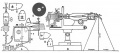
File:Gage1912 fig01.jpg ...roscope; O objective, Fl, I, El, the field lens, the real image and the eye lens of the ocular (Oc); M2 the prism or 45 degree mirror for reflecting the im(1,000 × 438 (49 KB)) - 15:47, 4 October 2017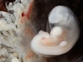
File:Stage13 bf5.jpg * Head: 1st, 2nd and 3rd pharyngeal arch, forebrain, site of lens placode, site of otic placode, stomodeum(901 × 676 (65 KB)) - 08:30, 28 August 2013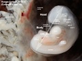
File:Stage13 bf6.jpg * Head: 1st, 2nd and 3rd pharyngeal arch, forebrain, site of lens placode, site of otic placode, stomodeum(901 × 676 (80 KB)) - 14:49, 28 February 2014
File:Stage13 bf6.gif * Head: 1st, 2nd and 3rd pharyngeal arch, forebrain, site of lens placode, site of otic placode, stomodeum(901 × 676 (463 KB)) - 08:33, 28 August 2013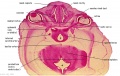
File:Stage 22 image 057.jpg ...ryo_Stages/Stage22/08/Stage22-08.html?zoom=6&lat=-1611.5&lon=1761&layers=B Lens and Cornea](1,000 × 632 (135 KB)) - 04:12, 10 December 2019
File:Stage13 bf5.gif * Head: 1st, 2nd and 3rd pharyngeal arch, forebrain, site of lens placode, site of otic placode, stomodeum(600 × 450 (197 KB)) - 08:24, 12 August 2011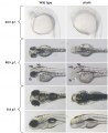
File:Chokh.png ...rphology of the head and trunk is normal in mutants. At 48 h.p.f., a small lens is visible in chk mutants (arrow). fb, forebrain; hb, hindbrain; mb, midbra(748 × 916 (534 KB)) - 10:20, 26 October 2017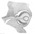
File:Keibel Mall 2 173.jpg ...der the magnification (X 100) employed. The thickened proximal wall of the lens vesicle fills about the half of the lumen of this structure, and in the lum(818 × 800 (142 KB)) - 11:28, 21 February 2014
File:Stage14 bf27.jpg * Head: 1st, 2nd and 3rd pharyngeal arch, forebrain, site of lens placode, site of otic placode, stomodeum(1,000 × 1,291 (208 KB)) - 22:07, 14 June 2016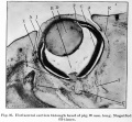
File:Brown016.jpg At F, Fig. 16, is shown a band of tissue connecting the lens with the retina. This will eventually form the suspensory ligament or the Z(800 × 742 (124 KB)) - 13:13, 30 January 2016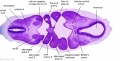
File:Stage 13 image 057.jpg ...egion of the developing eyes [[:File:Stage 13 image 057.jpg|B1L - L optic (lens) placode]] | [[:File:Stage 13 image 058.jpg|B2L - L placode and optic vesic(1,000 × 511 (99 KB)) - 07:59, 6 October 2011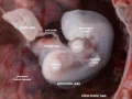
File:Stage14 bf24.jpg * Head: 1st, 2nd and 3rd pharyngeal arch, forebrain, site of lens placode, site of otic placode, stomodeum(1,231 × 923 (156 KB)) - 06:54, 7 April 2016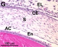
File:Mouse cornea P0.jpg ...ma (S) Anterior chamber (AC). eyelid (EL) corneal endothelial (En) layer. lens (L) and the corneal epithelium (CE)(703 × 561 (101 KB)) - 11:52, 24 January 2015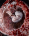
File:Stage14 bf20.jpg * Head: 1st, 2nd and 3rd pharyngeal arch, forebrain, site of lens placode, site of otic placode, stomodeum(800 × 1,000 (158 KB)) - 14:43, 28 February 2014
File:Stage14 bf19.jpg * Head: 1st, 2nd and 3rd pharyngeal arch, forebrain, site of lens placode, site of otic placode, stomodeum(1,200 × 1,500 (287 KB)) - 14:44, 28 February 2014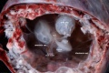
File:Stage14 bf25.jpg * Head: 1st, 2nd and 3rd pharyngeal arch, forebrain, site of lens placode, site of otic placode, stomodeum(2,000 × 1,334 (337 KB)) - 16:02, 16 March 2014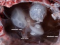
File:Stage14 bf26.jpg * Head: 1st, 2nd and 3rd pharyngeal arch, forebrain, site of lens placode, site of otic placode, stomodeum(1,263 × 947 (150 KB)) - 16:02, 16 March 2014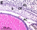
File:Mouse cornea E16.5.jpg Anterior chamber (AC). eyelid (EL) corneal endothelial (En) layer. lens (L) and the corneal epithelium (CE)(701 × 562 (113 KB)) - 11:51, 24 January 2015
File:Stage14 bf4.jpg Features: midbrain, nasal placode, lens pit, 1,2,3 pharyngeal arches, fourth ventricle of brain, 1st pharyngeal gro(2,650 × 2,000 (352 KB)) - 17:55, 30 May 2011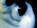
File:Stage14 bf4a.jpg Features: midbrain, nasal placode, lens pit, 1,2,3 pharyngeal arches, fourth ventricle of brain, 1st pharyngeal gro(1,325 × 1,000 (137 KB)) - 17:56, 30 May 2011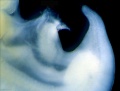
File:Stage14 bf4b.jpg Features: midbrain, nasal placode, lens pit, 1,2,3 pharyngeal arches, fourth ventricle of brain, 1st pharyngeal gro(1,060 × 800 (99 KB)) - 17:56, 30 May 2011File:Day 11.5 Lens Vesicle completely separated from surface.JPG Day 11.5: the lens vesicle is completely separated from the surface.(511 × 484 (29 KB)) - 13:01, 25 June 2014
File:Stage14 bf21.jpg * Head: 1st, 2nd and 3rd pharyngeal arch, forebrain, site of lens placode, site of otic placode, stomodeum(2,000 × 1,334 (323 KB)) - 14:49, 28 February 2014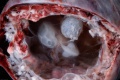
File:Stage14 bf22.jpg * Head: 1st, 2nd and 3rd pharyngeal arch, forebrain, site of lens placode, site of otic placode, stomodeum(1,500 × 1,001 (214 KB)) - 14:48, 28 February 2014
File:Stage14 bf23.jpg * Head: 1st, 2nd and 3rd pharyngeal arch, forebrain, site of lens placode, site of otic placode, stomodeum(1,000 × 667 (122 KB)) - 14:48, 28 February 2014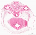
File:Stage 22 image 008.jpg ...ryo_Stages/Stage22/08/Stage22-08.html?zoom=6&lat=-1611.5&lon=1761&layers=B Lens and Cornea](1,200 × 1,161 (322 KB)) - 20:03, 11 September 2014File:Day 11 Closure of lens vesicle.JPG Day 11: Closure of the lens vesicle occurs.(560 × 528 (27 KB)) - 13:19, 18 August 2014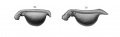
File:Keibel1910 fig28-29.jpg ...iking downward bend of the tail. The epiphysis shows in surface views. The lens is are well defined. The eye is now prominent and the lens better defined. The ear is no longer visible in(1,700 × 520 (51 KB)) - 15:03, 10 January 2015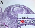
File:Mouse cornea E12.5.jpg At E12.5, ocular mesenchymal cells migrated into the space between the lens (L) and the corneal epithelium (CE, arrow in enlarged inset) in eyes.(700 × 558 (123 KB)) - 11:50, 24 January 2015
File:Stage12 bf3a.jpg Features: day 26, 27 somites, forebrain, site of lens placode, site of otic placode , stomodeum, 1st pharyngeal arch, 2nd pharyng(373 × 800 (31 KB)) - 12:29, 7 September 2009
File:Stage12 bf3b.jpg Features: day 26, 27 somites, forebrain, site of lens placode, site of otic placode , stomodeum, 1st pharyngeal arch, 2nd pharyng(280 × 600 (21 KB)) - 12:29, 7 September 2009
File:Stage12 bf3c.jpg Features: day 26, 27 somites, forebrain, site of lens placode, site of otic placode , stomodeum, 1st pharyngeal arch, 2nd pharyng(187 × 400 (11 KB)) - 12:30, 7 September 2009
File:Stage12 bf3.jpg Features: day 26, 27 somites, forebrain, site of lens placode, site of otic placode , stomodeum, 1st pharyngeal arch, 2nd pharyng(466 × 1,000 (43 KB)) - 12:29, 7 September 2009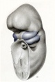
File:Streeter1922-fig09.jpg No. {{CE1787}} Carnegie Collection. X 22. The olfactory disk and the lens of the eye are outlined by dots.(667 × 1,000 (85 KB)) - 04:57, 17 August 2017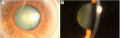
File:Images of congenital hereditary cataracts due to mutations of crystallin genes.png ..., showing cataract phenotype. B: Slit lamp view of the len of the proband. Lens opacities were mainly located in the nuclear area of lenses as well as in t(594 × 190 (118 KB)) - 11:06, 26 September 2012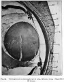
File:Brown022.jpg ...re the branches of the hyaloid artery which furnishes the nutrition to the lens during its development, and it will be remembered that this artery atrophie(625 × 800 (93 KB)) - 13:07, 30 January 2016
File:Stage12 bf4.jpg Features: day 26, 27 somites, forebrain, site of lens placode, site of otic placode , stomodeum, 1st pharyngeal arch, 2nd pharyng(933 × 1,000 (97 KB)) - 12:39, 7 September 2009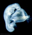
File:Stage12 bf4a.jpg Features: day 26, 27 somites, forebrain, site of lens placode, site of otic placode , stomodeum, 1st pharyngeal arch, 2nd pharyng(746 × 800 (67 KB)) - 12:39, 7 September 2009
File:Stage12 bf4b.jpg Features: day 26, 27 somites, forebrain, site of lens placode, site of otic placode , stomodeum, 1st pharyngeal arch, 2nd pharyng(560 × 600 (41 KB)) - 12:39, 7 September 2009
File:Stage12 bf4c.jpg Features: day 26, 27 somites, forebrain, site of lens placode, site of otic placode , stomodeum, 1st pharyngeal arch, 2nd pharyng(373 × 400 (10 KB)) - 12:40, 7 September 2009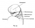
File:Kollmann344.jpg ...rd. The place where the eye emerges later is identified as a small bump. A lens plate has not yet been created.(639 × 516 (31 KB)) - 14:40, 17 October 2011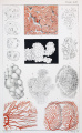
File:Hassall1849 plate41.jpg Fig. 2. Vesicles of slightly enlarged thyroid, viewed with a lens only.(1,280 × 2,075 (857 KB)) - 11:38, 22 January 2019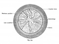
File:Kollmann719.jpg ...moved. From all over the area to push the vessel back to the center of the lens lateral pole which is itself free of the vessels is a sign of incipient rec(775 × 579 (87 KB)) - 17:28, 18 August 2012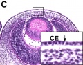
File:Mouse cornea E13.5.jpg * At E12.5, ocular mesenchymal cells migrated into the space between the lens (L) and the corneal epithelium (CE, arrow in enlarged inset) in eyes.(700 × 550 (103 KB)) - 11:50, 24 January 2015
File:Stage13 bf4.jpg * Head: 1st, 2nd and 3rd pharyngeal arch, forebrain, site of lens placode, site of otic placode, stomodeum(1,200 × 800 (132 KB)) - 16:38, 11 September 2014
File:Stage14 bf18.jpg * Head: 1st, 2nd and 3rd pharyngeal arch, forebrain, site of lens placode, site of otic placode, stomodeum(1,800 × 2,250 (549 KB)) - 17:28, 11 September 2014
File:Stage19 bf17.jpg * Ectoderm: sensory placodes, lens pit, otocyst, nasal pits moved ventrally, fourth ventricle of brain(1,041 × 1,000 (68 KB)) - 10:53, 3 February 2014
File:Lewis1903 plate01.jpg ...Gf. 5, Gasserian ganglion. H, Cerebral hemisphere. ;, Jugular ganglion. L, Lens. M. b, Mid-brain. Mdb., Mandibular process. Md. ob, Medulla oblongata. Mic,(1,280 × 1,639 (328 KB)) - 13:00, 2 August 2019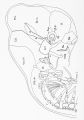
File:Lewis1903 plate01L.jpg ...Gf. 5, Gasserian ganglion. H, Cerebral hemisphere. ;, Jugular ganglion. L, Lens. M. b, Mid-brain. Mdb., Mandibular process. Md. ob, Medulla oblongata. Mic,(1,280 × 1,822 (246 KB)) - 13:24, 2 August 2019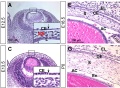
File:Mouse cornea development 01.jpg * At E12.5, ocular mesenchymal cells migrated into the space between the lens (L) and the corneal epithelium (CE, arrow in enlarged inset) in eyes.(1,200 × 880 (325 KB)) - 11:48, 24 January 2015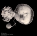
File:Stage19 bf16.jpg * Ectoderm: sensory placodes, lens pit, otocyst, nasal pits moved ventrally, fourth ventricle of brain(1,041 × 1,000 (99 KB)) - 10:52, 3 February 2014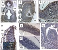
File:Mouse head TFII-I expression 01.jpg ...nstrates TFII-I expression in the inner and outer retinal layer as well as lens primordium. ...Future Cerebral Cortex, IL-Inner Retinal Layer, IR-Intra-Retinal Space, LP-Lens Primordium, M-Mesenchyme, NE-Neuroepithelium, NL-Neural Lumen, NT-Neural Tu(1,000 × 862 (298 KB)) - 12:09, 8 July 2011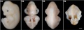
File:Bat - Miniopterus schreibersii fuliginosus Stage 17.jpg * lv, lens vesicle(1,200 × 427 (50 KB)) - 14:56, 4 July 2012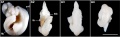
File:Bat - Miniopterus schreibersii fuliginosus Stage 13.jpg * lv, lens vesicle(1,200 × 367 (47 KB)) - 14:57, 4 July 2012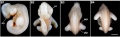
File:Bat - Miniopterus schreibersii fuliginosus Stage 14.jpg * lv, lens vesicle(1,200 × 367 (51 KB)) - 14:57, 4 July 2012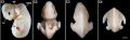
File:Bat - Miniopterus schreibersii fuliginosus Stage 15.jpg * lv, lens vesicle(1,200 × 367 (48 KB)) - 14:57, 4 July 2012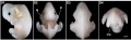
File:Bat - Miniopterus schreibersii fuliginosus Stage 16.jpg * lv, lens vesicle(1,200 × 367 (47 KB)) - 14:57, 4 July 2012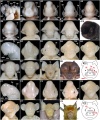
File:Bat-craniofacial development.jpg ...ga, glossopharyngeal arch; ha, hyoid arch; hr, hair; lp, lens placode; lv, lens vesicle; md, mandible; mx, maxilla; ma, mandibular arch; mnl, main nose-lea(600 × 720 (140 KB)) - 15:26, 25 June 2015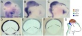
File:Chicken brain gene expression.jpg ...chematic summary of the expression patterns. di: diencephalic vesicle; le: lens; mes: mesencephalic vesicle; met: metencephalic vesicle; rt: retina. The ar(1,200 × 530 (138 KB)) - 13:33, 7 August 2012
File:Wen1928-Fig10.jpg ...en the number of crest cells in each section was counted with an immersion lens and plotted in appropriate position. The stippled area accordingly indicate(495 × 1,200 (108 KB)) - 15:34, 21 April 2016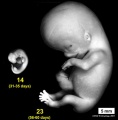
File:Stage14compare23.jpg * Features: midbrain, nasal placode, lens pit, 1,2,3 pharyngeal arches, fourth ventricle of brain, 1st pharyngeal gro(593 × 601 (32 KB)) - 00:12, 11 December 2013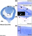
File:Mouse eye E18.jpg ...ller size with no anterior chamber and an infiltration of cells behind the lens in Tgfbr2-mutant embryos as compared with control embryos. Boxes indicate m(884 × 977 (169 KB)) - 15:44, 30 August 2014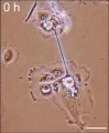
File:Bronchial epithelial bridge.jpg ...frame was taken using a Nikon Diaphot inverted microscope with a 10x phase lens.(397 × 482 (28 KB)) - 12:32, 14 April 2011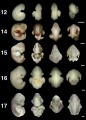
File:Bat embryo stage 12 to 17.jpg Hand plate and footplate form; lens vesicle; auditory hillocks; premaxillary centers.(800 × 1,120 (91 KB)) - 12:22, 3 July 2012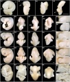
File:Bat-embryonic stages 11-22.jpg ...exure; cvf, cervical flexure; eam, external auditory meatus; h, heart; lv, lens vesicle; np, nasal pit; plp, plagiopatagium; pr, pigmented retina; prp, pro(600 × 703 (108 KB)) - 12:42, 3 July 2012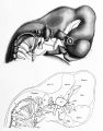
File:Johnston1907 fig020.jpg ...misphere. Ven. IV., roof of the {{fourth ventricle}}. Op., optic cup. L, {{lens}}. Na., nasal pit. Ot., otocyst.(1,280 × 1,632 (277 KB)) - 07:58, 24 February 2020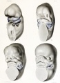
File:Streeter1922-plate01.jpg No. 1787 Carnegie Collection. X 22. The olfactory disk and the lens of the eye are outlined by dots.(871 × 1,200 (174 KB)) - 04:54, 17 August 2017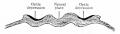
File:Bailey456.jpg ...entral nervous system fiber tracts. Of the accessory optic structures, the lens, the epithelium of the lids and conjunctiva, the eyelashes, the Meibomian g(591 × 168 (20 KB)) - 06:33, 31 August 2011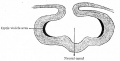
File:Bailey457.jpg ...entral nervous system fiber tracts. Of the accessory optic structures, the lens, the epithelium of the lids and conjunctiva, the eyelashes, the Meibomian g(659 × 335 (40 KB)) - 06:33, 31 August 2011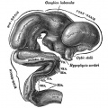
File:Gray0652.jpg ...ts peripheral extremity is expanded into a vesicle, in which a rudimentary lens and retina are formed; the stalk becomes solid and nerve fibers make their(698 × 700 (103 KB)) - 14:14, 26 May 2014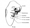
File:Kollmann349.jpg ...pendages are well developed, partly hidden behind the eye, the through the lens ectoderm is visible through the wide slit-like mouth opening is above the h(709 × 657 (51 KB)) - 15:13, 17 October 2011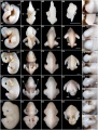
File:Bat-Miniopterus schreibersii fuliginosus Stages 13-17.jpg * lv, lens vesicle(600 × 790 (120 KB)) - 12:55, 3 July 2012
File:Gray0654.jpg ...ts peripheral extremity is expanded into a vesicle, in which a rudimentary lens and retina are formed; the stalk becomes solid and nerve fibers make their(402 × 500 (39 KB)) - 14:16, 26 May 2014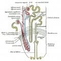
File:Gray1128.jpg ...vis of the kidney. If the surface of one of the papillæ be examined with a lens, it will be seen to be studded over with minute openings, the orifices of t(700 × 698 (114 KB)) - 09:55, 4 June 2013
File:Braune 1877 plate 3.jpg ...though the external form of the globe be established, the relations of the lens and iris must be rendered after further sections. The fine dust which even(1,012 × 1,200 (242 KB)) - 09:14, 10 November 2012
File:Gray0653.jpg ...ts peripheral extremity is expanded into a vesicle, in which a rudimentary lens and retina are formed; the stalk becomes solid and nerve fibers make their(698 × 700 (108 KB)) - 16:40, 26 May 2014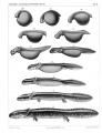
File:Keibel1910 plate03.jpg ...iking downward bend of the tail. The epiphysis shows in surface views. The lens is are well defined. The eye is now prominent and the lens better defined. The ear is no longer visible in(1,891 × 2,476 (425 KB)) - 15:07, 10 January 2015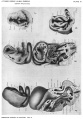
File:Gage1905-plate03.jpg ...pore; the olfactory region and its extension over the cerebrum; the future lens; the mandible and gill-cleft-like pocket at the corner of the mouth.(1,042 × 1,500 (238 KB)) - 14:33, 18 August 2016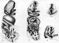
File:Gage1905-plate3.jpg ...pore; the olfactory region and its extension over the cerebrum; the future lens; the mandible and gill-cleft-like pocket at the corner of the mouth.(2,048 × 1,500 (392 KB)) - 14:33, 18 August 2016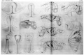
File:Locy1895 plate27.jpg ...ng the rudiments of the seventh, eighth ninth, and tenth next-es. Note the lens and choroid Hssure in the eye vesic1e.(4,342 × 2,850 (477 KB)) - 15:46, 27 August 2018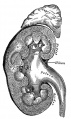
File:Gray1127.jpg ...indicated between A and A’ in Fig. 1127). If the cortex be examined with a lens, it will be seen to consist of a series of lighter-colored, conical areas,(417 × 700 (108 KB)) - 23:14, 17 September 2012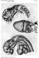
File:Gage1905-plate02.jpg ...ending also over the cerebral region; the large neuroporic thickening; the lens epithelium with a tract extending along the lachrymal furrow.(1,007 × 1,500 (221 KB)) - 14:32, 18 August 2016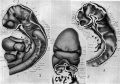
File:Gage1905-plate2.jpg ...ending also over the cerebral region; the large neuroporic thickening; the lens epithelium with a tract extending along the lachrymal furrow.(2,149 × 1,500 (392 KB)) - 14:33, 18 August 2016
File:Marcello Malpighi.jpg ...ll if yoii examine the turgid lung of a frog with a microscope of a single lens against the horizontal sun." ...optic vesicles with their stalk, the cleft in the vesicles, and later the lens formation are traced out. The twisting of the body and the appearance and d(652 × 869 (418 KB)) - 13:49, 20 February 2020


