Search results
From Embryology
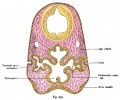
File:Kollmann697.jpg ...h increases the glass body and later allowed to develop. Law is made the lens pouch only in passing, left through the middle.(731 × 612 (94 KB)) - 19:34, 22 October 2012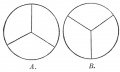
File:Gray0885.jpg ==Lens== ...and arrangement of the radiating lines on the front and back of the fetal lens.(667 × 400 (20 KB)) - 22:29, 19 August 2012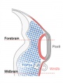
File:Lens-neural crest signaling 01.jpg ==Wnt mediates lens repression by neural crest cells and Transforming growth factor-β== # NCCs (blue) secrete TGF-βs, which signal to the non-lens ectoderm and dorsal optic vesicle.(300 × 400 (24 KB)) - 17:25, 31 August 2011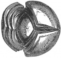
File:Gray0884.jpg ==Lens Structure== The crystalline lens, hardened and divided. (Enlarged.)(419 × 400 (58 KB)) - 15:12, 19 August 2012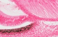
File:Stage 22 image 211.jpg ==Developing Lens and Iris - Human Embryo Carnegie stage 22== * lens(1,200 × 760 (242 KB)) - 11:41, 14 June 2016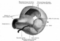
File:Bailey464.jpg ==Fig. 464. Model showing lens and formation of optic cup== ...oted the fact that typical optic cup formation may occur in cases where no lens is developed. The optic cup when first formed is not a complete cup, for th(869 × 592 (67 KB)) - 06:44, 31 August 2011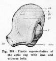
File:Hertwig265.jpg ==Fig. 265. Plastic representation of the optic cup with lens and vitreous body== ...its lower surface.) aus, Optic [choroid] fissure ; yl, vitreous body ; I, lens.(460 × 500 (36 KB)) - 21:49, 28 March 2012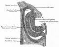
File:Bailey466.jpg ==Fig. 466. Section through optic cup and lens invagination of chick of fifty-four hours' incubation== Between the lens anlage and the pigmented layer of the retina is the broad inner layer of th(859 × 683 (139 KB)) - 15:54, 1 February 2011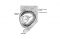
File:Streeter1957 fig06-19.jpg ...reous humor of the eye and forms a network on the posterior surface of the lens. X60.(1,280 × 834 (120 KB)) - 08:03, 18 April 2018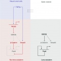
File:Lens-neural crest signaling 02.jpg ==Wnt mediates lens repression by neural crest cells and Transforming growth factor-β== Proposed molecular model to explain TGF-β- and Wnt-mediated lens restriction. Broken lines: interactions inferred from the literature.(521 × 522 (22 KB)) - 14:10, 29 April 2011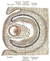
File:Gray0867.jpg ...enters the cup through the choroidal fissure and around the equator of the lens becomes intimately united with this reticular tissue, and contributes to fo(495 × 600 (131 KB)) - 08:59, 19 August 2012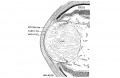
File:Streeter1957 fig06-23.jpg ...reous humor of the eye and forms a network on the posterior surface of the lens. X60. [[Category:Week 8]][[Category:Vision]][[Category:Lens]][[Category:Carnegie Embryo 4570]](1,280 × 834 (161 KB)) - 10:27, 9 May 2018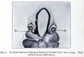
File:Brown006.jpg ...into the secondary optic vesicle (see A, Fig. 7), and eventually forms the lens, which will be described later. As the lens vesicle passes into the secondary optic vesicle, some of the mesoblastic ce(800 × 543 (63 KB)) - 07:12, 31 August 2011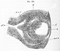
File:Foster128.jpg # the absence at this stage of mesoblast between the lens and the epiblast ; the interval between the two has however been made too g # the arteria centralis retinae forming the vascular capsule of the lens and continuous with vascular structures round the edges of the optic cup.(998 × 859 (223 KB)) - 07:05, 17 March 2012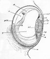
File:Foster051.jpg ...e elongated cells are shewn at ril, now forms nearly the whole mass of the lens, the front wall being reduced to a layer of flattened cells el. ...with the mesoblast m, and appears to be the rudiment of the capsule of the lens and suspensory ligament.(722 × 847 (112 KB)) - 10:24, 11 January 2011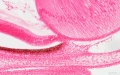
File:Stage 22 image 212.jpg ==Developing Lens and Iris - Human Embryo Carnegie stage 22== * lens(1,200 × 753 (269 KB)) - 11:41, 14 June 2016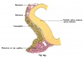
File:Kollmann695.jpg ==Figure 695 The lens unit with a rabbit embryo== ...of capillaries. In the whole extent of the lateral surface considered, the lens plate has a recess. It therefore describes it as an Linsengrübchen (foveol(734 × 519 (45 KB)) - 10:53, 18 May 2014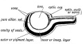
File:Keith1902 fig146.jpg ==Fig. 146. Diagrammatic Section of the Optic Cup and Lens==(900 × 480 (68 KB)) - 09:22, 8 January 2014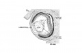
File:Streeter1957 fig06-20.jpg ...reous humor of the eye and forms a network on the posterior surface of the lens. X60.(1,280 × 834 (133 KB)) - 21:14, 15 March 2017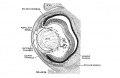
File:Streeter1957 fig06-22.jpg ...reous humor of the eye and forms a network on the posterior surface of the lens. X60.(1,280 × 834 (181 KB)) - 21:13, 15 March 2017


