Search results
From Embryology
Page title matches
File:Fetus week 9-10-icon.jpg (220 × 147 (12 KB)) - 12:57, 24 April 2014
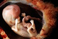
File:Human week 10 fetus 01.jpg ==Human Female Fetus (10 week)== {{Human Female Fetus Week 10 gallery}}(2,300 × 1,327 (448 KB)) - 11:57, 30 May 2016
File:Human week 10 fetus 12.jpg ==Human Female Fetus - Olfactory Nerve (10 week)== {{Human Female Fetus Week 10 gallery}}(1,200 × 900 (349 KB)) - 14:38, 25 May 2016
File:Human week 10 fetus 02.jpg ==Human Female Fetus (10 week)== See also [[:File:Human week 10 fetus 01.jpg|'''Large Image Version''']](800 × 462 (83 KB)) - 14:38, 11 October 2015
File:Human week 10 fetus 03.jpg ==Human Female Fetus - Pelvic Region (10 week)== {{Human Female Fetus Week 10 gallery}}(1,600 × 1,200 (370 KB)) - 17:21, 25 May 2016
File:Human week 10 fetus 04.jpg ==Human Female Fetus - Oral Cavity (10 week)== {{Human Female Fetus Week 10 gallery}}(1,600 × 1,200 (534 KB)) - 15:48, 25 May 2016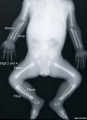
File:Human fetus skeleton x-ray 01.jpg ==Human Fetus Skeleton X-ray== {{Human fetus skeleton x-ray links}}(662 × 910 (54 KB)) - 17:05, 22 April 2014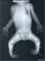
File:Human fetus skeleton x-ray 02.jpg ==Human Fetus Skeleton X-ray== Fetus with limb deformities including bowing of the digits, bowing and hypoplasia(686 × 928 (70 KB)) - 17:05, 22 April 2014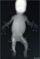
File:Human fetus skeleton x-ray 03.jpg ==Human Fetus Skeleton X-ray== Fetus with limb deformities (indicated by arrows), which include bowing of the ra(628 × 934 (81 KB)) - 17:04, 22 April 2014
File:2015BGDA Lecture - Development of the Embryo-Fetus 1.pdf ==BGDA Lecture - Development of the Embryo-Fetus 1== [[BGDA Lecture - Development of the Embryo/Fetus 1|Lecture Page]](2.65 MB) - 11:15, 7 May 2015File:2017 BGDA Lecture - Development of the Embryo-Fetus 1.pdf (3.39 MB) - 16:38, 3 May 2017File:2018 BGDA Lecture - Development of the Embryo-Fetus 1.pdf (8.38 MB) - 08:08, 4 May 2018File:2017 BGDA Lecture - Development of the Embryo-Fetus 2.pdf PDF version of 2017 lecture page: [[BGDA Lecture - Development of the Embryo/Fetus 2]](3.23 MB) - 08:44, 15 May 2017File:2018 BGDA Lecture - Development of the Embryo-Fetus 2.pdf [[BGDA Lecture - Development of the Embryo/Fetus 2]](5.67 MB) - 21:58, 13 May 2018File:2019 BGDA Lecture - Development of the Embryo-Fetus 1.pdf ==BGDA Lecture - Development of the Embryo/Fetus 1== [[BGDA Lecture - Development of the Embryo/Fetus 1]](1.93 MB) - 22:19, 2 May 2019File:BGDA Lecture 2014 - Development of the Embryo-Fetus 1.pdf ==BGDA Lecture - Development of the Embryo/Fetus 1==(869 KB) - 14:24, 7 May 2014File:2019 BGDA Lecture - Development of the Embryo-Fetus 2.pdf (3.59 MB) - 09:35, 13 May 2019


Page text matches
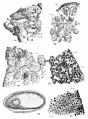
File:Simkins1928 plate09.jpg 38 Peripheral portion of an ovary from a seven-month fetus. X 750. 39 Peripheral portion of an ovary from a full-term fetus. cl.g., genitaloid cell. X 7 50.(1,548 × 2,096 (263 KB)) - 16:58, 31 January 2018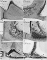
File:Anson1948 fig01.jpg (a) Base at posterior crus, left ear; 167 mm. fetus (Wisconsin series 105, slide 19, section 9). (b) Base and anterior crus, left ear; 175 mm. fetus (Wisconsin series 104, slide 17, section 5).(1,280 × 1,635 (310 KB)) - 17:02, 13 October 2017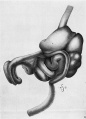
File:Bardeen1914-fig06.jpg ==Fig. 6. Lateral view fetus 40 mm== ...he stomach, small and large intestines, bursa omentalis and mesentery of a fetus 40 mm. long (No. 8, Wisconsin Collection).(721 × 1,000 (88 KB)) - 09:52, 3 October 2017File:2019 BGDA Lecture - Development of the Embryo-Fetus 1.pdf ==BGDA Lecture - Development of the Embryo/Fetus 1== [[BGDA Lecture - Development of the Embryo/Fetus 1]](1.93 MB) - 22:19, 2 May 2019File:2015BGDA Lecture - Development of the Embryo-Fetus 1.pdf ==BGDA Lecture - Development of the Embryo-Fetus 1== [[BGDA Lecture - Development of the Embryo/Fetus 1|Lecture Page]](2.65 MB) - 11:15, 7 May 2015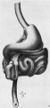
File:Bardeen1914-fig05.jpg ==Fig. 5. Ventral view fetus 40 mm== ...he stomach, small and large intestines, bursa omentalis and mesentery of a fetus 40 mm. long (No. 8, Wisconsin Collection). In [[:File:Bardeen1914-fig06.jpg(465 × 1,000 (61 KB)) - 07:05, 15 November 2015
File:Wallaby embryo 05.jpg ==Tammar Wallaby Embryo and Fetus== ...ld within the folds of the yolk sac, which has become highly vascular. The fetus has well developed fore limbs ready for the climb to the pouch and the tong(996 × 1,100 (127 KB)) - 11:06, 29 July 2019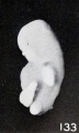
File:Mall Meyer1921 fig133.jpg ==Fig. 133. Marked macerated and deformed fetus from the same case== Fig. 133. Marked macerated and deformed fetus from the same case. X4.(365 × 610 (40 KB)) - 09:12, 6 December 2012
File:Mall Meyer1921 plate19.jpg Fig. 218. A fetus and cord showing marked maceration changes. No. 797. X1.35. Fig. 219. An older fetus, showing bleb-formation and curvature in extremities, due to maceration and(939 × 1,200 (237 KB)) - 18:44, 23 November 2012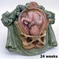
File:Galletti1770 week 24.jpg Fetus 24 weeks(450 × 450 (40 KB)) - 15:12, 13 October 2009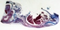
File:B050966-01.jpg ==Female Human Fetus 14cm==(2,000 × 992 (681 KB)) - 11:22, 25 June 2015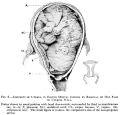
File:Frazer006 bw600.jpg ==Fetus during Third Trimester in Uterus compared to Non-pregnant Uterus== [[Category:Historic Embryology]] [[Category:Human Fetus]] [[Category:Third Trimester]] [[Category:Cartoon]](600 × 575 (47 KB)) - 17:30, 1 June 2013File:Human fetus skeleton x-ray 02.jpg ==Human Fetus Skeleton X-ray== Fetus with limb deformities including bowing of the digits, bowing and hypoplasia(686 × 928 (70 KB)) - 17:05, 22 April 2014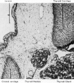
File:Boyd1950 fig09.jpg ==Fig. 9. Section fetus 145 mm to show aberrant portion of thyroid tissue lying between thyroid and {{Online Editor}} - [[Fetal Development|Fetus]] 145mm is about 16 weeks ({{GA}} 18 weeks).(801 × 900 (163 KB)) - 08:29, 21 March 2017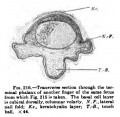
File:Keibel Mall 216.jpg ==Fig. 216 Human Fetus Finger== Transverse section through the terminal phalanx of another finger of the same fetus from which [[:File:Keibel Mall 215.jpg|Fig. 215]] is taken. The basal cell(366 × 356 (30 KB)) - 12:43, 13 January 2019File:Human week 10 fetus 01.jpg ==Human Female Fetus (10 week)== {{Human Female Fetus Week 10 gallery}}(2,300 × 1,327 (448 KB)) - 11:57, 30 May 2016File:Human fetus skeleton x-ray 03.jpg ==Human Fetus Skeleton X-ray== Fetus with limb deformities (indicated by arrows), which include bowing of the ra(628 × 934 (81 KB)) - 17:04, 22 April 2014
File:Mall Meyer1921 plate14.jpg Fig. 145. Enlarged fetus from same, showing maceration effects. X2. Fig. 150. Macerated, distorted normal cat fetus 10 mm. long. (After Kunz.)(941 × 1,200 (260 KB)) - 19:21, 23 November 2012
File:Gray0179.jpg ...outer aspect 95mm fetus]] | [[:File:Gray0181.jpg|Image - inner aspect 95mm fetus]](617 × 368 (47 KB)) - 18:34, 27 August 2012
File:Lineback1920 fig04-5.jpg ==Cross-section of the colon of a human fetus== Fig. 4. Cross-section of the colon of a human fetus 52 mm. CR. length.(1,200 × 756 (150 KB)) - 09:16, 17 January 2013


