Search results
From Embryology
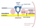
File:Mesoderm-cartoon4.jpg ==Mesoderm and Ectoderm Development cartoon== ...s a section through the trunk of the trilaminar embryo showing the further development of the 3 layers and the space (coelom) that forms in the mesoderm (only the(400 × 300 (20 KB)) - 10:09, 23 April 2020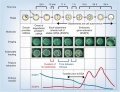
File:Model human blastocyst development.jpg ==Proposed Model for Human Embryo Development== ...gg (ESSP1) must be degraded as the transition from oocyte to embryo begins embryonic genome activation (EGA).(946 × 726 (84 KB)) - 14:47, 6 October 2015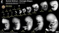
File:Human Carnegie stage 1-23.jpg ==Human Embryonic Development== ..., showing human external appearance and growth during the first 8 weeks of development.(1,000 × 563 (98 KB)) - 11:32, 14 May 2017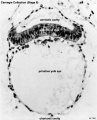
File:Stage6 bf03.jpg ...howing development of the bilaminar (two layer) embryo and the early extra-embryonic coeloms (spaces). Extra-embryonic coelom(646 × 800 (65 KB)) - 14:48, 1 October 2018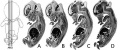
File:Human- fetal week 10 sagittal planes.jpg ...he embryonic period (up to week 8) but still only 2 weeks into early fetal development.(600 × 250 (29 KB)) - 15:39, 27 April 2010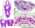
File:Stage13 and 22 thyroid development a.jpg ==Thyroid Gland Embryonic Development== ...e developing thyroid at the beginning of week 5 and at week 8 of embryonic development.(800 × 640 (94 KB)) - 08:50, 22 May 2012
File:Lung development overiview.png Summary of the lung formation from the embryonic stage to the fetal stage. Original Image is adapted from: Figure 1 from Embryonic Development of the Respiratory System page(717 × 549 (178 KB)) - 14:59, 9 November 2014
File:Bat embryo stage 12-17.jpg ==Bat Embryonic Development (stage 12-17)== :'''Links:''' [[Bat Development]] | [[:File:Bat embryo stage 10-13.jpg|stage 10 to 13]] | [[:File:Bat embry(548 × 767 (30 KB)) - 01:16, 29 April 2011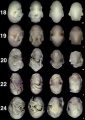
File:Bat embryo stage 18-24.jpg ==Bat Embryonic Development (stage 18-24)== :'''Links:''' [[Bat Development]] | [[:File:Bat embryo stage 10-13.jpg|stage 10 to 13]] | [[:File:Bat embry(518 × 734 (32 KB)) - 01:16, 29 April 2011
File:Thymic Epithelial Cell Development and Function.png ==Thymic Epithelial Cell Development and Function== '''Image shows Thymic Epithelial Cell Development and Function in fetal period'''(2,028 × 823 (3.17 MB)) - 11:37, 9 November 2014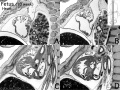
File:Human- fetal week 10 heart ABCD.jpg ...he embryonic period (up to week 8) but still only 2 weeks into early fetal development.(600 × 450 (133 KB)) - 16:10, 27 April 2010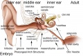
File:Adult hearing embryonic origins.jpg ==Adult Hearing Embryonic Origins== ...nd inner), each of which have their own separate components from different embryonic origins.(1,000 × 675 (80 KB)) - 14:26, 8 May 2018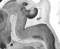
File:Human- fetal week 10 cerebellum C.jpg ...he embryonic period (up to week 8) but still only 2 weeks into early fetal development.(347 × 284 (25 KB)) - 18:53, 20 September 2012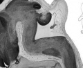
File:Human- fetal week 10 cerebellum D.jpg ...he embryonic period (up to week 8) but still only 2 weeks into early fetal development.(347 × 284 (23 KB)) - 18:53, 20 September 2012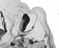
File:Human- fetal week 10 cerebellum B.jpg ...he embryonic period (up to week 8) but still only 2 weeks into early fetal development.(347 × 284 (21 KB)) - 18:53, 20 September 2012
File:BGDA PracManual 2011 Practical 7.pdf ==BGDA Practical Manual 2011 Practical 7 - Embryonic Development==(203 KB) - 14:43, 22 April 2011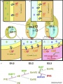
File:Mouse distal visceral endoderm 01.jpg ...MP signaling promotes the differentiation of primitive endoderm (PrE) into embryonic visceral endoderm (VE) until E4.5. (B) Embryonic visceral endoderm (VE) differentiates as a result of the concerted action o(959 × 1,280 (208 KB)) - 13:16, 3 May 2013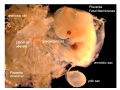
File:Placental membranes.jpg * '''amniotic sac''' - formed by the amniotic membrane (ectoderm and extra-embryonic mesoderm) completely surrounding the surrounding the embryo. * '''yolk sac''' - the yolk membrane (endoderm and extra-embryonic mesoderm) attached to the embryo at the umbilicus and continuous with the m(600 × 450 (99 KB)) - 07:46, 15 May 2014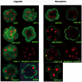
File:Antibody immunostaining of ligands and receptors in triploid human embryos.png ...mbryonic nuclei. Furthermore, growth factors were used to aid in embryonic development and promote blastocyst growth as well as IgG being applied to the negative ..., Shu Y, Cheng Y, Qiao J, et al. (2012) Promotion of Human Early Embryonic Development and Blastocyst Outgrowth In Vitro Using Autocrine/Paracrine Growth Factors.(3,439 × 3,444 (7.13 MB)) - 00:21, 20 August 2014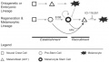
File:Zebrafish melanocyte development model.jpg ==Model for the parallel establishment of the zebrafish embryonic melanocyte lineage and the adult melanocyte stem cell lineage== ...and melanocytes during metamorphosis or regeneration, when inhibition from embryonic melanocytes (block arrow) is relieved. The MSC–derived melanocytes are se(484 × 277 (40 KB)) - 14:50, 20 February 2011


