Search results
From Embryology
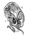
File:Mall1906-fig06.jpg (No. 12 of my collection). X 50. 03 third occipital myotome; coe, coelom; v, vein; st, septum transversum; l, liver; ph, pharynx; uv, umbilical vesi(600 × 716 (111 KB)) - 18:55, 20 September 2015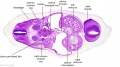
File:Stage 13 image 068.jpg ...the most cranial extension. This attachment now divides the intraembryonic coelom around the trachea into two canals, the L and R pleuro (pericardio-peritone ** Canals are lined by coelomic mesothelium and are continuous with whole I-E coelom (referred to hereafter simply as coelomic canals).(1,000 × 557 (97 KB)) - 10:00, 9 June 2011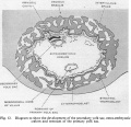
File:Hamilton1949 fig12.jpg ...Diagram to show the development of the secondary yolk sac, extra-embryonic coelom and remnant of the primary yolk sac==(1,312 × 1,265 (267 KB)) - 15:30, 15 May 2018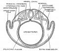
File:Keith1921 fig039.jpg ...ction across a vertebrate embryo to show the parts of the mesoderm, of the coelom==(815 × 706 (128 KB)) - 11:42, 23 December 2014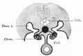
File:Bailey306.jpg * Coel - Coelom(624 × 419 (35 KB)) - 17:51, 19 September 2011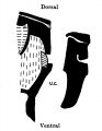
File:Wyburn1939-text-fig06.jpg ...gut. Oblique line = blood vessels. Black = mesoderm. U.C. =umbilical cord coelom.(579 × 750 (42 KB)) - 13:25, 15 September 2015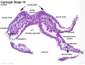
File:Stage10 K12202-04.jpg ...e 3 germ cell layers and the lateral plate divided into the intraembryonic coelom and associated mesoderm developmental regions. See [[:File:Stage10 K12202-0(1,434 × 1,105 (227 KB)) - 14:47, 15 August 2016
File:Stage10 K12202-03.jpg ...e 3 germ cell layers and the lateral plate divided into the intraembryonic coelom and associated mesoderm developmental regions. See [[:File:Stage10 K12202-0(1,434 × 1,105 (191 KB)) - 14:46, 15 August 2016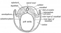
File:Keith1902 fig069.jpg ...otochord, (3) the" ingrowth of the mesoblast, and (4) the formation of the coelom.(1,000 × 548 (93 KB)) - 09:57, 7 January 2014
File:1901 Comparative development of the coelom.pdf ==Comparative Development of the Coelom==(5.49 MB) - 10:49, 20 February 2020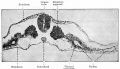
File:Bailey052.jpg The parietal mesoderm (lying above the coelom) is not labeled.(895 × 515 (99 KB)) - 23:41, 13 February 2011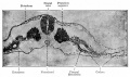
File:Bailey101.jpg The parietal mesoderm (lying above the coelom) is not labeled.(900 × 533 (110 KB)) - 12:45, 18 January 2011
File:Dandy1910-plate06.jpg ...All - Allantois Ch - Chorda; Ht - Heart; Coe - Coelom; P.C. — Pericardial coelom; Ca - Capillary, but no connection; Pl — Plexus of lateral aortic branche(1,000 × 2,166 (265 KB)) - 12:12, 28 May 2017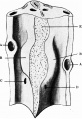
File:Waterston1915 fig04.jpg ==Fig. 4. Dorsal wall of the pericardial and upper peritoneal portions of coelom with pericardio-peritoneal passages and commencing lung buds==(633 × 919 (123 KB)) - 16:56, 24 August 2015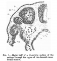
File:Low 01.jpg * Coe., coelom(502 × 535 (60 KB)) - 02:44, 29 May 2017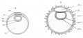
File:Minot1897 fig053.jpg Am.c, Amniotic cavity. Coe, Coelom. Ec, Ectoderm, in B, bearing the anlages of villi. Ent, Entoderm.(1,363 × 631 (94 KB)) - 17:11, 27 July 2015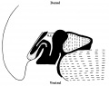
File:Wyburn1939-text-fig04.jpg ...liver. small dots = gut and allantoic diverticulum. U.C. = umbilical cord coelom. Black line indicates the plane of the umbilical veins.(1,117 × 882 (83 KB)) - 13:12, 15 September 2015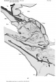
File:Bremer1914 plate01.jpg ...nections with the mesothelium. All., allantois; bl.i.. blood-island; Coe., coelom; Ect., ectoderm of chorion; f. , funnel-shaped connections, with unconnecte(684 × 1,000 (102 KB)) - 14:35, 12 August 2019
File:Odgers1937 plate02fig03.jpg ...he yolk sac surrounded on either side and ventrally by the extra-embryonic coelom.(1,000 × 692 (167 KB)) - 21:24, 29 June 2015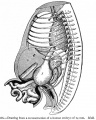
File:Bailey266.jpg The intestinal coils lie for the most part in the umbilical coelom. ...e and form secondary loops, all of which push their way into the umbilical coelom where they remain until the embryo reaches a length of 40 mm (compare [[Boo(731 × 913 (178 KB)) - 16:31, 15 April 2014


